Article

Carnosol and Related Substances Modulate Chemokine and Cytokine Production in Macrophages and Chondrocytes

DSM Nutritional Products, Wurmisweg 576, P. O. Box 2676, Basel 4002, Switzerland
*
Author to whom correspondence should be addressed.
Molecules 2016, 21(4), 465; https://doi.org/10.3390/molecules21040465
Submission received: 4 March 2016 / Revised: 5 April 2016 / Accepted: 6 April 2016 / Published: 8 April 2016
(This article belongs to the Section Natural Products Chemistry)

Abstract

:
Phenolic diterpenes present in Rosmarinus officinalis and Salvia officinalis have anti-inflammatory and chemoprotective effects. We investigated the in vitro effects of carnosol (CL), carnosic acid (CA), carnosic acid-12-methylether (CAME), 20-deoxocarnosol and abieta-8,11,13-triene-11,12,20-triol (ABTT) in murine macrophages (RAW264.7 cells) and human chondrocytes. The substances concentration-dependently reduced nitric oxide (NO) and prostaglandin E2 (PGE2) production in LPS-stimulated macrophages (i.e., acute inflammation). They significantly blunted gene expression levels of iNOS, cytokines/interleukins (IL-1α, IL-6) and chemokines including CCL5/RANTES, CXCL10/IP-10. The substances modulated the expression of catabolic and anabolic genes in chondrosarcoma cell line SW1353 and in primary human chondrocytes that were stimulated by IL-1β (i.e., chronic inflammation In SW1353, catabolic genes like MMP-13 and ADAMTS-4 that contribute to cartilage erosion were down-regulated, while expression of anabolic genes including Col2A1 and aggrecan were shifted towards pre-pathophysiological homeostasis. CL had the strongest overall effect on inflammatory mediators, as well as on macrophage and chondrocyte gene expression. Conversely, CAME mainly affected catabolic gene expression, whereas ABTT had a more selectively altered interleukin and chemokine gene exprssion. CL inhibited the IL-1β induced nuclear translocation of NF-κBp65, suggesting that it primarily regulated via the NF-κB signalling pathway. Collectively, CL had the strongest effects on inflammatory mediators and chondrocyte gene expression. The data show that the phenolic diterpenes altered activity pattern of genes that regulate acute and chronic inflammatory processes. Since the substances affected catabolic and anabolic gene expression in cartilage cells in vitro, they may beneficially act on the aetiology of osteoarthritis.

1. Introduction

Chronic inflammatory processes are implicated in the etiology of many diseases, including arthritis, atherosclerosis, diabetes and neurodegenerative disorders. Natural substances including phytochemicals can modulate a variety of these processes by interacting with receptors and signaling pathways, modulating enzyme activities or regulating gene expression via transcription factors. These phytochemicals may thereby modulate cell proliferation and differentiation and potentially influence disease development. Carnosol (CL) and structurally related diterpenes are major secondary plant metabolites in Lamiaceae spp. such as Salvia officinalis, Rosmarinus officinalis and Origanum vulgare that exhibit numerous biological features (Figure 1). Anti-oxidant properties of CL, carnosic acid (CA) or carnosic acid-12 methylether (CAME) have been demonstrated by different physicochemical methods [1,2,3,4,5,6]. CL and CA act on cell proliferation and may be therefore chemopreventive and anti-tumorigenic [4,7,8,9,10,11,12]. Anti-inflammatory effects were substantiated in various cellular systems [13,14,15,16,17,18,19] and initially shown by the inhibition of nitric oxide (NO) production [3,4]. At the molecular level, CL activates genes through anti-oxidant response elements [20,21], regulating gene expression along signaling pathways [4,22] and binding of transcription factors to promoter elements [23].
We investigated anti-inflammatory effects of CL and structurally related abietane diterpenes in macrophages and chondrocytes and thus significantly extend previous studies [3,4,24]. Most of them potently inhibited LPS-induced NO production and COX-2 dependent prostaglandin E2 (PGE2) production. Here, we demonstrate that abietane diterpenes modulated the expression level of cytokine and chemokine genes in macrophages (reflecting both acute and chronic inflammation) and chondrocytes, where pathophysiological conditions related to chronic inflammation and osteoarthritis (OA) were induced [25,26,27,28,29]. Therefore these molecules could offer novel perspectives in the treatment of diseases where inflammation and cartilage destruction are predominant feature like in OA [30].

2. Results

2.1. Anti-Inflammatory Effects Measured in Macrophages

Murine macrophage RAW264.7 cells were stimulated with lipopolysaccharide (LPS) to induce inflammatory mediators including NO and PGE2 and the inhibitory effects of CL and related abietane diterpenes were determined. CL potently reduced the production of both NO and PGE2, with IC50 of 5.0 ± 2.8 and 9.4 ± 2.1 μM, respectively (Table 1) (see also [4]). Related compounds such as CA, carnosic acid 12-methyl ether (CAME), 20-deoxocarnosol and abieta-8,11,13-triene-11,12,20 triol (ABTT) had comparable effects on NO production (except for 20-deoxocarnosol and ABTT), yet they inhibited PGE2 production less efficiently. It should be noted that the substances outperformed NG-nitro-l-arginine methyl ester (l-NAME), which is an inhibitor of iNOS-2 and thus blocks the production of NO in macrophages. None of the tested substances significantly affected cell viability at any concentrations as determined by the LDH release assay (data not shown). We also observed that CL, 20-deoxocarnosol and ABTT inhibited the in vitro enzyme activity of cyclooxygenase (COX)-1 and/or COX-2 with IC50 > 200 μM and thus at physiologically irrelevant concentrations (A.F. data not shown).

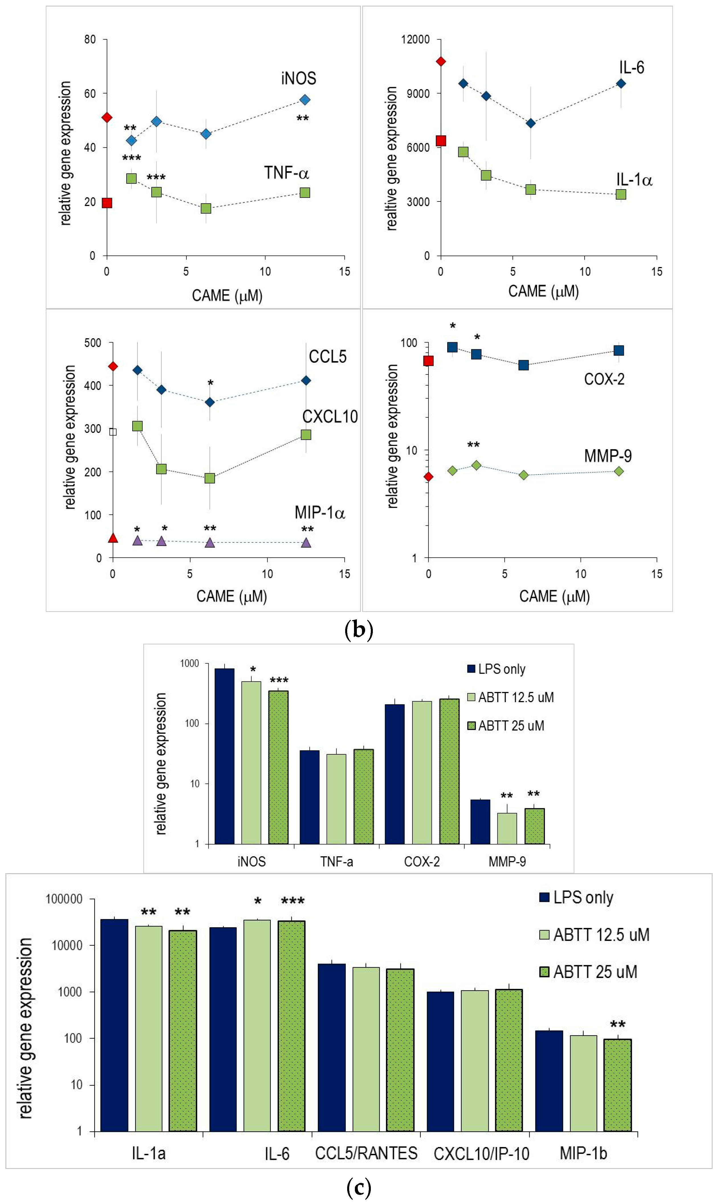
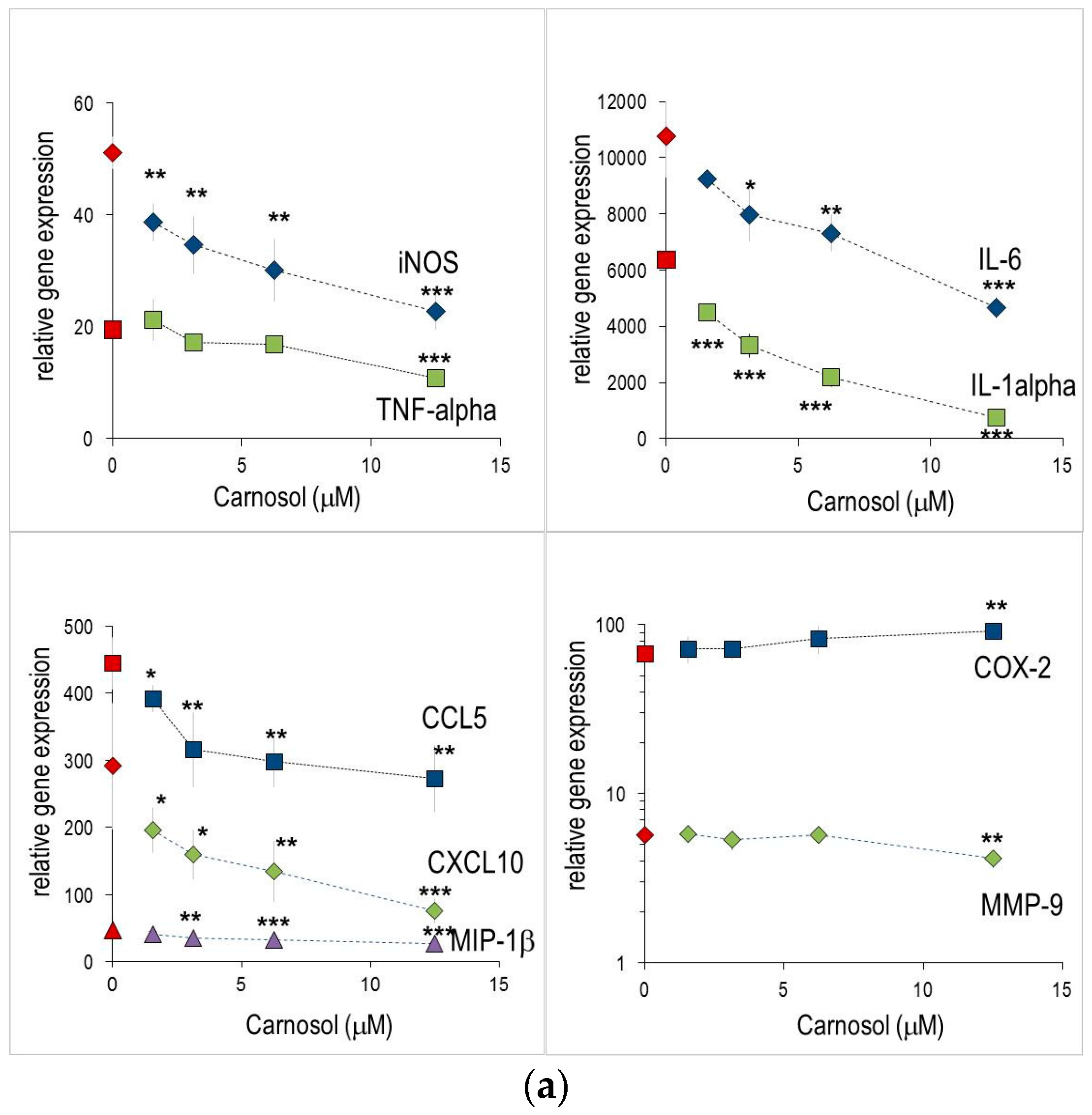
2.2. Abietane Diterpenes Modulate the Expression of Cytokine and Chemokine Genes in Macrophages

We investigated whether CL, CAME and ABTT affected the production of inflammatory mediators at the transcriptional level and thus changing the respective gene expression. To this aim, mRNA levels were measured in macrophages, which were treated for 4 h. All substances were tested at a range that encompassed the IC50 values established for NO or PGE2 production. The substances per se did not influence basal mRNA levels in unstimulated cells. LPS treatment significantly increased the expression of inflammatory genes such as COX-2, inducible nitric oxide synthase (iNOS), TNF-α, IL-1α, CCL4/MIP-1β, CCL5/RANTES and CXCL10/IP-10 (Supplementary Material Table S1). CL concentration-dependently diminished the expression levels of several genes (Figure 2a). For instance, iNOS, IL-1α, IL-6 or CXCL10/IP-10 were significantly reduced even at the lowest CL concentration tested (1.56 μM); at higher concentrations mRNA levels were reduced by up to 90% (e.g., IL-6). Conversely, CL did not alter expression level of e.g., COX-2, CCL4/MIP-1β or MMP-9 in LPS-stimulated murine macrophages. While CAME had small effects on IL-6 and IL-1α (Figure 2b), it failed to modulate expression of iNOS, TNF-α and CCL5/RANTES and CXCL10/IP-10 genes. Consistent with the observed effects on NO production, ABTT also impaired iNOS mRNA levels, but had only marginal effects on other inflammatory gene, and therefore displayed similar activities as CAME (Figure 2c). It should be noted, that CAME and ABTT increased expression of IL-6 or MMP-9. CL and CAME (at 12.5 μM) induced COX2 expression and iNOS expression, respectively.

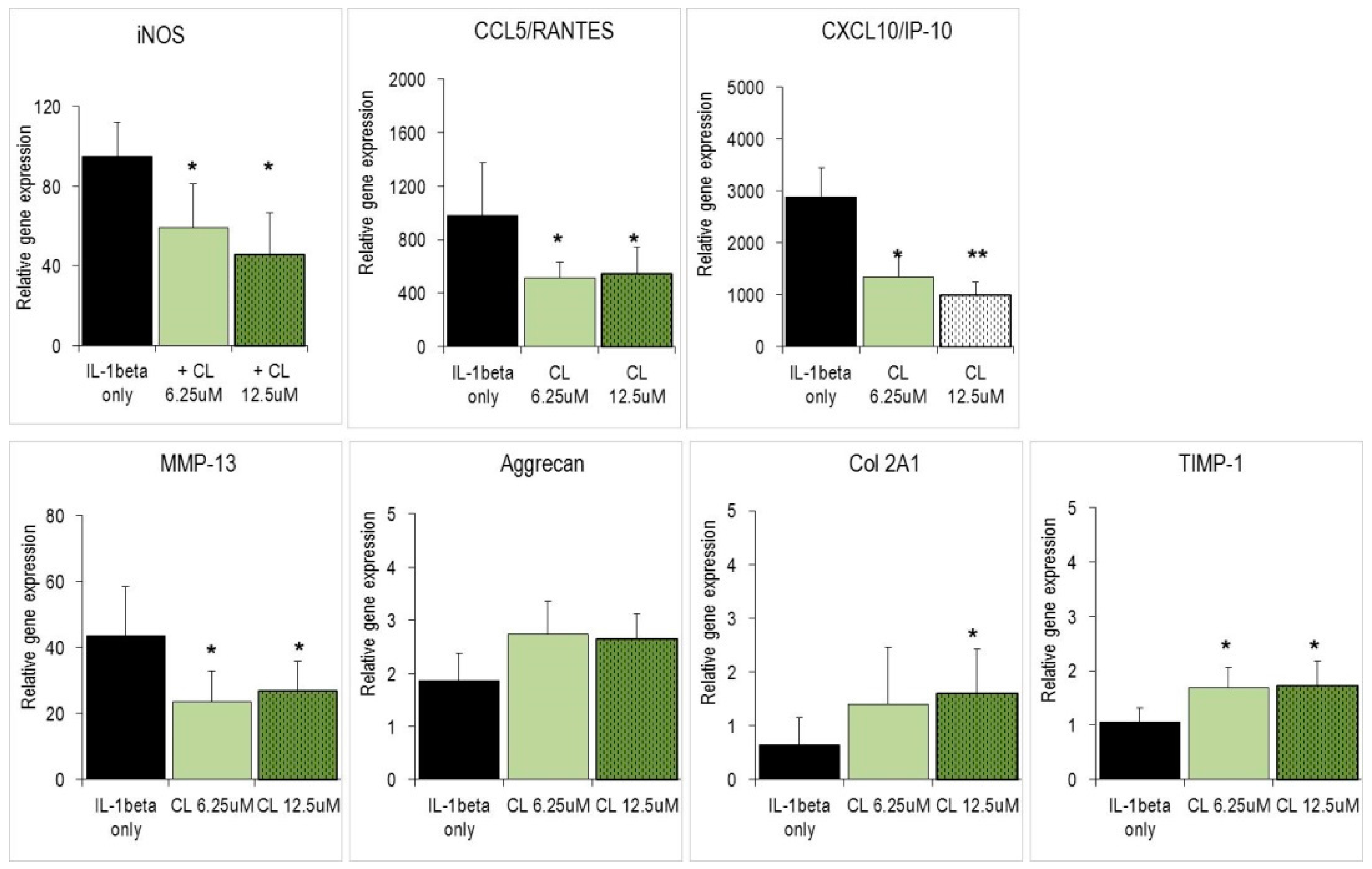
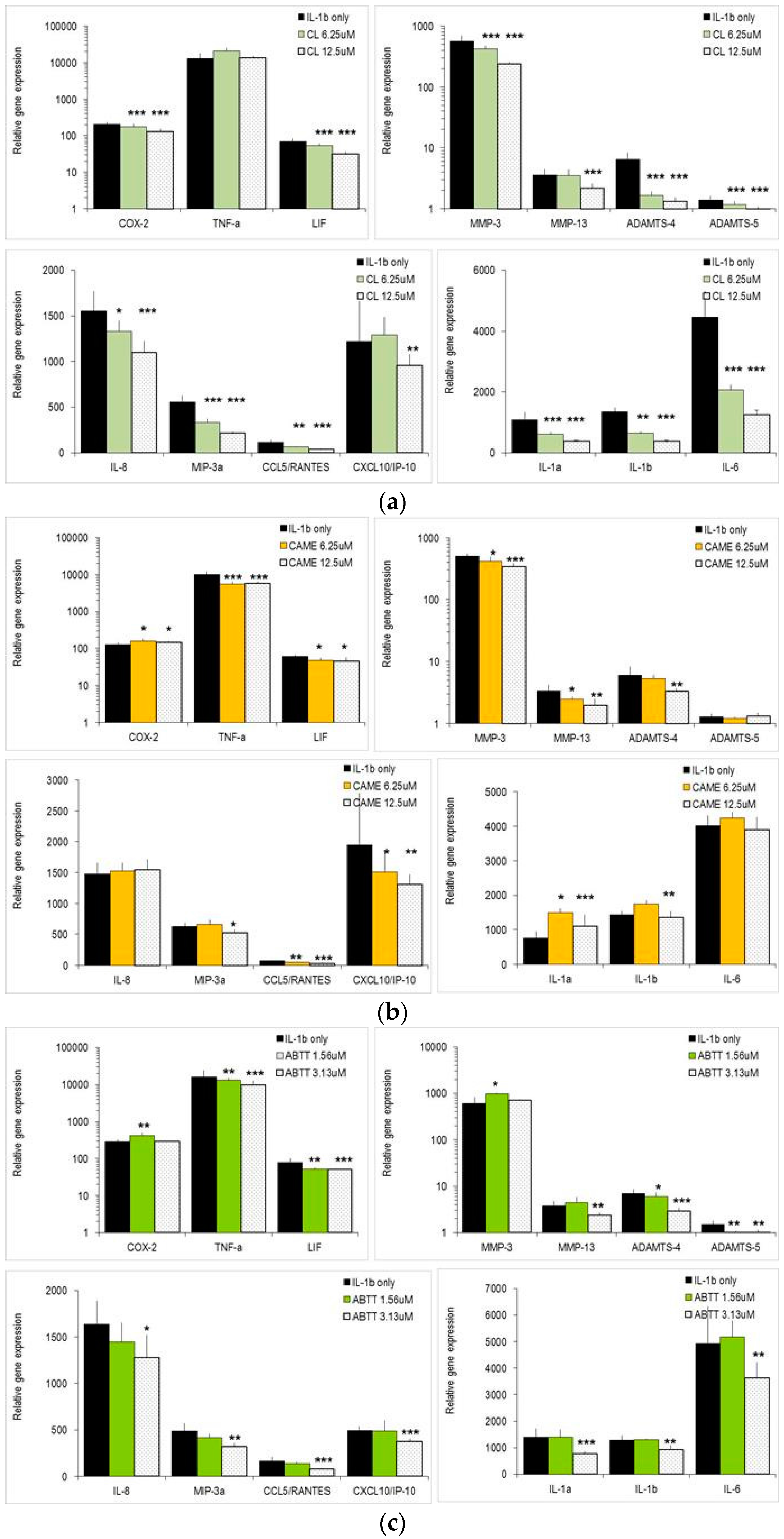
2.3. Carnosol and Related Diterpenes Impair Expression of Chemokine and Catabolic Genes in Chondrosarcoma Cells and Primary Chondrocytes

Since CL is a potent anti-inflammatory substance, it might induce changes in biological systems where chronic inflammation impairs cell and tissue homeostasis. Thus, we explored effects of CL on the chondrosarcoma SW1353 cells, which are a substitute for primary human chondrocytes and an in vitro surrogate for osteoarthritic tissue. SW1353 cells expressed a comparable although not identical set of genes as primary chondrocytes when they were activated with IL-1β [25,28,29]. Specifically, activated SW1353 cells strongly augmented gene expression levels of catabolic genes, i.e., matrix metalloproteinase (MMP)-3 and MMP-13, which are critically involved in erosion of the extracellular matrix (ECM) (Supplementary Material Table S2). Whereas IL-1β treatment only slightly affected expression levels of anabolic genes (i.e., aggrecan, collagen), it triggered a strong increase of chemokine gene expression, with CCL5/RANTES and CXCL10/IP-10 being the most responsive members (Supplementary Material Table S2). CL significantly diminished the expression levels of these genes (Figure 3). In contrast, expression of anabolic genes including aggrecan and Col2A1 as well as the anti-catabolic TIMP-1 was increased by CL.
Next, we studied the effects of CL on normal human articular chondrocytes from knee (NHAC-kn), which were activated with IL-1β. The IL-1β induced changes in NHAC-kn gene expression levels were comparable to those observed in SW1353 cells [28,29]. IL-1β significantly up-regulated catabolic genes (MMP-3, MMP-13, ADAMTS-4), interleukins (IL-1α, IL-1β, IL-6), chemokines (CXCL8/IL-8, CCL20/MIP-3α, CCL5/RANTES, CXCL10/IP-10) but also COX-2, TNF-α and LIF (Figure 4). CL significantly impaired the expression of three chemokine genes (CXCL8/IL-8, CCL20/MIP-3α, CCL5/RANTES) (Figure 4a). Similarly, IL-1α, IL-1β and IL-6 were concentration-dependently down-regulated by CL. It had no significant effect on TNF-α expression and only slightly down-regulated COX-2 and LIF. With regard to catabolic genes, MMP-3, MMP-13, ADAMTS-4 and ADAMTS-5 were robustly reduced by CL at 6.25 μM and 12.5 μM. In contrast, CAME impaired chondrocyte gene expression in a more restricted way (Figure 4b): it rather increased the expression levels of IL-1α, IL-1β and did not change IL-6 or chemokine genes (CXCL8/IL-8, CCL20/MIP-3α, CCL5/RANTES). However, it affected CXCL10/IP-10 and the catabolic genes MMP-3, MMP-13 and ADAMTS-4. ABTT exerted still another activity pattern (Figure 4c): three of four chemokine genes were concentration-dependently reduced by ABTT, while interleukins including IL-1α, IL-1β, and IL-6 were only affected by high concentrations of ABTT (25 μM). Unlike CL or CAME, ABTT had no major impact on mRNA levels of most catabolic genes. Collectively, these findings show that CL reduced the expression of genes involved in erosion of ECM and increased the expression levels of anabolic genes (in SW1353 chondrosarcoma cells). Furthermore it modulated gene expression of different chemokines and pro-inflammatory interleukins, whereas CAME and ABTT affected gene expression of cartilage-degrading enzymes and chemokines, respectively.

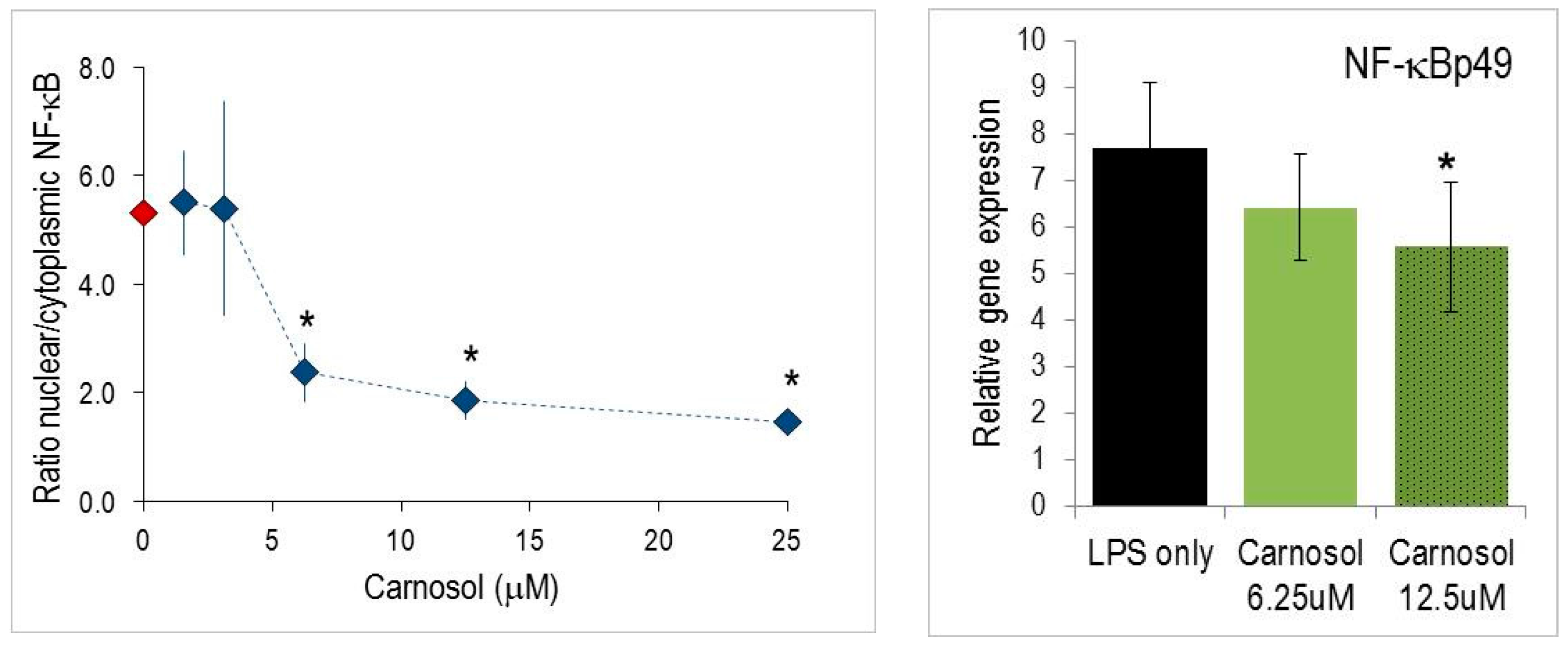
2.4. Nuclear Translocation of NF-κB

Gene activation is dependent on early events in the NF-κB signaling pathway. In order to further investigate the mode of action of CL in chondrocytes, the nuclear translocation of NF-kB was measured by Arrayscan™ cytometry (see Materials and Methods) [31]. IL-1β activated cells responded within 20 min by a substantial shift of NF-κBp65 to the cell nucleus, which was reflected by the appropriate shift in the ratio of nuclear/cytoplasmic fluorescence (Figure 5). The translocation of NF-κBp65 in the nucleus was significantly reduced by increasing concentrations of CL.

3. Discussion

The range of biological properties of CL, CA, CAME and ABTT described in the present study underscores their multiple effects on acute and chronic inflammation. These substances, which form a significant part of the dry plant mass in rosemary, oregano, and sage [4,5,32,33], influence both the production of inflammatory metabolites and the expression of inflammatory genes. It should be emphasized that the biological activities of the substances were analyzed in two diverse cellular models, i.e., macrophages and chondrocytes, the latter being critically involved in OA. More specifically, the study shows for the first time the effects of abietane diterpenes on activated chondrocytes and point at their possible use in OA. As convincingly shown in a recent study [30], carnosol potently inhibited pro-inflammatory cytokines and chemokines and reduced the production of mediators involved in ECM breakdown.
NO and PGE2 have important functions in acute and chronic inflammatory processes. In general, their inhibition is considered as a means of disease modification and pain relief, respectively. The IC50 values established for CL in this study are in good agreement with the reported data on NO production in macrophages [3,4,16] and extend the findings to CA, CAME, ABTT, 20-deoxocarnosol, whose effects were comparable to those of CL (Table 1). Part of this effect might be due to the NO scavenging activity of the substances when they are added to the cellular systems [2,4]. Moreover, most of the substances markedly reduced the production of COX-2 dependent PGE2 in murine macrophages. Similar effects had been reported before in epithelial cells [23] but not in macrophages. With regard to eicosanoid production, CL may interact with the thromboxane (TX) A2 receptor [34] without impairing the biosynthesis of TXA nor cyclooxygenase activity [35].
We have investigated the effect of substances on the inflammatory response at the transcriptional level, since CL is expected to influence gene expression regulated by NF-κB [4] or AP-1 [23]. Nuclear translocation of NF-κBp65 was markedly impeded by CL (Figure 5 and [4,22]. As a consequence, it concentration-dependently reduced iNOS gene expression and some other (but not all) inflammatory genes that are under the control of the NF-κB signaling pathway. On the other side, COX-2 mRNA levels in murine macrophages were not impaired (Figure 2). Presumably, prostaglandin E2 synthase is targeted by the substances rather than COX-2 and thus accounts for reduced PGE2 production (Table 1). Indeed, unchanged or even increased COX-2 expression might be beneficial since COX-2 activity is required during the resolution of the acute inflammatory response (for a review see [36]). It should be emphasized that in chronic inflammation, COX-2 inhibitors are a paramount pharmacological target of widely proven efficacy for pain relief. CL had pleiotropic effects on gene expression, since pro-inflammatory cytokine expression levels (i.e., IL-1α and IL-6) were also altered. During the inflammatory response to LPS, macrophages also up-regulated numerous members of other gene families, including chemokines. CXCL10/IP-10, CCL5/RANTES and CCL4/MIP-1β were among the most responsive (Supplementary Material Table S1). CL significantly reduced mRNA levels of CXCL10/IP-10 and CCL5/RANTES, whereas CCL4/MIP-1β expression was refractory to CL. Thus, the observed modulation of gene expression levels does not correlate with the presence of one common regulatory element (such as NF-κB binding element), suggesting complex interactions of CL and different transcription factors (NF-κB, AP-1) and signaling pathways (MAPK) as described for epithelial cells [23]. Also, via its genuine anti-oxidant properties, CL influences cellular redox potential and thus the glutathione and superoxide dismutase activities. This represents an additional means to control expression levels of genes involved in the inflammatory response [15,18].
Among the tested substances CL was the most efficacious, both in its effect on the production of inflammatory metabolites and the modulation of gene expression. Presumably, the five tested diterpenes (Figure 1 and Table 1) differ in their anti-oxidant properties [6,18]. In line with this, CL modulated the cellular redox potential and thus the glutathione and superoxide dismutase activities [18,20]. This represents an additional means to control expression levels of genes involved in the inflammatory response. It should be noted that the mode of action of the diterpenes is idiosyncratic, since the extent and changes of inflammatory gene expression is only partially overlapping (see Figure 2). Plausibly, the features of each substance (Figure 1) shape substance-specific interactions with factors that control gene expression.
The in vivo relevance of these data has to be examined in the light of achievable plasma concentrations and in extenso systemic levels of the substances. In a murine colonic carcinogenesis model, the inclusion of 0.1% CL in the diet reduced adenoma formation [11] and a comparable dietary intake significantly changed liver glutathione-S-transferase and NAD(P)H-quinone reductase [37]. In mice fed CL, steady-state plasma CL concentrations reached 4.2 μM (H. Mohajeri, personal communication); this is close to the in vitro IC50 for NO (Table 1). Intra-gastrically applied CA attained plasma concentrations of 42.5 mg/L CA (i.e., 130 μM) within ~2 h [36] in rats. Hence, CL and CA plasma concentrations were well in the range of IC50 values that are needed to significantly modulate molecular and cellular parameters.
Finally, IL-1β is considered the key molecule that triggers osteoarthritis (OA), while both NO and PGE2 play a role in the OA development [27,38,39,40]. Increased production of NO in OA tissue contributes to a slowly progressing inflammation [40,41]. Here we show for the first time, that the phenolic diterpene CL concentration-dependently reduced IL-1β expression levels in macrophages and chondrocytes. This suggests that these substances might prevent or delay disease initiation and progression. More importantly, CL significantly modulated the expression of different genes that are pivotal in OA etiology: Catabolic genes (MMPs, ADAMTS) encode for enzymes that erode the extracellular matrix (ECM) in articulate tissue. As shown in Figure 3 and Figure 4, gene expression of cartilage-degrading enzymes (MMP-3, MMP-13, ADAMTS-4) was repressed by various abietane diterpenes, while anabolic genes like Col2A1 and aggrecan were up-regulated. It should be noted that other MMPs like MMP-9 and MMP-2 were not affected by CL (Figure 2, and data not shown). Hence, CL has an activity pattern that fulfils most of the requirements for a chondro-protective and cartilage-regenerating substance. Also, chondrocytes responded to IL-1β stimulation by expressing chemokine genes that are crucial for the recruitment of different cell populations to the sites of inflammation [28]. A CL-dependent reduction of CCL5/RANTES and CXCL10/IP-10 is expected to impair the recruitment of neutrophils and activated T lymphocytes, respectively, to sites of inflammation. The same cell populations are targeted by two other chemokines, CXCL8/IL-8 and CCL20/MIP-3α, which are also down-regulated in chondrocytes by the tested substances. Via these subtle changes of various chemokines the cell migration in response to inflammatory stimuli may be reduced. As a corollary of the multiple biological effects, dietary supplementation with abietane diterpenes is expected to significantly modify OA disease development and severity.

4. Materials and Methods

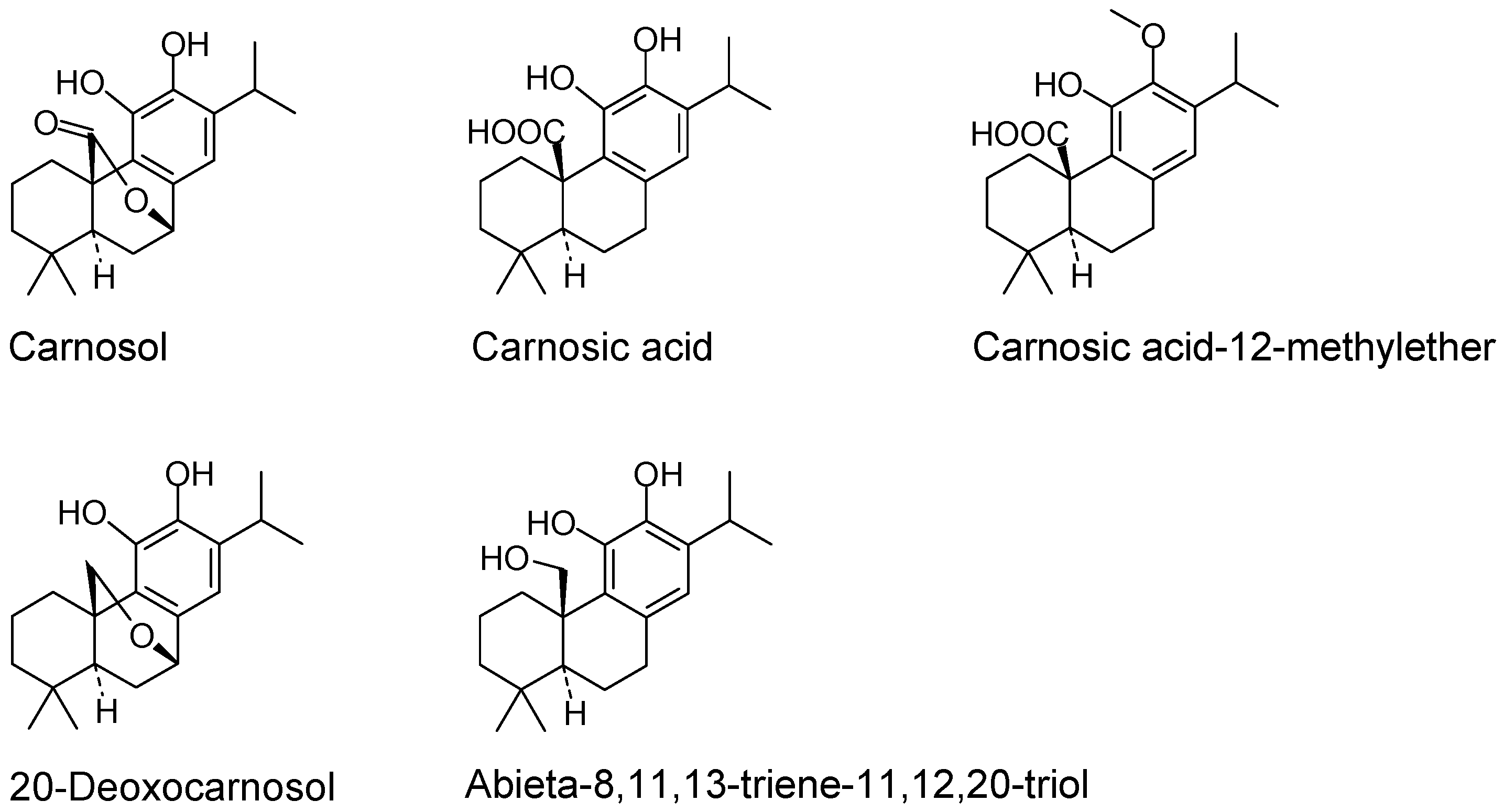
4.1. Phytochemicals and Reagents

Carnosol (CL), carnosic acid (CA), carnosic acid 12-methyl ester (CAME), 20-deoxocarnosol and abieta-8,11,13-triene-11,12,20 triol (ABTT) (Figure 1) were from Cayman Chemicals (Ann Arbor, MI, USA) or Fluorochem Ltd. (Glossop, Hadfield, UK) and also isolated from Salvia officinalis or Rosmarinus officinalis applying solvent extraction and reverse-phased chromatography. Structure elucidation of defined peaks of the resulting chromatogram was done by 1H-NMR and 2D-NMR. Compounds were dissolved in DMSO and added to the culture medium concomitantly with the stimulus. Final DMSO concentration in culture medium was 0.5%. E. coli LPS (serotype 055:B5) and fetal bovine serum (FBS) were from Sigma (St. Louis, MO, USA). DMEM and non-essential amino acids (NEAA) were from Invitrogen (Carlsbad, CA, USA). N(G)-nitro-l-arginine methyl ester (l-NAME) was from Sigma. Human IL-1β was from PeproTech EC (London, UK).

4.2. Cell Culture

RAW264.7 macrophage cells were from ATCC (Manassas, VA, USA) and cultured in DMEM supplemented with 50 U/mL penicillin, 50 μg/mL streptomycin, 0.1 mM NEAA (DMEM-C) and 10% FBS. Cells were seeded into 12-well or 96-well plates at 1 and 0.05 × 10⁶ cells per well, respectively, and used after 2 days of pre-culture. Cells were starved for 18 h in DMEM-C containing 0.25% FBS before the start of treatment and stimulated with lipopolysaccharide (LPS) (1 μg/mL) for 4–24 h in phenol red-free DMEM-C containing 0.25% FBS. Test substances were added concomitantly with the stimulus.
SW1353 chondrosarcoma cells were from ATCC and cultured in DMEM-C containing 10% FBS [29]. Cells were seeded into 6-well plates at 0.5 × 10⁶ cells per well. Sub-confluent cell monolayers were washed and incubated overnight in DMEM-C containing 0.25% FBS and 0.2% lactalbumin hydrolysate (Bacto™ LC, Becton Dickinson, Franklin Lakes, NJ, USA). Cells were activated with 10 ng/mL IL-1β in phenol-red free DMEM-C supplemented with 0.25% FBS and 0.2% lactalbumin hydrolysate without or with test compounds for 4–24 h. Normal human articular chondrocytes obtained from knee (NHAC-kn) were from Lonza (Basel, Switzerland) and cultured in chondrocyte growth medium (Lonza). Cells were seeded into 6-well plates at 0.5 × 10⁶ cells per well and activated with IL-1β (10 ng/ml) for 4–24 h. Cells were lysed in RLT buffer (Qiagen, Hilden, Germany) after 2–4 h of culture and total RNA was extracted. Culture supernatants were harvested after 24 h of culture and stored at −80°C.

4.3. RNA Isolation, cDNA Synthesis and RT-PCR

Total RNA was isolated using the RNeasy Mini Kits (Qiagen) as described [42]. RNA quality and quantity was assessed by Nanodrop® ND-1000 and evaluated by the ND-1000 3.2.1 software (Witec AG, Littau, Switzerland). Total RNA was transcribed into first strand cDNA using the Superscript™ First-Strand Synthesis System for RT-PCR from Invitrogen. Real-time PCR analysis was performed with the ABI PRISM® 7700 Sequence Detection System or the ABI 7900HT Fast Real-Time PCR System (Applied Biosystems (ABI), Foster City, CA, USA). Primers and probes were designed with the Primer Express™ software purchased from ABI. PCR was done using the Taqman® universal PCR Master Mix (ABI). 18S rRNA primers and probes were used as internal standards. Relative gene expression quantification was done by subtracting threshold cycles (CT) for ribosomal RNA from the CT of the targeted gene (ΔCT). Relative mRNA levels were then calculated as 2 −ΔΔCT (fold change), where ΔΔCT refers to the ΔCT of unstimulated minus treated cells. The indicated values were obtained from at least three independent series of experiments, where each treatment was done in duplicates with each being analyzed twice in RT-PCR.

4.4. Measurement of Nitric Oxide and PGE2 Determination

The concentration of NO in culture supernatants was measured using the Griess Reaction [43]. Secreted PGE2 was determined by Enzyme Immuno Assay (EIA) (Cayman Chemicals).

4.5. NF-κB Translocation Experiments in Chondrocytes

NHAC-kn chondrocytes were grown in 96-well plates to sub-confluence. They were pre-incubated with various concentrations of CL for 1 h. NHAC-kn were activated with IL-1β for 20 min. Thereafter, cells were washed, fixed and permeabilized as described [31]. Immunostaining for NF-κBp65 was performed using the NF-κB Activation HitKit™ (Cellomics™ Inc., Pittsburgh, PA, USA). Nuclei were counter-stained with Hoechst dye. Immunofluorescence was measured by quantitative cytometric technique, Arrayscan™, with the Cellomics instrumentation (Cellomics™ Inc.) using the instruments settings as detailed before [27] and expressed as Mean_CircRingAvgIntenRatioCh2 (for detail see: Cellomics HCS application guide at http://www.cellomics.com). All treatments were done in triplicates.

4.6. Statistical Analysis

Data were obtained from at least three independent series of experiments and presented as means +/− standard deviation. p values <0.05 (calculated by Student’s t test or one way ANOVA) were considered statistically significant.

5. Conclusions

Carnosol and related abietanes from Salvia and Rosmarinus potently modulated cytokines, chemokines and genes implicated in cartilage erosion. Hence they beneficially influenced acute and chronic inflammatory processes in macrophages and chondrocytes.

Supplementary Materials

Supplementary materials can be accessed at: https://www.mdpi.com/1420-3049/21/4/465/s1.

Acknowledgments

We thank Göde Schüler (DNP Nutritional Products, Basel, Switzerland) and Grit Kluge (Analyticon, Berlin, Germany) for providing and isolating substances from natural sources, Claus Kilpert for analytical data and Hasan Mohajeri for data on plasma concentrations of carnosol. We are grateful to Christof Wehrli for structural formula of abietanes and Giorgio LaFata for critical reading of the manuscript and insightful comments.

Author Contributions

J.S. and D.R. conceived and designed the study; A.F. contributed materials and reagents; N.R. and N.S. performed the experimental work and compiled the data; J.S. analyzed and interpreted the data and wrote the paper.

Conflicts of Interest

The authors are employees of DSM Nutritional Products, which funded this study and covered costs for open access publications.

Abbreviations

The following abbreviations are used in this manuscript:
ABTT8,11,13-Abietatriene-11,12,20-triol
CACarnosic acid
CLCarnosol
CAMECarnosic acid 12-methyl-ether
NHAC-knNormal human articulocytes-knee
NONitric oxide
PGE2Prostaglandin E2

References

  1. Aruoma, O.I.; Halliwell, B.; Aeschbach, R.; Loligers, J. Antioxidant and pro-oxidant properties of active rosemary constituents: Carnosol and carnosic acid. Xenobiotica 1992, 22, 257–268. [Google Scholar] [CrossRef] [PubMed]
  2. Aruoma, O.I.; Spencer, J.P.; Rossi, R.; Aeschbach, R.; Khan, A.; Mahmood, N.; Munoz, A.; Murcia, A.; Butler, J.; Halliwell, B. An evaluation of the antioxidant and antiviral action of extracts of rosemary and provencal herbs. Food Chem. Toxicol. 1996, 34, 449–456. [Google Scholar] [CrossRef]
  3. Chan, M.M.; Ho, C.T.; Huang, H.I. Effects of three dietary phytochemicals from tea, rosemary and turmeric on inflammation-induced nitrite production. Cancer Lett. 1995, 96, 23–29. [Google Scholar] [CrossRef]
  4. Lo, A.H.; Liang, Y.C.; Lin-Shiau, S.Y.; Ho, C.T.; Lin, J.K. Carnosol, an antioxidant in rosemary, suppresses inducible nitric oxide synthase through down-regulating nuclear factor-kappab in mouse macrophages. Carcinogenesis 2002, 23, 983–991. [Google Scholar] [CrossRef] [PubMed]
  5. Miura, K.; Kikuzaki, H.; Nakatani, N. Antioxidant activity of chemical components from sage (Salvia officinalis L.) and thyme (Thymus vulgaris L.) measured by the oil stability index method. J. Agric. Food Chem. 2002, 50, 1845–1851. [Google Scholar] [CrossRef] [PubMed]
  6. Zeng, H.H.; Tu, P.F.; Zhou, K.; Wang, H.; Wang, B.H.; Lu, J.F. Antioxidant properties of phenolic diterpenes from rosmarinus officinalis. Acta Pharmacol. Sin. 2001, 22, 1094–1098. [Google Scholar] [PubMed]
  7. Dorrie, J.; Sapala, K.; Zunino, S.J. Carnosol-induced apoptosis and downregulation of BcL-2 in B-lineage leukemia cells. Cancer Lett. 2001, 170, 33–39. [Google Scholar] [CrossRef]
  8. Huang, M.T.; Ho, C.T.; Wang, Z.Y.; Ferraro, T.; Lou, Y.R.; Stauber, K.; Ma, W.; Georgiadis, C.; Laskin, J.D.; Conney, A.H. Inhibition of skin tumorigenesis by rosemary and its constituents carnosol and ursolic acid. Cancer Res. 1994, 54, 701–708. [Google Scholar] [PubMed]
  9. Johnson, J.J.; Syed, D.N.; Heren, C.R.; Suh, Y.; Adhami, V.M.; Mukhtar, H. Carnosol, a dietary diterpene, displays growth inhibitory effects in human prostate cancer PC3 cells leading to G2-phase cell cycle arrest and targets the 5′-AMP-activated protein kinase (AMPK) pathway. Pharm. Res. 2008, 25, 2125–2134. [Google Scholar] [CrossRef] [PubMed]
  10. Mace, K.; Offord, E.A.; Harris, C.C.; Pfeifer, A.M. Development of in vitro models for cellular and molecular studies in toxicology and chemoprevention. Arch. Toxicol. Suppl. 1998, 20, 227–236. [Google Scholar] [PubMed]
  11. Moran, A.E.; Carothers, A.M.; Weyant, M.J.; Redston, M.; Bertagnolli, M.M. Carnosol inhibits beta-catenin tyrosine phosphorylation and prevents adenoma formation in the C57bl/6j/min/+ (min/+) mouse. Cancer Res. 2005, 65, 1097–1104. [Google Scholar] [PubMed]
  12. Visanji, J.M.; Thompson, D.G.; Padfield, P.J. Induction of G2/M phase cell cycle arrest by carnosol and carnosic acid is associated with alteration of cyclin a and cyclin b1 levels. Cancer Lett. 2006, 237, 130–136. [Google Scholar] [CrossRef] [PubMed]
  13. Johnson, J.J. Carnosol: A promising anti-cancer and anti-inflammatory agent. Cancer Lett. 2011, 305, 1–7. [Google Scholar] [CrossRef] [PubMed]
  14. Kim, S.J.; Kim, J.S.; Cho, H.S.; Lee, H.J.; Kim, S.Y.; Kim, S.; Lee, S.Y.; Chun, H.S. Carnosol, a component of rosemary (Rosmarinus officinalis L.) protects nigral dopaminergic neuronal cells. Neuroreport 2006, 17, 1729–1733. [Google Scholar] [CrossRef] [PubMed]
  15. Park, K.W.; Kundu, J.; Chae, I.G.; Kim, D.H.; Yu, M.H.; Kundu, J.K.; Chun, K.S. Carnosol induces apoptosis through generation of ROS and inactivation of STAT3 signaling in human colon cancer HCT116 cells. Int. J. Oncol. 2014, 44, 1309–1315. [Google Scholar] [PubMed]
  16. Peng, C.H.; Su, J.D.; Chyau, C.C.; Sung, T.Y.; Ho, S.S.; Peng, C.C.; Peng, R.Y. Supercritical fluid extracts of rosemary leaves exhibit potent anti-inflammation and anti-tumor effects. Biosci. Biotechnol. Biochem. 2007, 71, 2223–2232. [Google Scholar] [CrossRef] [PubMed]
  17. Poeckel, D.; Greiner, C.; Verhoff, M.; Rau, O.; Tausch, L.; Hornig, C.; Steinhilber, D.; Schubert-Zsilavecz, M.; Werz, O. Carnosic acid and carnosol potently inhibit human 5-lipoxygenase and suppress pro-inflammatory responses of stimulated human polymorphonuclear leukocytes. Biochem. Pharmacol. 2008, 76, 91–97. [Google Scholar] [CrossRef] [PubMed]
  18. Wijeratne, S.S.; Cuppett, S.L. Potential of rosemary (Rosemarinus officinalis L.) diterpenes in preventing lipid hydroperoxide-mediated oxidative stress in Caco-2 cells. J. Agric. Food Chem. 2007, 55, 1193–1199. [Google Scholar] [CrossRef] [PubMed]
  19. Yao, H.; Chen, Y.; Zhang, L.; He, X.; He, X.; Lian, L.; Wu, X.; Lan, P. Carnosol inhibits cell adhesion molecules and chemokine expression by tumor necrosis factor-alpha in human umbilical vein endothelial cells through the nuclear factor-κb and mitogen-activated protein kinase pathways. Mol. Med. Rep. 2014, 9, 476–480. [Google Scholar] [PubMed]
  20. Martin, D.; Rojo, A.I.; Salinas, M.; Diaz, R.; Gallardo, G.; Alam, J.; De Galarreta, C.M.; Cuadrado, A. Regulation of heme oxygenase-1 expression through the phosphatidylinositol 3-kinase/AKT pathway and the Nrf2 transcription factor in response to the antioxidant phytochemical carnosol. J. Biol. Chem. 2004, 279, 8919–8929. [Google Scholar] [CrossRef] [PubMed]
  21. Takahashi, T.; Tabuchi, T.; Tamaki, Y.; Kosaka, K.; Takikawa, Y.; Satoh, T. Carnosic acid and carnosol inhibit adipocyte differentiation in mouse 3T3-L1 cells through induction of phase2 enzymes and activation of glutathione metabolism. Biochem. Biophys. Res. Commun. 2009, 382, 549–554. [Google Scholar] [CrossRef] [PubMed]
  22. Huang, S.C.; Ho, C.T.; Lin-Shiau, S.Y.; Lin, J.K. Carnosol inhibits the invasion of B16/F10 mouse melanoma cells by suppressing metalloproteinase-9 through down-regulating nuclear factor-κb and c-jun. Biochem. Pharmacol. 2005, 69, 221–232. [Google Scholar] [CrossRef] [PubMed]
  23. Subbaramaiah, K.; Cole, P.A.; Dannenberg, A.J. Retinoids and carnosol suppress cyclooxygenase-2 transcription by CREB-binding protein/p300-dependent and -independent mechanisms. Cancer Res. 2002, 62, 2522–2530. [Google Scholar] [PubMed]
  24. Bai, N.; He, K.; Roller, M.; Lai, C.S.; Shao, X.; Pan, M.H.; Ho, C.T. Flavonoids and phenolic compounds from Rosmarinus officinalis. J. Agric. Food Chem. 2010, 58, 5363–5367. [Google Scholar] [CrossRef] [PubMed]
  25. Gebauer, M.; Saas, J.; Sohler, F.; Haag, J.; Soder, S.; Pieper, M.; Bartnik, E.; Beninga, J.; Zimmer, R.; Aigner, T. Comparison of the chondrosarcoma cell line SW1353 with primary human adult articular chondrocytes with regard to their gene expression profile and reactivity to IL-1beta. Osteoarthr. Cartil. 2005, 13, 697–708. [Google Scholar] [CrossRef] [PubMed]
  26. Goldring, M.B.; Berenbaum, F. The regulation of chondrocyte function by proinflammatory mediators: Prostaglandins and nitric oxide. Clin. Orthop. Relat. Res. 2004, S37–S46. [Google Scholar] [CrossRef]
  27. Goldring, S.R.; Goldring, M.B. The role of cytokines in cartilage matrix degeneration in osteoarthritis. Clin. Orthop. Relat. Res. 2004, S27–S36. [Google Scholar] [CrossRef]
  28. Sandell, L.J.; Xing, X.; Franz, C.; Davies, S.; Chang, L.W.; Patra, D. Exuberant expression of chemokine genes by adult human articular chondrocytes in response to IL-1β. Osteoarthr. Cartil. 2008, 16, 1560–1571. [Google Scholar] [CrossRef] [PubMed]
  29. Schwager, J.; Hoeller, U.; Wolfram, S.; Richard, N. Rose hip and its constituent galactolipids confer cartilage protection by modulating cytokine, and chemokine expression. BMC Complement. Altern. Med. 2011, 11, 105. [Google Scholar] [CrossRef] [PubMed]
  30. Sanchez, C.; Horcajada, M.N.; Membrez Scalfo, F.; Ameye, L.; Offord, E.; Henrotin, Y. Carnosol inhibits pro-inflammatory and catabolic mediators of cartilage breakdown in human osteoarthritic chondrocytes and mediates cross-talk between subchondral bone osteoblasts and chondrocytes. PLoS ONE 2015, 10, e0136118. [Google Scholar] [CrossRef] [PubMed]
  31. Ding, G.J.; Fischer, P.A.; Boltz, R.C.; Schmidt, J.A.; Colaianne, J.J.; Gough, A.; Rubin, R.A.; Miller, D.K. Characterization and quantitation of nf-kappab nuclear translocation induced by interleukin-1 and tumor necrosis factor-alpha. Development and use of a high capacity fluorescence cytometric system. J. Biol. Chem. 1998, 273, 28897–28905. [Google Scholar] [CrossRef] [PubMed]
  32. Del Bano, M.J.; Lorente, J.; Castillo, J.; Benavente-Garcia, O.; del Rio, J.A.; Ortuno, A.; Quirin, K.W.; Gerard, D. Phenolic diterpenes, flavones, and rosmarinic acid distribution during the development of leaves, flowers, stems, and roots of rosmarinus officinalis. Antioxidant activity. J. Agric. Food Chem. 2003, 51, 4247–4253. [Google Scholar] [CrossRef] [PubMed]
  33. Guerrero, I.C.; Andres, L.S.; Leon, L.G.; Machin, R.P.; Padron, J.M.; Luis, J.G.; Delgadillo, J. Abietane diterpenoids from salvia pachyphylla and S. clevelandii with cytotoxic activity against human cancer cell lines. J. Nat. Prod. 2006, 69, 1803–1805. [Google Scholar] [CrossRef] [PubMed]
  34. Lee, J.J.; Jin, Y.R.; Lim, Y.; Hong, J.T.; Kim, T.J.; Chung, J.H.; Yun, Y.P. Antiplatelet activity of carnosol is mediated by the inhibition of TXA2 receptor and cytosolic calcium mobilization. Vasc. Pharmacol. 2006, 45, 148–153. [Google Scholar] [CrossRef] [PubMed]
  35. Lee, J.J.; Jin, Y.R.; Lee, J.H.; Yu, J.Y.; Han, X.H.; Oh, K.W.; Hong, J.T.; Kim, T.J.; Yun, Y.P. Antiplatelet activity of carnosic acid, a phenolic diterpene from Rosmarinus officinalis. Planta Med. 2007, 73, 121–127. [Google Scholar] [CrossRef] [PubMed]
  36. Gilroy, D.W.; Lawrence, T.; Perretti, M.; Rossi, A.G. Inflammatory resolution: New opportunities for drug discovery. Nat. Rev. Drug Discov. 2004, 3, 401–416. [Google Scholar] [CrossRef] [PubMed]
  37. Singletary, K.W. Rosemary extract and carnosol stimulate rat liver glutathione-s-transferase and quinone reductase activities. Cancer Lett. 1996, 100, 139–144. [Google Scholar] [CrossRef]
  38. Amin, A.R.; Attur, M.; Patel, R.N.; Thakker, G.D.; Marshall, P.J.; Rediske, J.; Stuchin, S.A.; Patel, I.R.; Abramson, S.B. Superinduction of cyclooxygenase-2 activity in human osteoarthritis-affected cartilage. Influence of nitric oxide. J. Clin. Investig. 1997, 99, 1231–1237. [Google Scholar] [CrossRef] [PubMed]
  39. Murrell, G.A.; Jang, D.; Williams, R.J. Nitric oxide activates metalloprotease enzymes in articular cartilage. Biochem. Biophys. Res. Commun. 1995, 206, 15–21. [Google Scholar] [CrossRef] [PubMed]
  40. Vuolteenaho, K.; Moilanen, T.; Knowles, R.G.; Moilanen, E. The role of nitric oxide in osteoarthritis. Scand. J. Rheumatol. 2007, 36, 247–258. [Google Scholar] [CrossRef] [PubMed]
  41. Melchiorri, C.; Meliconi, R.; Frizziero, L.; Silvestri, T.; Pulsatelli, L.; Mazzetti, I.; Borzi, R.M.; Uguccioni, M.; Facchini, A. Enhanced and coordinated in vivo expression of inflammatory cytokines and nitric oxide synthase by chondrocytes from patients with osteoarthritis. Arthritis Rheum. 1998, 41, 2165–2174. [Google Scholar] [CrossRef]
  42. Richard, N.; Porath, D.; Radspieler, A.; Schwager, J. Effects of resveratrol, piceatannol, tri-acetoxystilbene, and genistein on the inflammatory response of human peripheral blood leukocytes. Mol. Nutr. Food Res. 2005, 49, 431–442. [Google Scholar] [CrossRef] [PubMed]
  43. D’Acquisto, F.; Cicatiello, L.; Iuvone, T.; Ialenti, A.; Ianaro, A.; Esumi, H.; Weisz, A.; Carnuccio, R. Inhibition of inducible nitric oxide synthase gene expression by glucocorticoid-induced protein(s) in lipopolysaccharide-stimulated J774 cells. Eur. J. Pharmacol. 1997, 339, 87–95. [Google Scholar] [CrossRef]
  • Sample Availability: Samples of the compounds are available from the authors (limited stock).
Figure 1. Structures of the abietane diterpenes used in this study.
Molecules 21 00465 g001
Figure 2. Abietane diterpenes down-regulated gene expression in RAW264.7 cells. Cells were stimulated with LPS in the presence of indicated amounts of substances for 4 h and the levels of mRNA determined by quantitative RT-PCR. “Relative gene expression”, which indicates gene expression levels in stimulated relative to unstimulated cells, was calculated as described in Materials and Methods. Asterisks indicate statistical significant differences compared to LPS-stimulated cells (* p < 0.05, ** p < 0.01, *** p < 0.001; n = 3). Effect of carnosol (a); carnosic acid 12-methylether (CAME) (b) and abieta-8,11,13-triene-11,12,20 triol (ABTT) (c).
Molecules 21 00465 g002aMolecules 21 00465 g002b
Figure 3. Effect of substances on IL-1β activated chondrosarcoma SW1353 cells. Cells were stimulated with IL-1β for 4 h in the presence of indicated substances. Gene expression was determined by quantitative RT-PCR as described in Figure 2. Asterisks indicate statistical significant differences compared to IL-1β stimulated cells (* p < 0.05, ** p < 0.01, n = 4).
Molecules 21 00465 g003
Figure 4. Effect of substances on IL-1β activated NHAC-kn. Cells were stimulated with IL-1β for 4 h in the presence of indicated substances. Gene expression was determined by quantitative RT-PCR as described in Figure 2. Asterisks indicate statistical significant differences compared to IL-1β stimulated cells (* p < 0.05, ** p < 0.01, *** p < 0.001; n = 4). (a): Effect of carnosol (CL); (b): effect of carnosic acid 12-methylether (CAME); (c): effect of abieta-8,11,13-triene-11,12,20 triol (ABTT). Note log scale on the y axis in some panels.
Molecules 21 00465 g004
Figure 5. Carnosol altered the NF-kB signaling pathway. Left panel: Nuclear translocation of NF-κB. Chondrocytes (NHAC-kn) were pre-incubated with carnosol for 1 h and stimulated with IL-1β for 20 min. The relative nuclear immunofluorescence is indicated on the y-axis (see Materials and Methods for details). Asterisks indicate statistically significant differences between the different treatments (* p< 0.05). Right panel: Carnosol reduces expression of transcription factors of the NF-κB pathway. Cells were stimulated for 4 h and gene expression levels determined by RT-PCR (* p < 0.05).
Molecules 21 00465 g005
Table 1. IC50 values for abietane diterpene in NO and PGE2 production.
Carnosol (CL)Carnosic Acid 12-Methyl Ether (CAME)20-Deoxy-CarnosolCarnosic Acid (CA)Abieta-8,11,13-Triene-11,12,20 Triol (ABTT)l-NAME 1
NO5.0 ± 2.83.8 ± 0.611.9 ± 0.66.9 ± 2.212.5 ± 4.8150 ± 34
PGE29.4 ± 2.144.4 ± 6.118.8 ± 6.411.4 ± 0.97.8 ± 4.6>1000
1 NG-nitro-l-arginine methyl ester. RAW 264.7 cells were stimulated with LPS for 24 h in the presence of varying concentrations of compounds (0.16–50 μM). NO and PGE2 were measured in culture supernatants and the IC50 values were calculated. Values are means ± standard deviation of at least three independent experimental series. Numbers indicate means ± SD (in μM) of IC50 values for NO and PGE2 (n = 5).

