Article

Effects of Ursolic Acid Derivatives on Caco-2 Cells and Their Alleviating Role in Streptozocin-Induced Type 2 Diabetic Rats

Department of Pharmaceutical Engineering, Faculty of Chemical Engineering and Light Industry, Guangdong University of Technology, Guangzhou 510006, China
*
Authors to whom correspondence should be addressed.
These authors contributed equally to this work.
Molecules 2014, 19(8), 12559-12576; https://doi.org/10.3390/molecules190812559
Submission received: 24 June 2014 / Revised: 1 August 2014 / Accepted: 11 August 2014 / Published: 19 August 2014
(This article belongs to the Special Issue Prodrugs)

Abstract

:
In this study, the effect and mechanism of a series of ursolic acid (UA) derivatives on glucose uptake were investigated in a Caco-2 cells model. Their effect on hyperglycemia, hyperlipidemia and oxidative stress were also demonstrated in streptozocin (STZ)-induced diabetic rats. 2-[N-(7-nitrobenz-2-oxa-1,3-diazol-4-yl)amino]-2-deoxy-glucose (2-NBDG) was used as a fluorescein in Caco-2 cells model to screen UA derivatives by glucose uptake and expression of glucose transporter protein (SGLT-1, GLUT-2). Moreover, STZ-induced diabetic rats were administered with these derivatives for 4 weeks of treatment. The fasting blood glucose (FBG), insulin levels, biochemical parameters, lipid levels, and oxidative stress markers were finally evaluated. The results of this study indicated that compounds 10 and 11 significantly inhibited 2-NBDG uptake under both Na+-dependent and Na+-independent conditions by decreasing SGLT-1 and GLUT-2 expression in the Caco-2 cells model. Further in vivo studies revealed that compound 10 significantly reduced hyperglycemia by increasing levels of serum insulin, total protein, and albumin, while the fasting blood glucose, body weight and food intake were restored much closer to those of normal rats. Compounds 10 and 11 showed hypolipidemic activity by decreasing the total amounts of cholesterol (TC) and triglycerides (TG). Furthermore, compound 10 showed antioxidant potential which was confirmed by elevation of glutathione (GSH) and superoxide dismutase (SOD) and reduction of malondialdehyde (MDA) levels in the liver and kidney of diabetic rats. It was concluded that compound 10 caused an apparent inhibition of intestinal glucose uptake in Caco-2 cells and hypoglycemia, hypolipidemia and augmented oxidative stress in STZ-induced diabetic rats. Thus, compound 10 could be developed as a potentially complementary therapeutic or prophylactic agent for diabetics mellitus and its complications.

Graphical Abstract

1. Introduction

Currently, there are more than 347 million people worldwide that have diabetes, making diabetes mellitus (DM) one of the most serious chronic diseases in the world. The WHO projects that diabetes will be the 7th leading cause of death in 2030 [1]. DM is a metabolic disorder characterized by glucose intolerance and changes in protein and lipid metabolism; it has become more and more common because of unhealthy diet and sedentary lifestyle in the general public [2,3]. Long-term diabetic patients who are treated ineffectively suffer from complications such as nephropathy, retinopathy, heart disease, peripheral neuropathy and so on [4]. The risks of acquiring cardiovascular disease, stroke, and cancer are higher in DM patients [5,6,7]. In addition, the overall risk of dying among people with DM linked complications is at least double the risk of their peers without DM [8]. Hypoglycemic agents, including insulin and other modern oral drugs, can control the blood glucose levels, but they may also have limited efficacy and many other undesirable effects. Therefore, it is evident that there is an urgent need for more effective therapeutic agents.
Pentacyclic triterpenes are abundantly distributed in herbal medicine. Ursolic acid (UA, 3β-hydroxy-urs-12-en-28-oic, 1), which is a pentacyclic triterpene, has been reported to possess many bioactivities, such as antitumor [9,10], anti-inflammatory [11] anti-diabetic activities [12] and so on. The mechanism of its anti-diabetic activity has been proven by alleviating diabetic complications like atherosclerosis and nephropathy [13,14]. However, there are few studies focused on the properties of UA derivatives on glucose transport and streptozotocin (STZ)-induced DM rats. In light of this, we hypothesized that an additional mechanism of action might be implicated in the anti-diabetic activity of UA derivatives. In this study, we implemented a fluorometric method to evaluate a multitude of properties of glucose uptake in Caco-2 cells with 2-NBDG [15,16], which has several advantages over other available glucose tracers [17]. Glucose can be absorbed into blood capillaries when dietary carbohydrates are digested into monosaccharides [18]. Therefore, a strategy of managing diabetes is to inhibit the glucose uptake in the intestines to control the blood glucose level [19]. In the intestines, glucose is transported mainly by two transporters (SGLT-1, GLUT-2), depending on the luminal glucose concentration.
The detrimental effect of diabetic complications is complicated through oxidative stress [20]. Diabetes is usually associated with augmented production of molecules of reactive oxygen species (ROS) or abated antioxidant defense systems, both of which result in enhanced oxidative damage and lead to ROS-mediated diabetic pathogenesis [21]. Compared with healthy adults, type 2 DM patients have lower levels of glutathione (GSH) and superoxide dismutase (SOD), the primary endogenous antioxidants. In contrast, malondialdehyde (MDA), a highly toxic byproduct generated partially by lipid oxidation and ROS, is increased in patients with diabetes [22]. Thus, enhancing the antioxidant capacity is one method to relieve diabetes and its complications.
In the present study, an investigation was undertaken to evaluate the effects of UA and its derivatives shown in Figure 1 [23], which were synthesized in our laboratory, on the expression of glucose uptake and glucose transporter protein (SGLT-1, GLUT-2) in Caco-2 cells model. Furthermore, the anti-hyperglycemic, anti-hyperlipidemic, anti-oxidant inhibitory effects of these derivatives in STZ-induced DM rats were also investigated.
Figure 1. The structure of evaluated compounds 112.
Molecules 19 12559 g001

2. Results and Discussion

2.1. Cell Toxicity

As shown in Table 1, the cytotoxicity of UA and its derivatives against Caco-2 cells was studied. The results indicate that the viability of Caco-2 cells was greater than 90%, with the exception of cells treated with compound 6, for which the cell viability was 86.12%, thus, it is not suitable for in vivo studies.
Table 1. Cell toxicity of UA and its derivatives on Caco-2 cells.
CompoundInhibition Rate %CompoundInhibition Rate %
19.88 ± 6.5189.86 ± 2.68
25.02 ± 2.3894.40 ± 3.09
38.65 ± 3.49107.84 ± 4.15
48.53 ± 5.69112.29 ± 6.08
58.72 ± 1.18127.67 ± 5.36
613.88 ± 4.52Phlorizin7.70 ± 3.91
78.14 ± 3.81Phloretin4.70 ± 2.59
Values denote mean ± SD, n = 4. Inhibitory percentage of cells treated with each compound at a concentration of 100 μM for 24 h.

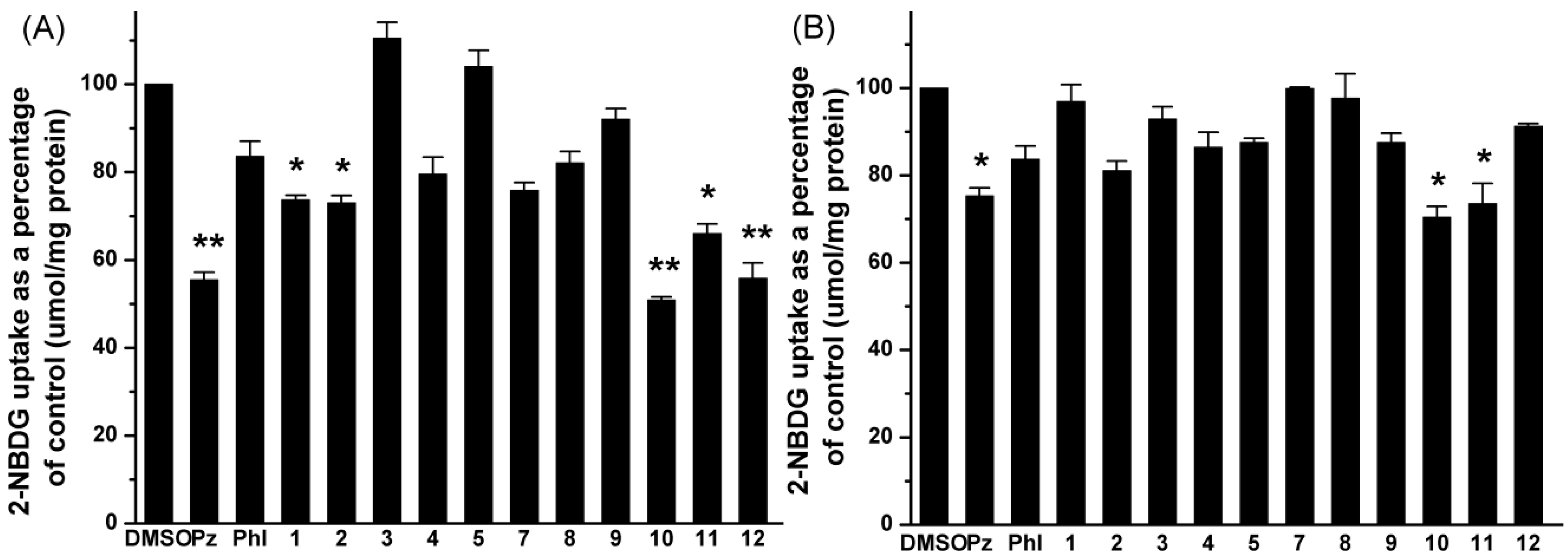
2.2. Effects of UA and Its Derivatives on Glucose Uptake by Caco-2 Cells

Caco-2 cell are widely used as a model for intestinal absorption studies [24]. As illustrated in the sodium-dependent glucose uptake study (Figure 2A), compounds 12 and 1012 caused a statistically significant reduction in glucose uptake, especially compounds 10 and 12 (** p < 0.01). Phlorizin and phloretin were used as positive controls. The effect of these compounds reducing 2-NBDG adsorption are as follows: phlorizin (56% reduction), compound 1 (74% reduction), compound 2 (73% reduction), compound 10 (51% reduction), compound 11 (66% reduction), compound 12 (56% reduction). The other compounds had no effect on glucose uptake under these conditions.
Figure 2. Effects of selected compounds on glucose uptake in Caco-2 cells under condition of (A) Na+-dependent and (B) Na+-independent. Cells were treated with vehicle, positive and the indicated compounds (100 μM). The data reported represent the means (n = 3) ± SD. * p < 0.05, ** p < 0.01.
Molecules 19 12559 g002
The sodium-independent glucose uptake study was also performed like the sodium-dependent one, but NaCl and Na2HPO4 were replaced by KCl and K2HPO4. As shown in Figure 2B, only phlorizin and compounds 10 and 11 had a significant reduction effect on glucose uptake (* p < 0.05), which was significantly decreased to 75%, 70%, 74% of the control value, whereas the other compounds had no detectable effect under these conditions.

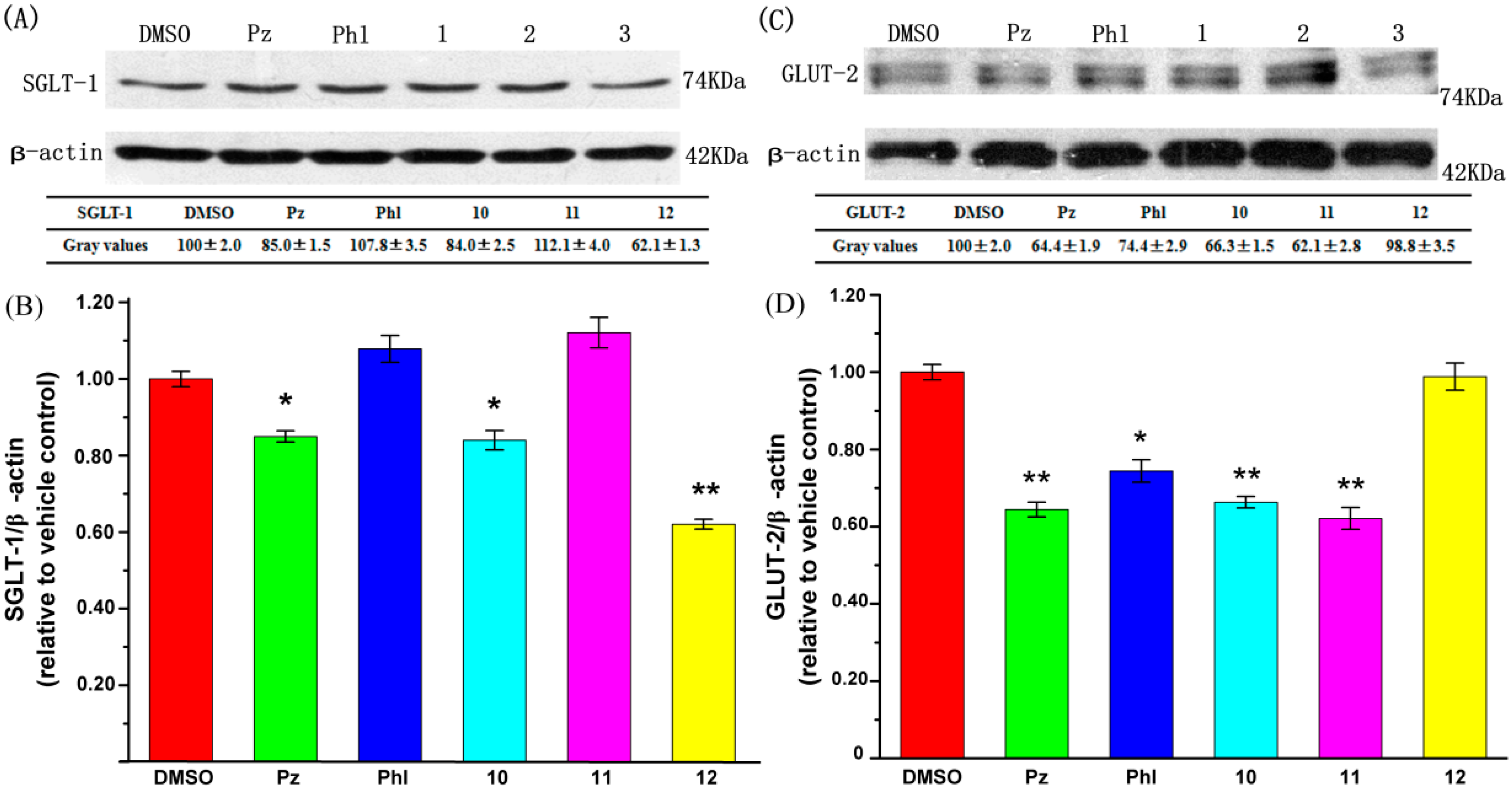
2.3. Effect of UA and Its Derivatives on Glucose Transporter Protein Expression

SGLT-1 and GLUT-2 are the most significant transporters involved in glucose transportation in the intestines [25,26]. Caco-2 cells were treated with 0.1% DMSO, 100 μM phlorizin, 100 μM phloretin, 100 μM compounds 1012 for 6 h, then Caco-2 cells were lysed and analysed by western blotting. The effect of compounds 1012 on glucose uptake suggested that these compounds may exert their effects through modulation of glucose transporter expression. As shown in Figure 3, compounds 10 and 12 are able to significantly reduce the protein expression of SGLT-1, particularly compound 12, whereas compounds 10 and 11 had significant effects on reducing the expression of protein GLUT-2.
Figure 3. Effects of selected compounds on protein expression level of SGLT-1 and GLUT-2. (A) Representive SGLT-1 immunoblots; (B) Quantitation of SGLT-1 immunoblots; (C) Representive SGLT-2 immunoblots; (D) Quantitation of SGLT-2 immunoblots. Protein expression levels of SGLT-1 and GLUT-2 were quantified with respect to the level of β-actin and expressed as relative changes in comparison to the vehicle control. The data reported represent the means (n = 3) ± SD. * p < 0.05, ** p < 0.01.
Molecules 19 12559 g003

2.4. Acute Oral Toxicity Study of UA and Its Derivatives

The acute oral toxicity study revealed these tested compounds’ non-toxic nature; no toxic reactions or lethality were observed at the doses of 100, 200 and 500 mg/kg. Based on the result, 100 mg/kg was selected as the maximum dose for oral administration.

2.5. Effects of UA and Its Derivatives on Fasting Blood Glucose Levels

During the 4 week treatment, the blood glucose levels were changed in normal, diabetic control, and diabetic treated rats. The results are presented in Table 2. The fasting blood glucose levels of normal rats were not changed until the end of the period, while the blood glucose were significantly increased in untreated diabetic rats as compared with normal control. However, the high levels of blood glucose decreased in diabetic rats treated with compound 10 and glibenclamide. At the end of the 4 week treatment, 100 mg/kg of compound 10 decreased blood glucose levels (49.6%) as compared with diabetic control. The glibenclamide group achieved 54.4% drop in blood glucose levels. In addition, compounds 1, 11 and 12 had no significant effect on lowing fasting blood glucose levels.
Table 2. Effects of glibenclamide and the selected compounds on fasting blood glucose levels of normal, diabetic control, and diabetic treated rats.
GroupFasting Blood Glucose Level (mg/dL)
Week 0Week 1Week 2Week 3Week 4
Normal control73.80 ± 4.5075.60 ± 7.2072.00 ± 6.3072.00 ± 5.4081.00 ± 2.16
Diabetic control320.40 ± 20.52401.40 ± 22.50428.40 ± 50.40433.80 ± 39.06446.40 ± 31.68
Glibenclamide (10 mg/kg)338.40 ± 16.38271.80 ± 18.36 *183.60 ± 11.70 *216.00 ± 9.72 *203.40 ± 13.50 *
Compound 1 (100 mg/kg)342.00 ± 19.08405.00 ± 17.46392.40 ± 13.32379.80 ± 19.44396.00 ± 16.92
Compound 10 (100 mg/kg)320.40 ± 12.96270.00 ± 16.74 *286.20 ± 18.90234.00 ± 14.58 *225.00 ± 11.70 *
Compound 11 (100 mg/kg)318.60 ± 17.64365.40 ± 20.34352.80 ± 27.36324.00 ± 23.04351.00 ± 26.64
Compound 12 (100 mg/kg)325.80 ± 14.58392.40 ± 36.54379.80 ± 30.60408.60 ± 20.52432.00 ± 28.26
The data reported represent the means (n = 6) ± SD; * Mean values that are significantly different from diabetic control group (p < 0.05).

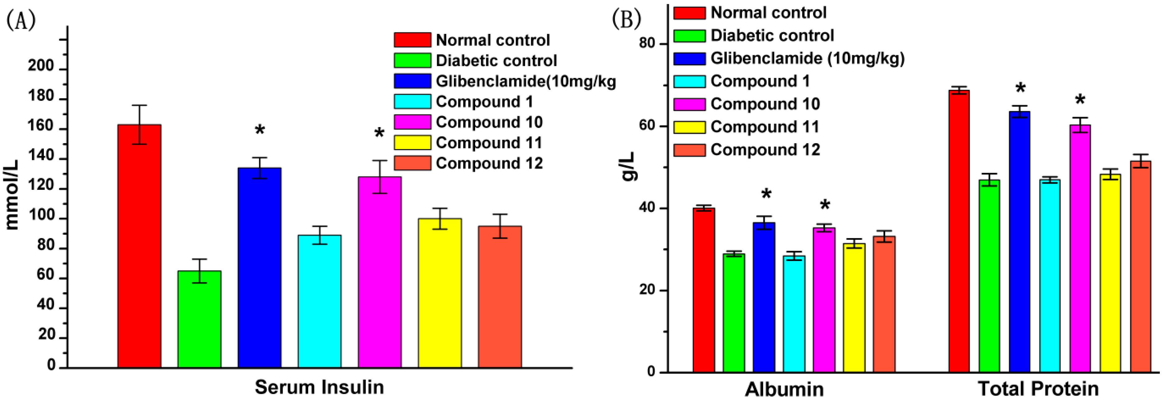
2.6. Effect of UA and Its Derivatives on Serum Biochemical Parameters

After 4 weeks of treatment, the serum insulin, total protein and albumin levels in untreated diabetic rats were significantly reduced compared to the normal control group. Figure 4 shows that after 4 weeks of administration of glibenclamide (10 mg/kg) and compound 10 (100 mg/kg), the serum insulin, total protein and albumin levels in diabetic rats were increased significantly as compared with the diabetic control. Compounds 1, 11 and 12 had a certain but non-significant effect of increasing insulin levels. In addition, compound 1, 11 and 12 caused no appreciable improvement in increasing total protein and albumin levels in diabetic rats.
Figure 4. Effects of compounds 1, 10, 11 and 12 on serum biochemical parameters of STZ-induced diabetic rats in comparison with normal and diabetic control rats after 4 weeks treatment. At the end of the treatment period, rats were fasted for 12 h and blood was drawn to collect the serum. Panels denote (A) serum insulin; (B) albumin and total protein levels. The data reported represent the means (n = 6) ± SD. * Mean values that are significantly different from diabetic control group (p < 0.05).
Molecules 19 12559 g004

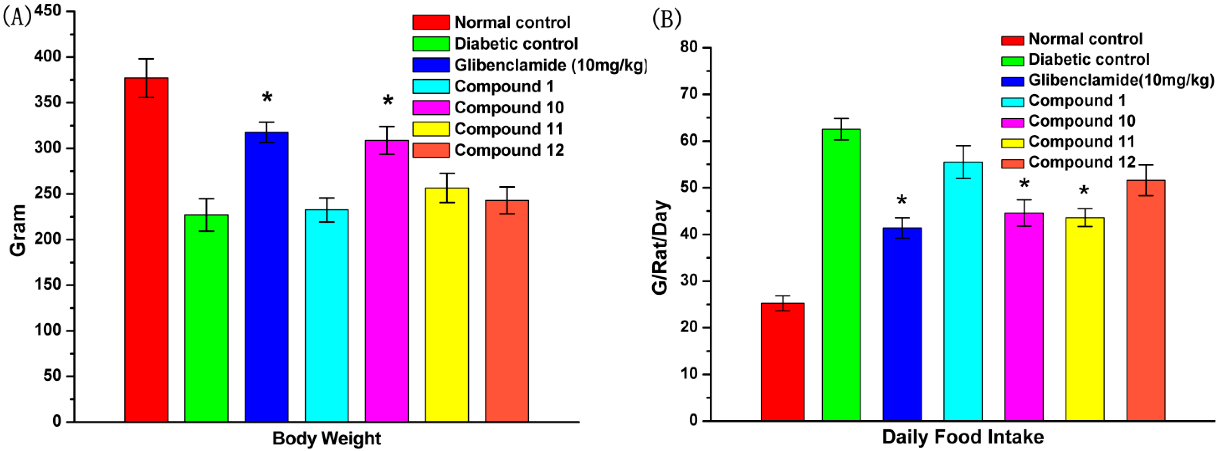
2.7. Effect of UA and Its Derivatives on Body Weight and Food Intake

The effect of tested compounds 1, 10, 11 and 12 on changes of body weight and daily food intake are presented in Figure 5. As shown in Figure 5A, it could be concluded that there was a significant increase in body weight for compound 10-treated diabetic rats when compared with the diabetic control and the glibenclamide treatment group by the end of the treatment period, while the other compounds had no such appreciable effect. Moreover, as shown in Figure 5B, there was a significant increase in food intake in diabetic rats. After oral administration of glibenclamide, compound 10 and 11, the three compounds could reduce daily food intake significantly compared with the untreated diabetic rats. In contrast, compounds 1 and 12 presented no obvious decrease.
Figure 5. Effects of compounds 1, 10, 11 and 12 on (A) body weight and (B) daily food intake of STZ-induced diabetic rats in comparison with normal and diabetic control rats. The data reported represent the means (n = 6) ± SD. * Significant difference compared to diabetic control (p < 0.05).
Molecules 19 12559 g005

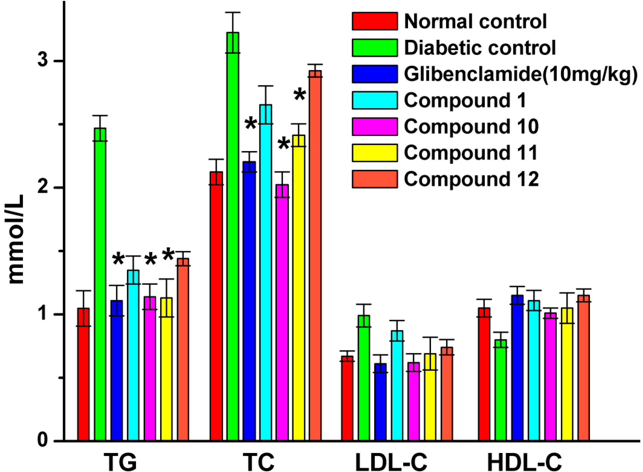
2.8. Effect of UA and Its Derivatives on Hyperlipidemia

Figure 6 shows the effect of glibenclamide and UA derivatives on serum TG, TC, LDL-C and HDL-C levels in diabetic rats and normal control.
Serum TG, TC and LDL-C levels were significantly elevated in diabetic control when compared with the normal control, while the HDL-C levels in diabetic control were significantly decreased when compared with those in normal control. However, Serum TC, TG levels were significantly decreased after treatment with glibenclamide, compounds 10 and 11. Compared with diabetic control, serum LDL-C levels were also lowered, while HDL-C levels were higher with the tested compound treatment but none of gave a significant effect (p < 0.05).

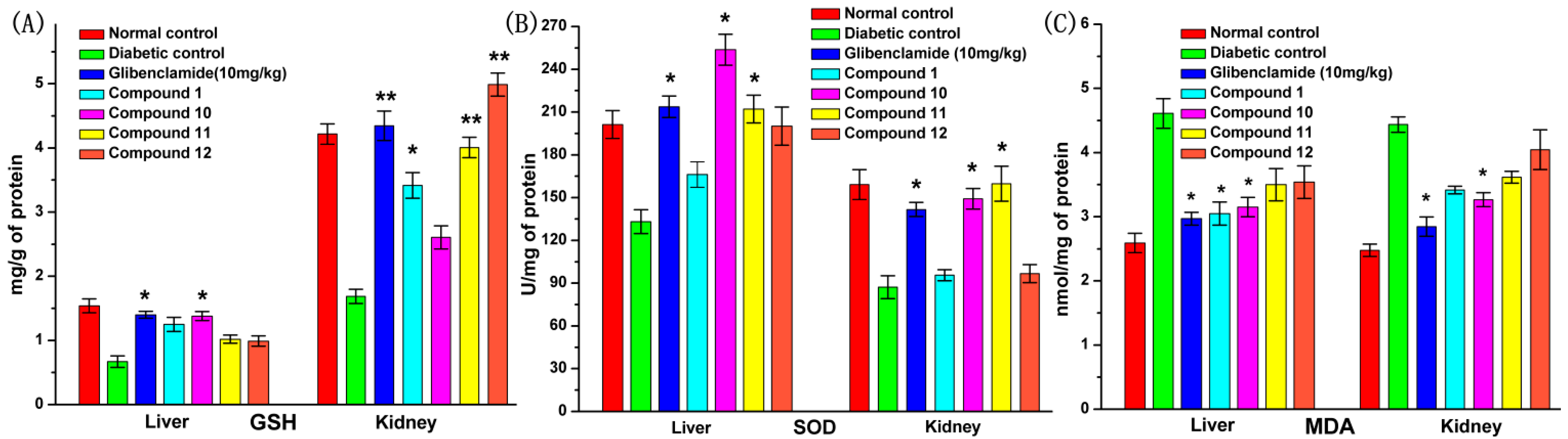
2.9. Effect of UA and Its Derivatives on Oxidative Stress

Figure 7 reveals that the levels of GSH and SOD in diabetic control were reduced while the levels of MDA were significantly increased when compared with those in normal control. All tested compounds produced an increase in GSH levels in liver and kidney. In particularly glibenclamide and compound 10 demonstrated significant effects on the liver, while compounds 1, 11 and 12 showed a great effect on kidney. There was a great improvement of SOD level in the liver and kidney of diabetic rats after treatment with glibenclamide, compounds 10 and 11. Compared with the diabetic control, the MDA levels in the liver of diabetic rats were obviously down-regulated by daily administration of glibenclamide, compounds 1 and 10, while the MDA levels were significantly reduced by glibenclamide and compound 10 (p < 0.05).
Figure 6. Effects of compounds 1, 10, 11 and 12 on serum lipid profiles of STZ-induced diabetic rats in comparison with normal and diabetic control rats after 4 weeks of treatment. At the end of the treatment period, rats were fasted for 12 h and blood was drawn to collect the serum. The data reported represent the means (n = 6) ± SD. * Significant difference compared to diabetic control (p < 0.05).
Molecules 19 12559 g006
Figure 7. Antioxidant effect of compounds 1, 10, 11 and 12 on the kidney and liver of STZ-induced diabetic rats. Panels denote (A) GSH, (B) SOD and (C) MDA levels. The data reported represent the means (n = 6) ± SD. * Significant difference compared to diabetic control (p < 0.05), ** Significant very difference compared to diabetic control (p < 0.01).
Molecules 19 12559 g007

2.10. Discussion

UA has been reported to possess a wide range of bioactivities, including positive effects in curing various complications of diabetes and lowing blood glucose levels [13,27,28]. However, the anti-diabetic potential of UA derivatives and their mechanism(s) of action have not been thoroughly investigated. In the present study, the activities of UA and some of its derivatives were assayed in a Caco-2 cell model. Moreover, the potential of these derivatives in STZ-induced diabetic rats model was also investigated.
The results of MTT assays indicated that at a concentration of 100 μM none of the derivatives showed toxicity to Caco-2 cells, except for compound 6, which is in line with the results of acute oral toxicity studies in vivo. The effects of these derivatives on glucose uptake under Na+-dependent and Na+-independent conditions in Caco-2 cells were assessed. The fluorescent glucose analog probe 2-NBDG was used to measure glucose uptake rates [29,30,31]. Specificity of glucose transporters to the analog probe was confirmed by inhibition of uptake of 2-NBDG by D-glucose and phlorizin [13,32].
Before a meal, glucose concentration in plasma is much higher than that in lumen [33]. Any glucose could be quickly captured by SGLT-1, because SGLT-1 is a low-capacity, high-affinity transporter and is the only transporter capable of moving glucose against a concentration gradient; GLUT-2 is a high-capacity, low-affinity facilitative transporter that equilibrates glucose between plasma and enterocytes [34,35]. To gain insight into the effects of UA and its derivatives on glucose uptake, we studied the most important glucose transporter proteins as the major transporters are responsible for the absorption of glucose by Caco-2 cells. Under sodium-dependent conditions, it is expected that both SGLT-1 and GLUT-2 would operated at the apical surface to absorb glucose, while under sodium-free conditions, only GLUT-2 worked to carry out glucose uptake [36]. As shown in Figure 2 and Figure 3, compound 10 could reduce the glucose uptake by the reduction of both SGLT-1 and GLUT-2 expression under Na+-dependent conditions. The effect of compound 11 relied on the decrease of GLUT-2, while compound 12 relied on the inhibition of the expression of SGLT-1. The results also suggested that the inhibition of GLUT-2 by the tested compounds was greater than that of SGLT-1 [37]. Phlorizin, compounds 10 and 11 all reduced glucose uptake under both Na+-dependent and Na+-free conditions, which meant that the inhibition of GLUT-2 by these three compounds was greater than that of SGLT-1. This was consistent with the results of a protein expression study.
According to the result of an in vitro study, the anti-diabetic potential of UA and its derivatives was investigated in STZ-induced DM rats. After daily administration of the tested compounds, blood glucose levels were significantly reduced by compound 10, whereas insulin levels were increased, and compound 10 (100 mg/kg) had a similar effect as glibenclamide (10 mg/kg) (Figure 5). Elevated insulin levels in diabetics usually normalize the serum and tissue proteins by improving protein synthesis and reducing protein degradation or protein glycosylation [38]. The characteristic loss of body weight associated with STZ-induced DM rats could be attributed to dehydration and catabolism of fat or breakdown of tissue proteins, which leads to muscle waste [29]. The recovery of body weight observed in the compound 10 treated DM rats could be the result of increased glucose uptake, insulin secretion and decreased fasting blood glucose levels, as an indication of improved glycemic control in the rats.
The dyslipidemia associated with diabetes is typically a combination of hyperlipidemia with insulin resistance and even modest abdominal obesity involves elevation of LDL cholesterol, an increase of TG and a decrease of HDL cholesterol [39]. The DM rats treated with compounds 10 and 11 showed that the two compounds have significantly effect in reducing serum TC and TG levels (Figure 6), which were demonstrated as the effectiveness of these compounds against experimental STZ-induced DM in rats.
Streptozotocin has been widely used to induce DM, which may result from an increase of reactive oxygen species (ROS) and the inhibition of free radicals defense system [40]. ROS may cause peroxidation by react with lipids, resulting in elevated lipid peroxidation. The increase of lipid peroxidation might be an indication of a decrease of enzymatic and non-enzymatic antioxidants in defense mechanisms [41]. It is well known that glutathione (GSH) is a major endogenous antioxidant, one of its major functions being that its sulfhydryl (SH) group is a strong nucleophile that confers antioxidant protection [42], so the depletion of liver and kidney GSH levels reflect augmented oxidative stress [43,44]. Compound 10 showed a significant restoration of GSH content in the liver of DM rats (Figure 7A). The natural cellular antioxidant enzyme superoxide dismutase (SOD) plays a pivotal role in oxygen defense metabolism by reducing superoxide to water and molecular oxygen [45,46]. Compounds 10 and 11 showed a significant increase in SOD levels in the liver and kidney in DM rats model (Figure 7B). Malondialdehyde (MDA) as the final product of lipid peroxidation is an indicator of peroxidation level. That is, the higher the concentration of the MDA, the more serious peroxide levels in the body are. Compound 10 showed a significant elevation of MDA levels in the liver and kidney in DM rats (Figure 7C). After treatment with compound 10, there was an increase of the activities of SOD and GSH, and a decrease in the activity of MDA. These results indicated that compound 10 could effectively protect cells against oxidative stress by scavenging free radicals.
Hyperlipidemia is characterized by the metabolic syndrome in addition to a disproportionate elevation of apo B levels. The measurement of fasting glucose and apo B in addition to the fasting lipid profile can help to estimate the risk of coronary artery disease and to guide treatment decisions in patients with the metabolic syndrome [47]. It is known that there are a certain number of physiological antioxidants in the type 2 DM model. The depletion of antioxidants in the diabetic condition is a main cause of diabetic pathogenesis. Present therapeutic strategies typically attempt to relieve the clinical manifestation of diabetes and its complications, which justifies the therapeutic use of anti-diabetic agents coupled with antioxidants in cases of fast emergence of diabetes [48].
In this study, the bioactivity of UA and its derivatives in Caco-2 cells model and STZ-induced diabetic rats were studied. Regarding the mechanism of action of compound 10 could be concluded that it played an important role in both Na+-dependent and Na+-independent conditions by inhibiting glucose transporter protein expression, and compound 10 has significant capability of reducing hyperglycemia, hyperlipidemia and oxidative stress in STZ-induced DM rats, so compound 10 not only can reduce the absorption of glucose in STZ-induced DM rats intestinal, but also has the effect of alleviating diabetic complications and may therefore contribute to effective diabetes management in the future.

3. Experimental Section

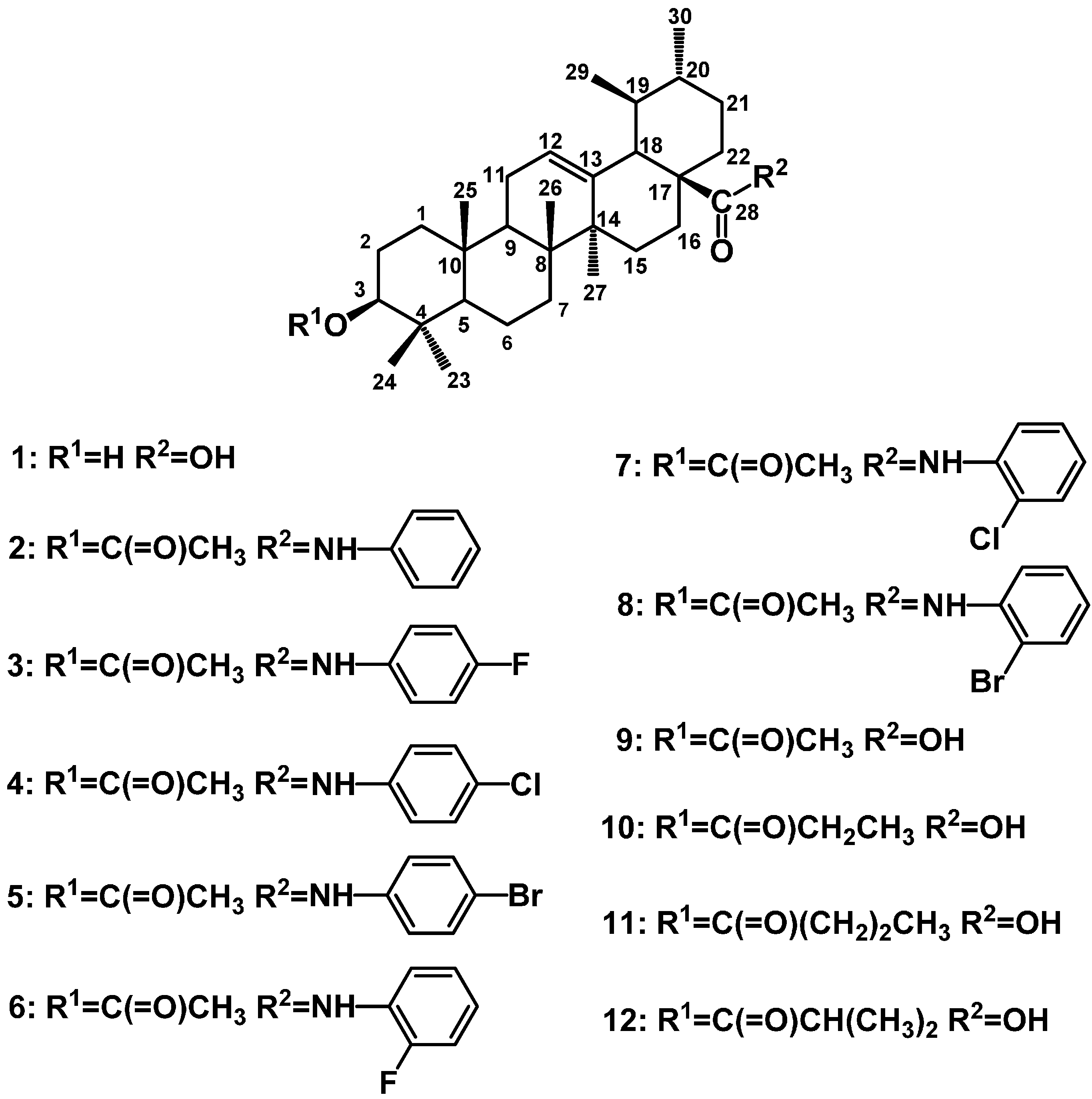
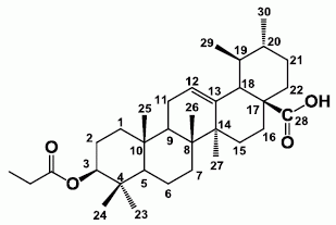
3.1. Preparation of UA and Its Derivatives

UA was purchased from Nanjing Zelang Medical Technology Co., Ltd. (Nanjing, China), with over 98% purity. UA derivatives (see Figure 1) were obtained from our research team [23]. These compounds were purified by column chromatography and their structures were all confirmed.

3.2. In vitro Cell Culture

Human intestinal Caco-2 cells (ATCC, Rockville, MD, USA) were incubated at 37 °C in a humidified atmosphere of 5% CO2 in air. The cells were cultured in Dulbecco’s modified Eagle’s modified Eagle’s medium (DMEM) with 10% heat-inactivated fetal bovine serum, 1% non-essential amino acids, 1% L-glutamine, 1% penicillin/streptomycin (Gibco Life Technologies, Grand Island, NY, USA). The cells were sub-cultured at confluence by 0.05% trypsin-0.5 mM EDTA treatment before they were applied in the experiment.

3.3. Cytotoxicity Assay

A MTT test was conducted to determine the possible toxicity of UA and its derivatives to Caco-2 cells. Briefly, cells were seeded at a density of 1 × 10⁴ cells/mL in a 96-well plate and incubated for 24 h. The next day, cells were incubated with vehicle (0.1% DMSO) and compounds at 100, 50, 25, 12.5 μmol/L for 24 h, and then 5 g/L MTT solution (20 μL) was added for 4 h. Absorbance at 570 nm was measured using a Multimodel Plate Reader (Infinite 200, Tecan, Mannedorf, Switzerland). The results were expressed as the percentage of control cells demonstrating cell viability after treated with the testing compounds.

3.4. Glucose Uptake by Caco-2 Cell Monolayers

Caco-2 cells, which were derived from human colon adenocarcinoma, were selected for intestinal absorption studies because these cells express the morphological characteristics and most of the functional properties of differentiated small-intestinal enterocytes [49]. Caco-2 cells were seeded on 24-well plates at the density of 2 × 10⁵ cells/well. The medium was changed every 1–2 days, and the culture was carried out for 13 days. Then Caco-2 cells were placed in serum-free media for 24 h and then they were washed twice with Hanks’ balanced salt solution (HBSS, pH 7.5, 140 mM NaCl, 5 mM KCl, 1.2 mM Na2HPO4, 2 mM CaCl2, 1.2 mM MgSO4, 20 mM HEPES, 0.2% bovine serum albumin) before the uptake studies. When a sodium free buffer was required, NaCl and Na2HPO4 were replaced with equal amounts of KCl and K2HPO4, respectively. After washing, the cells were incubated for 15 min at room temperature in HBSS before the commencement of this experiment. The uptake studies were initiated by adding HBSS containing either control or test solution (100 μM) and 2-BNDG (100 μM) for 30 min at 37 °C. Glucose uptake was stopped by adding twofold volume of ice-cold PBS and the wells were washed with ice-cold PBS three times. Fluorescent intensity was measured by a Multimodel Plate Reader (Tecan Infinity 200) with the 485 nmex and 535 nmemiss filter set before and after adding 2-NBDG. Then the cells were lysed in 200 μL lysis buffer (10 mM Tris-HCl pH = 7.4, 150 mM NaCl, 1% Triton-x-100, 1 mM EDTA, 0.1% SDS) and supplemented immediately before they were processed with 10 μg/mL PMSF. Cells were allowed to lyse on ice, and then vortexed and sonicated. The lysate was used for protein determination, protein concentrations were determined as described by BCA kit (Boster, Wuhan, China) with bovine serum albumin as standard.

3.5. Western Blot Analysis

Caco-2 cells were cultured on 6-well plates at a density of 1 × 10⁶ cells/well for 13 days. The cells were placed in serum-free medium for 24 h prior to the treatment. Then UA and its derivatives (100 μM) were added, after 6 h incubation, the cells were immediately washed three times with ice-cold PBS and lysed in 200 μL lysis buffer. Cells were allowed to lyse on ice, and then scraped, vortexed, sonicated and stored at −80 °C for further analysis. Protein concentrations were determined as described and cell lysate was subjected to western blot analysis [49]. Briefly, proteins with same amount were denatured in sample buffer, separated on 8% SDS-PAGE gel, and transferred onto a polyvinylidene diflouride membrane. The membranes were blocked for 1 h at 37 °C with 10% defatted milk in Tris-buffered saline (10 mM Tris-HCl, pH 7.5 and 137 mM NaCl) containing 0.05% Tween 20 (TBST). Membranes were then incubated overnight at 4 °C in blocking buffer with antibodies for GLUT-2, SGLT-1 and β-actin (EMD, Millipore, Billerica, MA, USA). Membranes were washed with TBST and incubated 1 h with appropriate secondary antibodies. Revelation was performed by using the enhanced chemiluminescence reagent and blue-light sensitive film. Densitometric analysis of films was performed with a Hewlett-Packard scanner equipped with a transparent adaptor and UN-SCAN-IT software.

3.6. In Vivo Experimental Animals

Adult male albino rats of Sprague-Dawley rats (180–220 g) were obtained from the Guangdong Medical Lab Animal Center, and maintained under standard laboratory conditions in a temperature (25–30 °C) and light-controlled (12-h light/dark cycle) room with 35%–60% humidity. The animals were acclimatized for 10 days before the experiments and provided with rodent chow and water ad libitum. The investigation was conducted in accordance with the Guide for the Care and Use of Laboratory Animals and approved by the institutional Animal Ethics Committee.

3.7. Oral Acute Toxicity Studies

Healthy adult SD rats were used for this test. The rats were fasted overnight and divided into 13 groups (n = 4), the rats were orally fed with compound 1, 10, 11 and 12 with an increasing dose of 100, 200 and 500 mg/kg body weight [50,51]. Control group was given vehicle solution (0.5% CMC-Na). These rats were continuously observed for 2 h for their behavioral, neurological, and autonomic symptoms and then observed once more after a period of 24 to 72 h for any sign of lethality or death [52].

3.8. Induction of Diabetes

Diabetes was induced by a single intraperitoneal injection of streptozotocin (Sigma-Aldrich, St. Louis, MO, USA) at a dose of 65 mg/kg body weight. STZ was dissolved in 0.1 M cold citrate buffer, pH 4.5. Control group were injected with citrate buffer alone. After 7 days for the development of diabetes, plasma glucose levels were determined of each rat. Rats with fasting blood glucose (FBG) range of above 16.7 mmol/L were considered as DM rats and were used for further study [53].

3.9. Experimental Procedure

According to the body weight and plasma glucose levels the rats were divided randomly into seven groups with six rats each and treated as follows:
  • Group I: Normal rats treated with 0.5% CMC-Na (2 mL/200 g body weight).
  • Group II: Diabetic rats treated with 0.5% CMC-Na.
  • Group III: Diabetic rats treated with 10 mg/kg of glibenclamide.
  • Group IV: Diabetic rats treated with 100 mg/kg of compound 1 (UA).
  • Group V: Diabetic rats treated with 100 mg/kg of compound 10.
  • Group VI: Diabetic rats treated with 100 mg/kg of compound 11.
  • Group VII: Diabetic rats treated with 100 mg/kg of compound 12.
The treatment was continued daily for 28 days. Blood glucose levels and body weight were measured every week to ascertain the status of diabetes. After 4 weeks of treatment, the animals were fasted for 12 h and sacrificed by cervical dislocation under mild anaesthetization, blood was collected into heparinized tubes and centrifuged at 2,000 rpm for 10 min. Livers and kidneys were removed, washed and homogenized in ice-cold normal saline. This homogenates were centrifuged at 3,000 rpm for 10 min at 4 °C, and the supernatants were collected for the estimate of antioxidant markers.

3.10. Biochemical Analysis

Fasting blood glucose levels were estimated on days 0, 7, 14, 28 with the single touch glucometer (Life Scan, Johnson & Johnson Company, New Brunswick, NJ, USA). Serum insulin level was measured by use of a commercial immunoassay kit by enzyme linked immunosorbent assay (Uscn. Life Science Inc., Wuhan, China). Serum total protein, albumin, triglycerides (TG), total cholesterol (TC), high-density lipoprotein cholesterol (HDL-C), and low-density lipoprotein cholesterol (LDL-C) were measured with an automatic biochemical analyzer (Sinnowa D240, Nanjing, China).

3.11. Assessment of Oxidative Stress Markers

The tissue homogenates of liver and kidney were used for the estimate of antioxidant markers. Glutathione (GSH) level was measured with 5, 5'-dithiobis-(2-nitrobenzoic acid) (DTNB) as described by Sedlak and Lindsay [54]. Superoxide dismutase (SOD) activity was assessed according to the method of Sinha [55]. Malondialdehyde (MDA) activity was estimated with the thiobarbituric acid method [56].

3.12. Statistical Analysis

All experiment results were expressed with mean ± standard deviation (SD). The significant differences between the means of the experimental groups were determined with the analysis of variance (ANOVA), followed by a Tukey–Kramer multiple comparisons test (Graph Pad version 5.0; Graph Pad Software Inc., San Diego, CA, USA). The values were considered significant when p < 0.05.

4. Conclusions

In summary, our study suggests that compound 10 displays an inhibitory effect on 2-NBDG uptake through inhibiting SGLT-1 and GLUT-2 transporter protein expression in Caco-2 cells. This observation was corroborated by its benefits in attenuating hyperglycemia, hyperlipidemia and oxidative stress in a DM rat model. Moreover, compound 10 is easily obtained from UA by one step structure modification [23]. These finding are supportive to the application of compound 10 as a potential source for the discovery of active new and active oral medicine for future therapy of diabetes and its complications.

Acknowledgments

This research was supported by National Natural Science Foundation of China (Grant No. 21172046). The authors are also grateful to the combined research project of Guangdong Province and Ministry of Education for financial Support (project No. 2011B090600033), Guangdong Province Higher Education “Qianbaishi Engineering”, Guangzhou Science and Technology Plan (project No. 3013Y2-00081). The authors also would like to thank the anonymous reviewers for their valuable comments.

Author Contributions

Suqing Zhao and Kun Zhang jointly suggested the research work and led the project. Panpan Wu and Ping He perform the experiment and analyzed the data. Panpan Wu, Tianming Huang and Yujing Lu led the writing, edited and revised the manuscript. All authors read and approved the manuscript.

Abbreviations

2-NBDG
2-[N-(7-nitrobenz-2-oxa-1, 3-diazol-4-yl) amino]-2-deoxyglucose
SGLT-1
Na+-dependent glucose transporter
GLUT-2
Na+-independent glucose transporter
DM
diabetes mellitus
UA
ursolic acid
STZ
streptozotocin
ROS
reactive oxygen species
GSH
glutathione
SOD
superoxide dismutase
MDA
malondialdehyde
TG
triglyceride
TC
total cholesterol
LDL-C
low density lipoprotein cholesterol
HDL-C
high density lipoprotein cholesterol

Conflicts of Interest

The authors declare no conflict of interest.

References

  1. WHO. “Fact Sheet # 312” to Be Found under. Available online: http://www.who.int/mediacentre/factsheets/fs312/en/ (accessed on 15 December 2013).
  2. Tuomilehto, J.; Lindström, J.; Eriksson, J.G.; Valle, T.T.; Hämäläinen, H.; Ilanne-Parikka, P.; Keinänen-Kiukaanniemi, S.; Laakso, M.; Louheranta, A.; Rastas, M. Prevention of type 2 diabetes mellitus by changes in lifestyle among subjects with impaired glucose tolerance. N. Engl. J. Med. 2001, 344, 1343–1350. [Google Scholar] [CrossRef]
  3. Zimmet, P.; Alberti, K.G.; Shaw, J. Global and societal implications of the diabetes epidemic. Nature 2001, 414, 782–787. [Google Scholar] [CrossRef]
  4. Lopez-Candales, A. Metabolic syndrome X: A comprehensive review of the pathophysiology and recommended therapy. J. Med. 2001, 32, 283–300. [Google Scholar]
  5. Hays, N.P.; Galassetti, P.R.; Coker, R.H. Prevention and treatment of type 2 diabetes: Current role of lifestyle, natural product, and pharmacological interventions. Pharmacol. Ther. 2008, 118, 181–191. [Google Scholar] [CrossRef]
  6. Hung, H.Y.; Qian, K.; Morris-Natschke, S.L.; Hsu, C.S.; Lee, K.H. Recent discovery of plant-derived anti-diabetic natural products. Nat. Prod. Rep. 2012, 29, 580–606. [Google Scholar] [CrossRef]
  7. Jia, W.; Gao, W.; Tang, L. Antidiabetic herbal drugs officially approved in China. Phytother. Res. 2003, 17, 1127–1134. [Google Scholar] [CrossRef]
  8. Roglic, G.; Unwin, N.; Bennett, P.H.; Mathers, C.; Tuomilehto, J.; Nag, S.; Connolly, V.; King, H. The burden of mortality attributable to diabetes: Realistic estimates for the year 2000. Diabetes Care 2005, 28, 2130–2135. [Google Scholar] [CrossRef]
  9. Harmand, P.O.; Duval, R.; Delage, C.; Simon, A. Ursolic acid induces apoptosis through mitochondrial intrinsic pathway and caspase-3 activation in M4Beu melanoma cells. Int. J. Cancer 2005, 114, 1–11. [Google Scholar] [CrossRef]
  10. Kim, D.K.; Baek, J.H.; Kang, C.M.; Yoo, M.; Sung, J.W.; Kim, D.K.; Chung, H.Y.; Kim, N.D.; Choi, Y.H.; Lee, S.H. Apoptotic activity of ursolic acid may correlate with the inhibition of initiation of DNA replication. Int. J. Cancer 2000, 87, 629–636. [Google Scholar]
  11. Baricevic, D.; Sosa, S.; Della, L.R.; Tubaro, A.; Simonovska, B.; Krasna, A.; Zupancic, A. Topical anti-inflammatory activity of Salvia officinalis L. leaves: The relevance of ursolic acid. J. Ethnopharmacol. 2001, 75, 125–132. [Google Scholar] [CrossRef]
  12. Kazmi, I.; Rahman, M.; Afzal, M.; Gupta, G.; Saleem, S.; Afzal, O.; Shaharyar, M.A.; Nautiyal, U.; Ahmed, S.; Anwar, F. Anti-diabetic potential of ursolic acid stearoyl glucoside: A new triterpenic gycosidic ester from Lantana camara. Fitoterapia 2012, 83, 142–146. [Google Scholar] [CrossRef]
  13. Ullevig, S.L.; Zhao, Q.; Zamora, D.; Asmis, R. Ursolic acid protects diabetic mice against monocyte dysfunction and accelerated atherosclerosis. Atherosclerosis 2011, 219, 409–416. [Google Scholar] [CrossRef]
  14. Zhou, Y.; Li, J.S.; Zhang, X.; Wu, Y.J.; Huang, K.; Zheng, L. Ursolic acid inhibits early lesions of diabetic nephropathy. Int. J. Mol. Med. 2010, 26, 565–570. [Google Scholar]
  15. Zou, C.; Wang, Y.; Shen, Z. 2-NBDG as a fluorescent indicator for direct glucose uptake measurement. J. Biochem. Biophys. Methods 2005, 64, 207–215. [Google Scholar] [CrossRef]
  16. Yamada, K.; Saito, M.; Matsuoka, H.; Inagaki, N. A real-time method of imaging glucose uptake in single, living mammalian cells. Nat. Protoc. 2007, 2, 753–762. [Google Scholar] [CrossRef]
  17. Hassanein, M.; Weidow, B.; Koehler, E.; Bakane, N.; Garbett, S.; Shyr, Y.; Quaranta, V. Development of high-throughput quantitative assays for glucose uptake in cancer cell lines. Mol. Imaging Biol. 2011, 13, 840–852. [Google Scholar] [CrossRef]
  18. Nistor, B.L.; Martineau, L.C.; Benhaddou-Andaloussi, A.; Arnason, J.T.; Levy, E.; Haddad, P.S. Inhibition of intestinal glucose absorption by anti-diabetic medicinal plants derived from the James Bay Cree traditional pharmacopeia. J. Ethnopharmacol. 2010, 132, 473–482. [Google Scholar] [CrossRef]
  19. Kim, H.K.; Baek, S.S.; Cho, H.Y. Inhibitory effect of pomegranate on intestinal sodium dependent glucose uptake. Am. J. Chin. Med. 2011, 39, 1015–1027. [Google Scholar] [CrossRef]
  20. Haidara, M.A.; Yassin, H.Z.; Rateb, M.; Ammar, H.; Zorkani, M.A. Role of oxidative stress in development of cardiovascular complications in diabetes mellitus. Curr. Vasc. Pharmacol. 2006, 4, 215–227. [Google Scholar] [CrossRef]
  21. Pitozzi, V.; Giovannelli, L.; Bardini, G.; Rotella, C.M.; Dolara, P. Oxidative DNA damage in peripheral blood cells in type 2 diabetes mellitus: Higher vulnerability of polymorphonuclear leukocytes. Mutat. Res. 2003, 529, 129–133. [Google Scholar] [CrossRef]
  22. Evans, J.L. Antioxidants: Do they have a role in the treatment of insulin resistance? Indian J. Med. Res. 2007, 125, 355–372. [Google Scholar]
  23. Wu, P.P.; Zhang, K.; Lu, Y.J.; He, P.; Zhao, S.Q. In vitro and in vivo evaluation of the antidiabetic activity of ursolic acid derivatives. Eur. J. Med. Chem. 2014, 80, 502–508. [Google Scholar] [CrossRef]
  24. Sambuy, Y.; de Angelis, I.; Ranaldi, G.; Scarino, M.L.; Stammati, A.; Zucco, F. The Caco-2 cell line as a model of the intestinal barrier: Influence of cell and culture-related factors on Caco-2 cell functional characteristics. Cell. Biol. Toxicol. 2005, 21, 1–26. [Google Scholar] [CrossRef]
  25. Delie, F.; Rubas, W. A human colonic cell line sharing similarities with enterocytes as a model to examine oral absorption: advantages and limitations of the Caco-2 model. Drug Carrier Syst. 1997, 14, 221–286. [Google Scholar]
  26. Mahraoui, L.; Rodolosse, A.; Barbat, A.; Dussaulx, E.; Zweibaum, A.; Rousset, M.; Brot-Laroche, E. Presence and differential expression of SGLT1, GLUT1, GLUT2, GLUT3 and GLUT5 hexose-transporter mRNAs in Caco-2 cell clones in relation to cell growth and glucose consumption. Biochem. J. 1994, 298, 629–633. [Google Scholar]
  27. Jang, S.M.; Yee, S.T.; Choi, J.; Choi, M.S.; Do, G.M.; Jeon, S.M.; Yeo, J.; Kim, M.J.; Seo, K.I.; Lee, M.K. Ursolic acid enhances the cellular immune system and pancreatic beta-cell function in streptozotocin-induced diabetic mice fed a high-fat diet. Int. Immunopharmacol. 2009, 9, 113–119. [Google Scholar] [CrossRef]
  28. Jang, S.M.; Kim, M.J.; Choi, M.S.; Kwon, E.Y.; Lee, M.K. Inhibitory effects of ursolic acid on hepatic polyol pathway and glucose production in streptozotocin-induced diabetic mice. Metabolism 2010, 59, 512–519. [Google Scholar] [CrossRef]
  29. Arya, A.; Looi, C.Y.; Cheah, S.C.; Mustafa, M.R.; Mohd, M.A. Anti-diabetic effects of Centratherum anthelminticum seeds methanolic fraction on pancreatic cells, beta-TC6 and its alleviating role in type 2 diabetic rats. J. Ethnopharmacol. 2012, 144, 22–32. [Google Scholar] [CrossRef]
  30. Poitout, V.; Stout, L.E.; Armstrong, M.B.; Walseth, T.F.; Sorenson, R.L.; Robertson, R.P. Morphological and functional characterization of beta TC-6 cells-an insulin-secreting cell line derived from transgenic mice. Diabetes 1995, 44, 306–313. [Google Scholar] [CrossRef]
  31. Yamada, K.; Nakata, M.; Horimoto, N.; Saito, M.; Matsuoka, H.; Inagaki, N. Measurement of glucose uptake and intracellular calcium concentration in single, living pancreatic beta-cells. J. Biol. Chem. 2000, 275, 22278–22283. [Google Scholar] [CrossRef]
  32. Masumoto, S.; Akimoto, Y.; Oike, H.; Kobori, M. Dietary phloridzin reduces blood glucose levels and reverses Sglt1 expression in the small intestine in streptozotocin-induced diabetic mice. J. Agric. Food. Chem. 2009, 57, 4651–4656. [Google Scholar] [CrossRef]
  33. Goto, T.; Horita, M.; Nagai, H.; Nagatomo, A.; Nishida, N.; Matsuura, Y.; Nagaoka, S. Tiliroside, a glycosidic flavonoid, inhibits carbohydrate digestion and glucose absorption in the gastrointestinal tract. Mol. Nutr. Food Res. 2012, 56, 435–445. [Google Scholar] [CrossRef]
  34. Kellett, G.L.; Brot-Laroche, E.; Mace, O.J.; Leturque, A. Sugar absorption in the intestine: The role of GLUT2. A. Leturque. Annu. Rev. Nutr. 2008, 28, 35–54. [Google Scholar] [CrossRef]
  35. Brown, G.K. Glucose transporters: Structure, function and consequences of deficiency. Metab. Dis. 2000, 23, 237–246. [Google Scholar]
  36. Manzano, S.; Williamson, G. Polyphenols and phenolic acids from strawberry and apple decrease glucose uptake and transport by human intestinal Caco-2 cells. Mol. Nutr. Food Res. 2010, 54, 1773–1780. [Google Scholar] [CrossRef]
  37. Kellett, G.L.; Brot-Laroche, E. Apical GLUT2: A major pathway of intestinal sugar absorption. Diabetes 2005, 54, 3056–3062. [Google Scholar] [CrossRef]
  38. Almdal, T.P.; Vilstrup, H. Strict insulin therapy normalises organ nitrogen contents and the capacity of urea nitrogen synthesis in experimental diabetes in rats. Diabetologia 1988, 31, 114–118. [Google Scholar] [CrossRef]
  39. Knopp, R.H.; Retzlaff, B.; Aikawa, K.; Kahn, S.E. Management of patients with diabetic hyperlipidemia. Am. J. Cardiol. 2003, 91 (Suppl. 1), 24–28. [Google Scholar] [CrossRef]
  40. Kavalali, G.; Tuncel, H.; Goksel, S.; Hatemi, H.H. Hypoglycemic activity of Urtica pilulifera in streptozotocin-diabetic rats. J. Ethnopharmacol. 2003, 84, 241–245. [Google Scholar] [CrossRef]
  41. Cemek, M.; Kaga, S.; Simsek, N.; Buyukokuroglu, M.E.; Konuk, M. Antihyperglycemic and antioxidative potential of Matricaria chamomilla L. in streptozotocin-induced diabetic rats. J. Nat. Med. 2008, 62, 284–293. [Google Scholar] [CrossRef]
  42. Fang, Y.Z.; Yang, S.; Wu, G. Free radicals, antioxidants, and nutrition. Nutrition 2002, 18, 872–879. [Google Scholar] [CrossRef]
  43. Griesmacher, A.; Kindhauser, M.; Andert, S.E.; Schreiner, W.; Toma, C.; Knoebl, P.; Pietschmann, P.; Prager, R.; Schnack, C.; Schernthaner, G.; et al. Enhanced serum levels of thiobarbituric-acid-reactive substances in diabetes mellitus. Am. J. Med. 1995, 98, 469–475. [Google Scholar] [CrossRef]
  44. Dewanjee, S.; Das, A.K.; Sahu, R.; Gangopadhyay, M. Antidiabetic activity of Diospyros peregrina fruit: effect on hyperglycemia, hyperlipidemia and augmented oxidative stress in experimental type 2 diabetes. Food Chem. Toxicol. 2009, 47, 2679–2685. [Google Scholar] [CrossRef]
  45. Blake, D.R.; Allen, R.E.; Lunec, J. Free radicals in biological systems-A review orientated to inflammatory processes. Br. Med. Bull. 1987, 43, 371–385. [Google Scholar]
  46. Lin, Y.F.; Tsai, H.L.; Lee, Y.C.; Chang, S.J. Maternal vitamin E supplementation affects the antioxidant capability and oxidative status of hatching chicks. J. Nutr. 2005, 135, 2457–2461. [Google Scholar]
  47. Carr, M.C.; Brunzell, J.D. Abdominal obesity and dyslipidemia in the metabolic syndrome: Importance of type 2 diabetes and familial combined hyperlipidemia in coronary artery disease risk. J. Clin. Endocrinol. Metab. 2004, 89, 2601–2607. [Google Scholar] [CrossRef]
  48. Yoshihara, T.; Kumashiro, N.; Kanazawa, Y.; Mita, T.; Sakurai, Y.; Kawai, J.; Abe, M.; Motojima, K.; Hara, K.; Yamazaki, Y.; et al. Therapeutic efficacy of mitiglinide combined with once daily insulin glargine after switching from multiple daily insulin regimen of aspart insulin and glargine in patients with type 2 diabetes mellitus. Endocr. J. 2006, 53, 67–72. [Google Scholar] [CrossRef]
  49. Burnette, W.N. “Western blotting”: Electrophoretic transfer of proteins from sodium dodecyl sulfate-Polyacrylamide gels to unmodified nitrocellulose and radiographic detection with antibody and radioiodinated protein A. Anal. Biochem. 1981, 112, 195–203. [Google Scholar] [CrossRef]
  50. Shirwaikar, A.; Rajendran, K.; Barik, R. Effect of aqueous bark extract of Garuga pinnata Roxb. in streptozotocin-nicotinamide induced type-II diabetes mellitus. J. Ethnopharmacol. 2006, 107, 285–290. [Google Scholar] [CrossRef]
  51. Ghosh, M.N. Fundamentals of experimental pharmacology. Indian J. Pharmacol. 2007, 39, 216. [Google Scholar] [CrossRef]
  52. Turner, R.A.; Hebborn, P. Screening Methods in Pharmacology; Academic Press: New York, NY, USA, 1965; Volume 1, pp. 129–135. [Google Scholar]
  53. Sarkar, S.; Pranava, M.; Marita, R. Demonstration of the hypoglycemic action of Momordica charantia in a validated animal model of diabetes. Pharmacol. Res. 1996, 33, 1–4. [Google Scholar] [CrossRef]
  54. Sedlak, J.; Lindsay, R.H. Estimation of total, protein-bound, and nonprotein sulfhydryl groups in tissue with Ellman’s reagent. Anal. Biochem. 1968, 25, 192–205. [Google Scholar] [CrossRef]
  55. Sinha, A.K. Colorimetric assay of catalase. Anal. Biochem. 1972, 47, 389–394. [Google Scholar] [CrossRef]
  56. Draper, H.H.; Hadley, M. Malondialdehyde determination as index of lipid peroxidation. Method. Enzymol. 1990, 186, 421–431. [Google Scholar] [CrossRef]
  • Sample Availability: Samples of the compounds are available from the authors.

