Differences in the Phenolic Composition and Antioxidant Properties between Vitis coignetiae and Vitis vinifera Seeds Extracts

 ,
, 
Abstract
:1. Introduction
2. Results and Discussion
2.1. Content of Total Phenolics

| Solvent used for extraction | Species | Total phenolics (mg/g FW) | TEAC (mmol Trolox/g FW) |
|---|---|---|---|
| 80% methanol | Vitis coignetiae | 8.99 ± 0.06 a | 0.071 ± 0.002 a |
| Vitis vinifera | 21.21 ± 0.05 b | 0.152 ±0.002 b | |
| 80% acetone | Vitis coignetiae | 12.81 ± 0.16 a | 0.094 ± 0.002 a |
| Vitis vinifera | 30.02 ± 0.99 b | 0.196 ± 0.002 b |
2.2. Content of Condensed Tannins
| Solvent used for extraction | Species | Protein precipitation method (Absorbance at 510 nm/mg) | Vanillin/HCl method (Absorbance at 500 nm/mg) |
|---|---|---|---|
| 80% methanol | Vitis coignetiae | 0.223 ± 0.002 a | 0.305 ± 0.004 a |
| Vitis vinifera | 0.467 ± 0.005 b | 0.697 ± 0.005 b | |
| 80% acetone | Vitis coignetiae | 0.288 ± 0.007 a | 0.371 ± 0.003 a |
| Vitis vinifera | 0.566 ± 0.005 b | 0.817 ± 0.005 b |
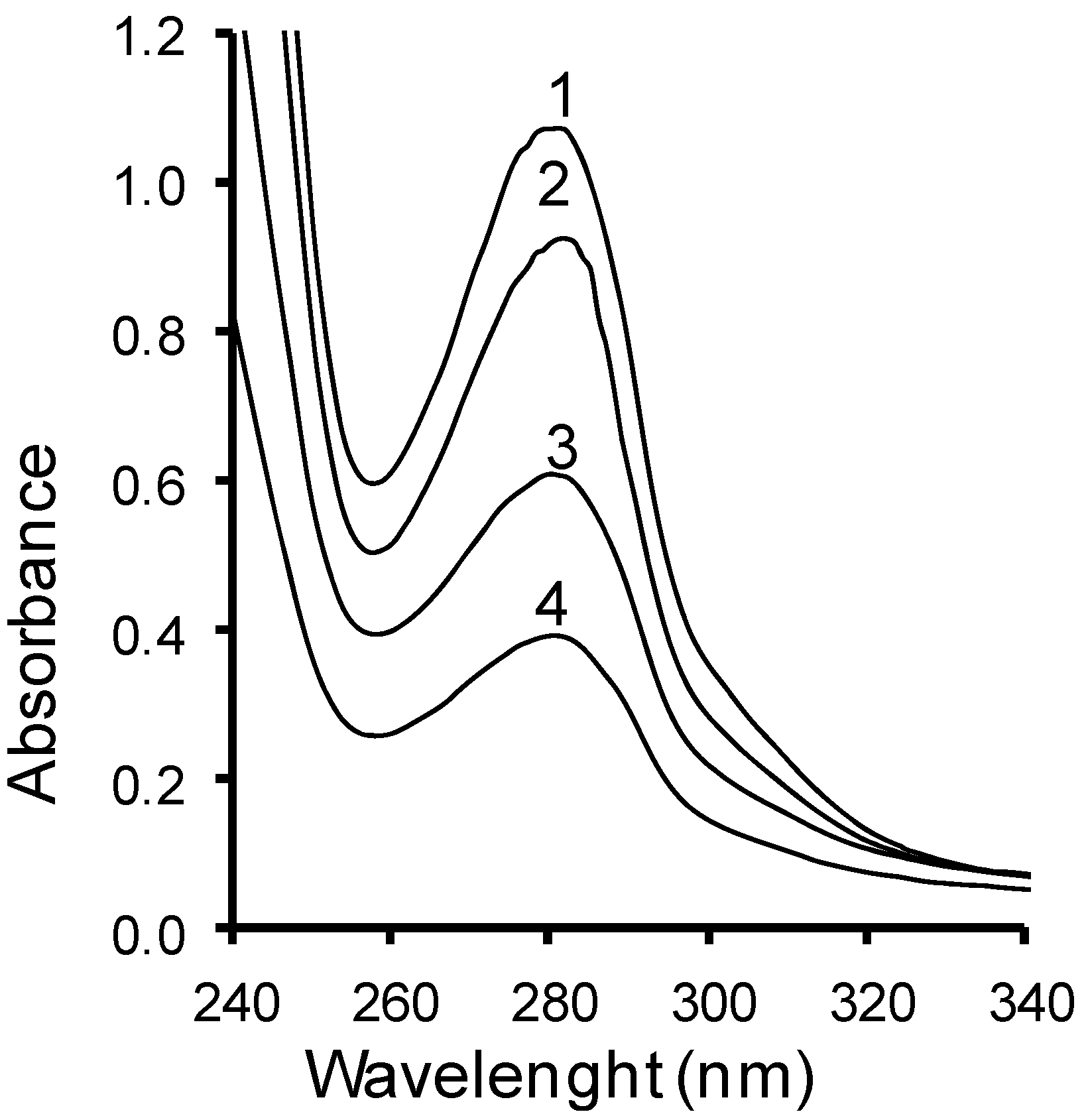
2.3. UV Spectra

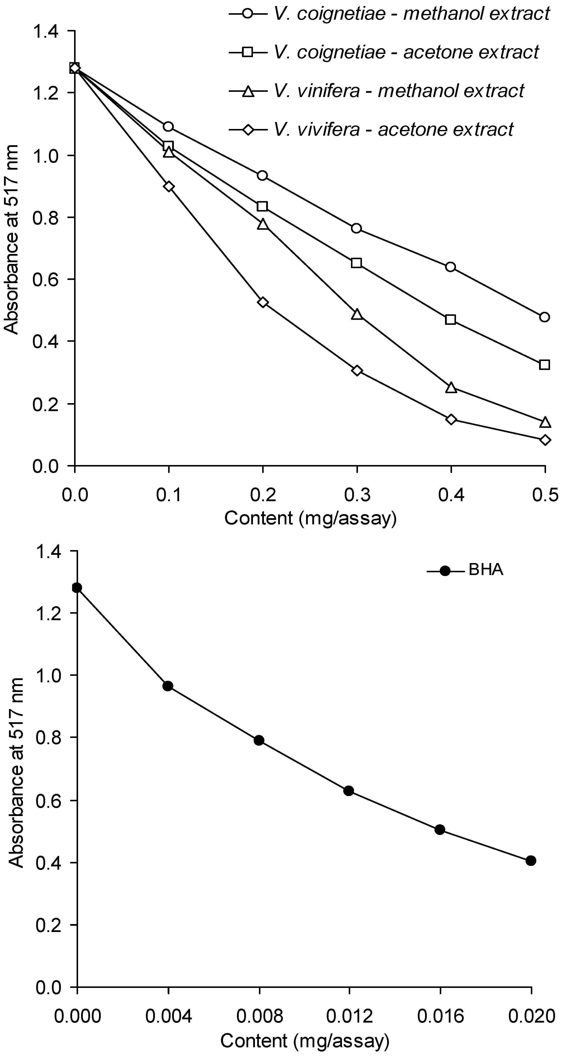
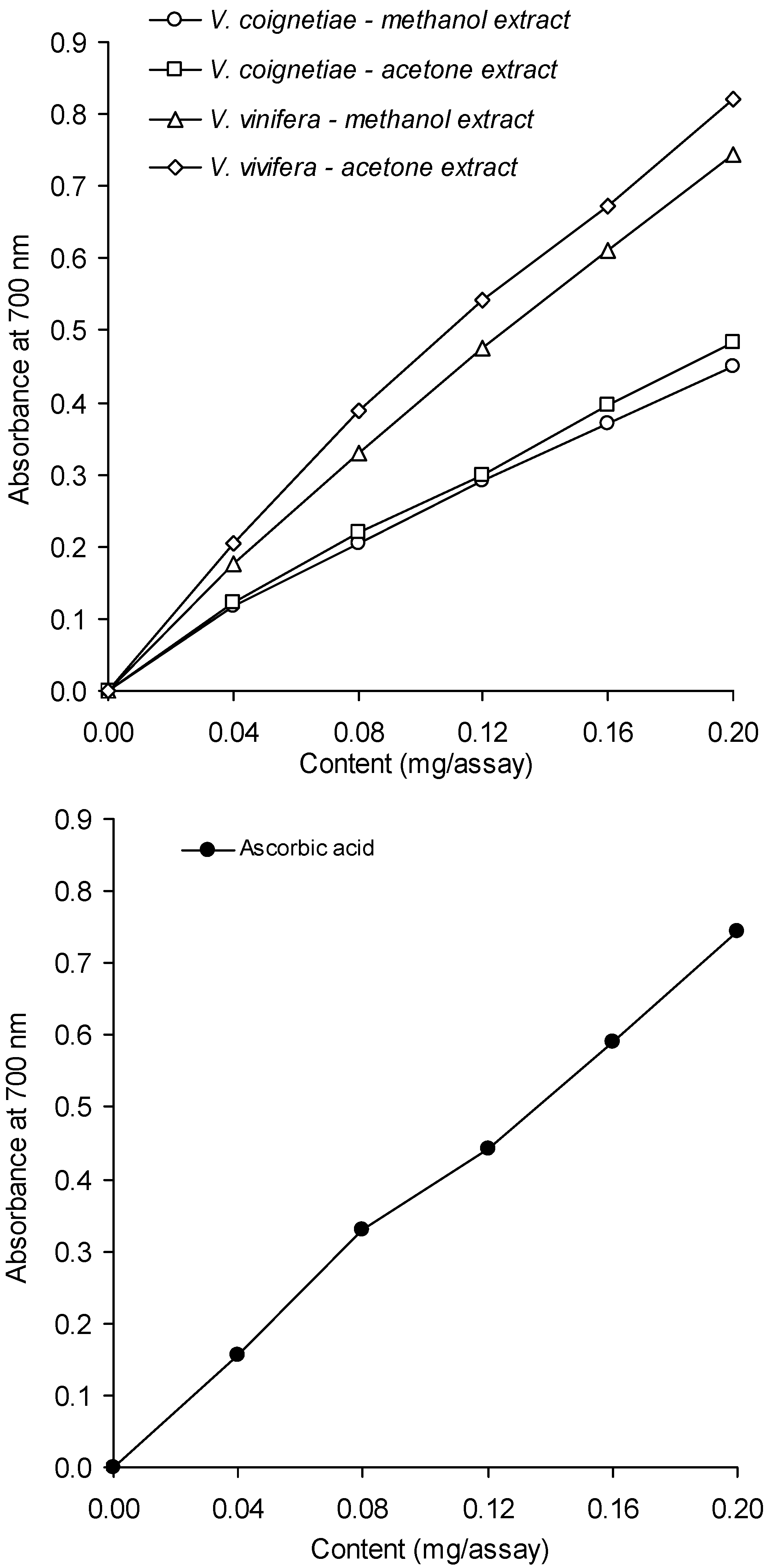
2.4. Antioxidant Activity


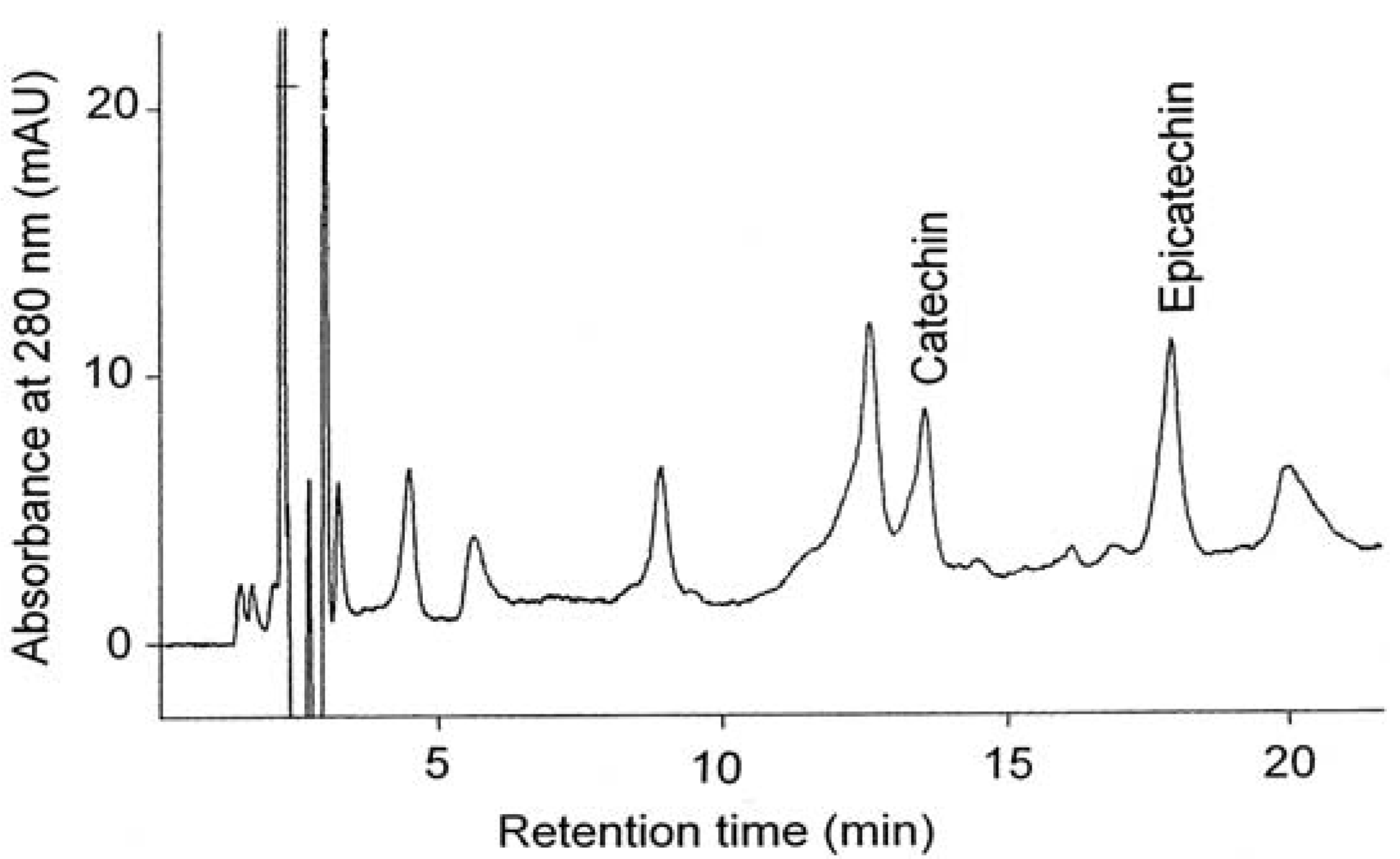
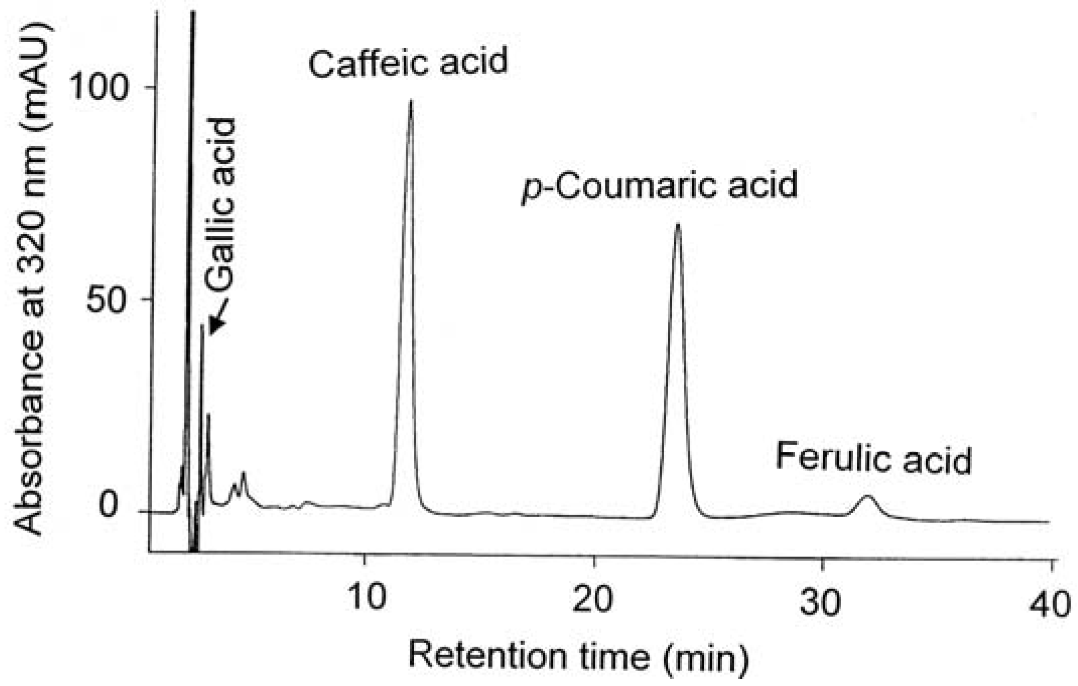
2.5. Content of Catechins and Phenolic Acids


| Solvent used for extraction | Species | (+)-Catechin (mg/g FW) | (−)-Epicatechin (mg/g FW) | (+)-Catechin + (−)-epicatechin (mg/g FW) |
|---|---|---|---|---|
| 80% methanol | Vitis coignetiae | 0.32 ± 0.02 a | 0.29 ± 0.01 a | 0.61 ± 0.18 a |
| Vitis vinifera | 0.53 ± 0.03 b | 0.45 ± 0.02 b | 0.98 ± 0.05 b | |
| 80% acetone | Vitis coignetiae | 0.38 ± 0.02 a | 0.36 ± 0.03 a | 0.74 ± 0.04 a |
| Vitis vinifera | 0.60 ± 0.03 b | 0.47 ± 0.03 b | 1.07 ± 0.06 b |
| Phenolic acid | Solvend used for extraction | Species | Form of phenolic acid | Content (mg/g FW) |
|---|---|---|---|---|
| Gallic | 80% methanol | Vitis coignetiae | Free | 35 ± 1 a |
| Esterified | 180 ± 6 a | |||
| Glucosided | 37 ± 1 a | |||
| Total | 252 ± 8 a | |||
| Vitis vinifera | Free | 51 ± 0.2 b | ||
| Esterified | 665 ± 19 b | |||
| Glucosided | 59 ± 2 b | |||
| Total | 774 ± 23 b | |||
| 80% acetone | Vitis coignetiae | Free | 38 ± 1 a | |
| Esterified | 355 ± 12 a | |||
| Glucosided | 47 ± 2 a | |||
| Total | 439 ± 13 a | |||
| Vitis vinifera | Free | 121 ± 3 b | ||
| Esterified | 860 ± 28 b | |||
| Glucosided | 76 ± 2 b | |||
| Total | 1057 ± 35 b | |||
| p-Coumaric | 80% methanol | Vitis coignetiae | Free | 1.56 ± 0.24 a |
| Esterified | 3.97 ± 0.26 a | |||
| Glucosided | 0.35 ± 0.05 a | |||
| Total | 5.88 ± 0.55 a | |||
| Vitis vinifera | Free | 3.25 ± 0.49 b | ||
| Esterified | 10.58 ± 0.53 b | |||
| Glucosided | traces | |||
| Total | 13.83 ± 1.02 b | |||
| 80% acetone | Vitis coignetiae | Free | - | |
| Esterified | 5.67 ± 0.53 a | |||
| Glucosided | 0.49 ± 0.09 a | |||
| Total | 6.16 ± 0.26 a | |||
| Vitis vinifera | Free | - | ||
| Esterified | 11.24 ± 0.87 b | |||
| Glucosided | traces | |||
| Total | 11.24 ± 0.87 b |
| Phenolic acid | Solvent used for extraction | Species | Content (μg/g FW) |
|---|---|---|---|
| Caffeic | 80% methanol | Vitis coignetiae | 3.78 ± 0.08 a |
| Vitis vinifera | 2.73 ± 0.42 a | ||
| 80% acetone | Vitis coignetiae | 3.36 ± 0.43 a | |
| Vitis vinifera | 2.35 ± 0.25 a | ||
| Ferulic | 80% methanol | Vitis coignetiae | 0.86 ± 0.14 a |
| Vitis vinifera | 1.95 ± 0.17 a | ||
| 80% acetone | Vitis coignetiae | trace | |
| Vitis vinifera | 2.35 ± 0.31 a |
3. Experimental
3.1. Chemicals
3.2. Plant Material
3.3. Extract Preparation
3.4. Content of Total Phenolics
3.5. Determination of Condensed Tannins by the Vanillin Method
3.6. Determination of Condensed Tannins by the Protein Precipitation Method
3.7. UV Spectra
3.8. Total Antioxidant Capacity (TAC)
3.9. Antiradical Activity of Seed Extracts against the DPPH Radical
3.10. Reducing Power of the Extracts
3.11. Separation and Analysis of Phenolic Acids by HPLC
3.12. Analysis of Catechins with HPLC
3.13. Statistical Analysis
4. Conclusions
References
- Shi, J.; Yu, J.; Pohorly, J.E.; Kakuda, Y. Polyphenolics in grape seeds—Biochemistry and functionality. J. Med. Food 2003, 6, 291–299. [Google Scholar] [CrossRef]
- Nunez, V.; Gomez-Cordoves, C.; Bartolome, B.; Hong, Y.; Mitchel, A.E. Non-galloylated and galloylated proanthocyanidin oligomers in grape seeds from Vitis vinifera L. cv. Graciano, Tempranillo and Cabernet Sauvignon. J. Sci. Agric. 2006, 86, 915–921. [Google Scholar] [CrossRef]
- Kennedy, J.A.; Troup, G.J.; Pilbrow, J.R.; Hutton, D.R.; Hewitt, D.; Hunter, C.R.; Ristic, R.; Iland, P.G.; Jones, G.P. Development of seed polyphenols in berries from Vitis vinifera L. CV. Shiraz. Aust. J. Grape Wine Res. 2000, 6, 244–254. [Google Scholar] [CrossRef]
- Guendez, R.; Kallithraka, S.; Makaris, D.P.; Kefalas, P. Determination of low molecular weight polyphenolic constituents in grape (Vitis vinifera sp.) seed extracts: Correlation with antiradical activity. Food Chem. 2005, 89, 1–9. [Google Scholar] [CrossRef]
- Simonetti, P.; Ciappellano, S.; Gardana, C.; Bramati, L.; Pietta, P. Procyanidins fro Vitis vinifera seeds: In vivo effects on oxidative stress. J. Agric. Food Chem. 2002, 50, 6217–6221. [Google Scholar] [CrossRef]
- Peng, Z.; Hayasaka, Y.; Iland, P.G.; Sefton, M.; Høj, P.; Waters, E.J. Quantitative analysis of polymeric procyanidins (tannins) from grape (Vitis vinifera) seeds by reverse phase high-performance liquid chromatography. J. Agric. Food Chem. 2001, 49, 26–31. [Google Scholar]
- Amarowicz, R.; Weidner, S. Biological activity of grapevine phenolic compounds. In Grapevine Molecular Physiology and Biochemistry, 2nd ed.; Roubelakis-Angelakis, K.A., Ed.; Springer Science + Business Media B.V.: Heidelberg, Germany, 2009; pp. 389–405. [Google Scholar]
- Saito, M.; Hosoyama, H.; Ariga, T.; Kataoka, S.; Yamaji, N. Antiulcer activity of grape seed extract and procyanidins. J. Agric. Food Chem. 1998, 46, 1460–1464. [Google Scholar] [CrossRef]
- Bonarska-Kujawa, D.; Sarapuk, J.; Bielecki, K.; Oszmiański, J.; Kleszczyńska, H. Antioxidant activity of extracts from apple, chokeberry and strawberry. Pol. J. Food Nutr. Sci. 2012, 62, 229–234. [Google Scholar]
- Ali, K.; Meltese, F.; Choi, Y.H.; Verporte, R. Metabolic constituents of grapevine and grape-derived products. Phytochem. Res. 2010, 9, 357–378. [Google Scholar] [CrossRef]
- Baydar, N.G.; Akkurt, M. Oil content and oil quality properties of some grape seeds. Turk. J. Agric. For. 2001, 25, 163–168. [Google Scholar]
- Narożna, D.; Mądrzak, C. Plant gens coding some enzymes of the phenylpropanoids. Biotechnologia 1999, 3, 154–165. [Google Scholar]
- Weidner, S.; Karolak, M.; Karamać, M.; Kosińska, A.; Amarowicz, R. Phenolic compounds and properties of antioxidants in grapevine roots (Vitis vinifera L.) under drought stress followed by recovery. Acta Soc. Bot. Pol. 2009, 78, 97–103. [Google Scholar] [CrossRef]
- Weidner, S.; Brosowska-Arendt, W.; Szczechura, W.; Karamać, M.; Kosińska, A.; Amarowicz, R. Effect of osmotic stress and post-stress recovery on the content of phenolics and properties of antioxidants in germinating seeds of grapevine Vitis californica. Acta Soc. Bot. Pol. 2011, 80, 11–19. [Google Scholar] [CrossRef]
- Dixon, R.A.; Paiva, N.L. Stress-induced phenylopropanoid metabolism. Plant Cell 1991, 7, 1085–1097. [Google Scholar]
- Janas, K.M.; Cvikrova, M.; Pałągiewicz, A.; Eder, J. Alternation in phenyl-propanoid content in soybean root during low temperature acclimation. Plant Physiol. Biochem. 2000, 38, 587–593. [Google Scholar] [CrossRef]
- Baydar, N.G.; Ozkan, G.; Sagdic, O. Total phenolic contents and antibacterial activities of grape (Vitis vinifera L.) extracts. Food Control 2004, 15, 335–339. [Google Scholar] [CrossRef]
- Weidner, S.; Powałka, A.; Karamać, M.; Amarowicz, R. Extracts of phenolic compounds from seeds of three wild grapevines—Comparison of their antioxidant activities and the content of phenolic compounds. Int. J. Mol. Sci. 2012, 13, 3444–3457. [Google Scholar] [CrossRef]
- Shafiee, M.; Carbonneau, M.A.; Urban, N.; Descomps, B.; Leger, C.L. Grape and grape seed extract capacities at protecting LDL against oxidation generated by Cu2+, AAPH or SIN-1 and decreasing superoxide THP-1 cell production. A comparison to other extracts or compounds. Free Radic. Res. 2003, 37, 573–584. [Google Scholar] [CrossRef]
- Frankel, E.N.; Waterhouse, A.L.; Teissedre, P.L. Principal phenolic phytochemicals in selected California wines and their antioxidant activity in inhibiting oxidation of human low-density lipoproteins. J. Agric. Food Chem. 1995, 43, 890–894. [Google Scholar] [CrossRef]
- Sanchez-Moreno, C.; Jimenez-Esrig, A.; Saura-Calixto, F. LDL oxidizability indexes in measurement of antioxidant activity in selected Spanish wines. Nutr. Res. 2002, 22, 507–517. [Google Scholar] [CrossRef]
- Weidner, S.; Karamać, M.; Amarowicz, R.; Szypulska, E.; Gołgowska, A. Changes in composition of phenolic compounds and antioxidant properties of Vitis amurensis seeds germinated under osmotic stress. Acta Physiol. Plant. 2007, 29, 283–290. [Google Scholar] [CrossRef]
- Da Silva, J.M.R.; Darmon, N.; Fernandez, Y.; Mitjavila, S. Oxygen free radical scavenger capacity in aqueous models of different procyanidins from grape seeds. J. Agric. Food Chem. 1991, 39, 1549–1552. [Google Scholar] [CrossRef]
- Pekić, B.; Kovać, V.; Alonso, E.; Revilla, E. Study of extraction of proanthocyanidins from grape seeds. Food Chem. 1998, 61, 201–206. [Google Scholar] [CrossRef]
- Kosińska, A.; Karamać, M. Antioxidant capacity of roasted health-promoting products. Pol. J. Food Nutr. Sci. 2006, 56, 80–84. [Google Scholar]
- Yen, G.-C.; Chen, H.-Y. Antioxidant activity of various tea extracts in relation to their antimutagenicity. J. Agric. Food Chem. 1995, 43, 27–32. [Google Scholar] [CrossRef]
- Escribano-Bailón, T.; Gutierrez-Fernandez, Y.; Rivas-Gonzolo, J.; Santos-Buelga, C. Characterization of procyanidins of Vitis vinifera v. Tinta Del Pais grape seeds. J. Agric. Food Chem. 1992, 40, 1794–1799. [Google Scholar] [CrossRef]
- Amarowicz, R.; Karamać, M.; Weidner, S.; Abe, S.; Shahidi, F. Antioxidant activity of wheat caryopses and embryos extracts. J. Food Lipids 2002, 9, 201–210. [Google Scholar] [CrossRef]
- Amarowicz, R.; Pegg, R.B.; Rahimi-Moghaddam, P.; Barl, B.; Weil, J.A. Free-radical scavenging capacity and antioxidant activity of selected plant species from the Canadian prairies. Food Chem. 2004, 84, 551–562. [Google Scholar] [CrossRef]
- Price, N.J.; van Scoyoc, S.; Butler, L.G. A critical evaluation of the vanillic reactions in an assay for tannin in sorghum grain. J. Agric. Food Chem. 1978, 26, 1214–1218. [Google Scholar] [CrossRef]
- Hagerman, A.; Butler, L. Protein precipitation method for quantitative determination of tannins. J. Agric. Food Chem. 1978, 26, 809–811. [Google Scholar] [CrossRef]
- Re, R.; Pellegrini, N.; Proteggente, A.; Pannala, A.; Yang, M.; Rice-Evans, C. Antioxidant activity applying an improved ABTS radical cation decolorization assay. Free Radic. Biol. Med. 1999, 26, 1231–1237. [Google Scholar] [CrossRef]
- Oyaizu, M. Studies on products of browning reaction—Antioxidative activities of products of browning reaction prepared from glucosamine. Jpn. J. Nutr. 1978, 44, 307–315. [Google Scholar] [CrossRef]
- Weidner, S.; Amarowicz, R.; Karamać, M.; Frączek, E. Changes in endogenous phenolic acids during development of Secale cereale caryopses and after dehydration treatment of unripe rye grains. Plant Physiol. Biochem. 2000, 38, 595–602. [Google Scholar] [CrossRef]
- Weidner, S.; Amarowicz, R.; Karamać, M.; Dąbrowski, G. Phenolic acids in caryopses of two cultivars of wheat, rye and triticale that display different resistance to pre-harvest sprouting. Eur. Food Res. Technol. 1999, 210, 109–113. [Google Scholar] [CrossRef]
- Oszmiański, J.; Bourzeix, M. Preparation of catechin and procyandidin standards from hawthorn (Crataegus azarolus L.) and pine (Pine mesogeensis fieschi) barks. Pol. J. Food Nutr. Sci. 1995, 45, 89–96. [Google Scholar]
- Sample Availability: Samples of the extracts are available from the authors.
© 2013 by the authors; licensee MDPI, Basel, Switzerland. This article is an open access article distributed under the terms and conditions of the Creative Commons Attribution license (http://creativecommons.org/licenses/by/3.0/).
Share and Cite
Weidner, S.; Rybarczyk, A.; Karamać, M.; Król, A.; Mostek, A.; Grębosz, J.; Amarowicz, R. Differences in the Phenolic Composition and Antioxidant Properties between Vitis coignetiae and Vitis vinifera Seeds Extracts. Molecules 2013, 18, 3410-3426. https://doi.org/10.3390/molecules18033410
Weidner S, Rybarczyk A, Karamać M, Król A, Mostek A, Grębosz J, Amarowicz R. Differences in the Phenolic Composition and Antioxidant Properties between Vitis coignetiae and Vitis vinifera Seeds Extracts. Molecules. 2013; 18(3):3410-3426. https://doi.org/10.3390/molecules18033410
Chicago/Turabian StyleWeidner, Stanisław, Anna Rybarczyk, Magdalena Karamać, Angelika Król, Agnieszka Mostek, Joanna Grębosz, and Ryszard Amarowicz. 2013. "Differences in the Phenolic Composition and Antioxidant Properties between Vitis coignetiae and Vitis vinifera Seeds Extracts" Molecules 18, no. 3: 3410-3426. https://doi.org/10.3390/molecules18033410