Bioengineering 2024, 11(5), 444; https://doi.org/10.3390/bioengineering11050444 - 30 Apr 2024
Cited by 19 | Viewed by 13759
Abstract
The field of peripheral nerve regeneration is a dynamic and rapidly evolving area of research that continues to captivate the attention of neuroscientists worldwide. The quest for effective treatments and therapies to enhance the healing of peripheral nerves has gained significant momentum in
[...] Read more.
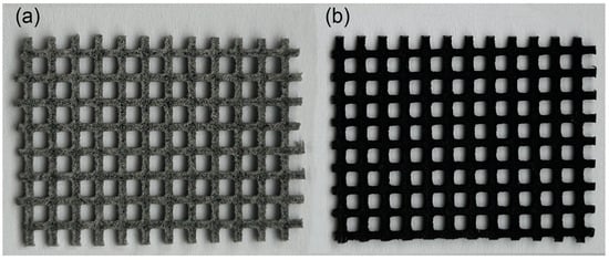
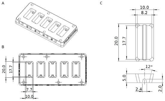
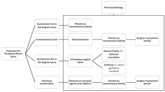
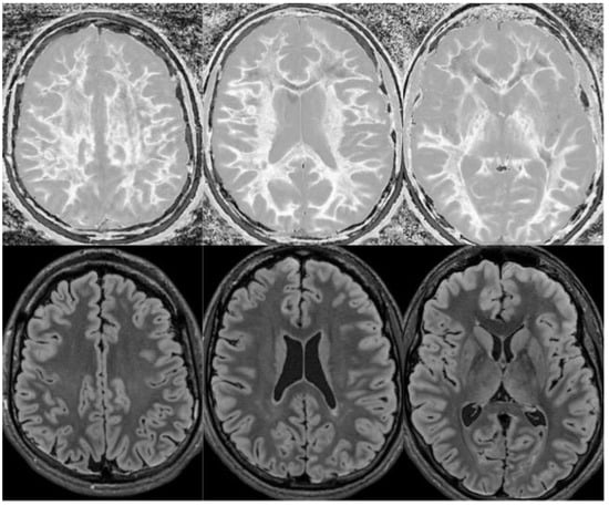
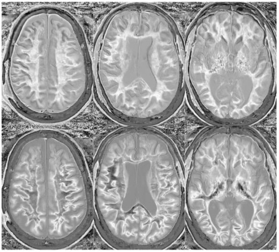
The field of peripheral nerve regeneration is a dynamic and rapidly evolving area of research that continues to captivate the attention of neuroscientists worldwide. The quest for effective treatments and therapies to enhance the healing of peripheral nerves has gained significant momentum in recent years, as evidenced by the substantial increase in publications dedicated to this field. This surge in interest reflects the growing recognition of the importance of peripheral nerve recovery and the urgent need to develop innovative strategies to address nerve injuries. In this context, this article aims to contribute to the existing knowledge by providing a comprehensive review that encompasses both biomaterial and clinical perspectives. By exploring the utilization of nerve guidance conduits and pharmacotherapy, this article seeks to shed light on the remarkable advancements made in the field of peripheral nerve regeneration. Nerve guidance conduits, which act as artificial channels to guide regenerating nerves, have shown promising results in facilitating nerve regrowth and functional recovery. Additionally, pharmacotherapy approaches have emerged as potential avenues for promoting nerve regeneration, with various therapeutic agents being investigated for their neuroprotective and regenerative properties. The pursuit of advancing the field of peripheral nerve regeneration necessitates persistent investment in research and development. Continued exploration of innovative treatments, coupled with a deeper understanding of the intricate processes involved in nerve regeneration, holds the promise of unlocking the complete potential of these groundbreaking interventions. By fostering collaboration among scientists, clinicians, and industry partners, we can accelerate progress in this field, bringing us closer to the realization of transformative therapies that restore function and quality of life for individuals affected by peripheral nerve injuries.
Full article
(This article belongs to the Special Issue Innovations in Nerve Regeneration)
►
Show Figures
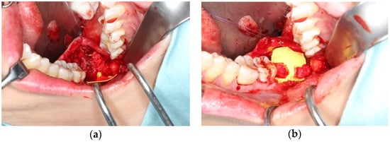
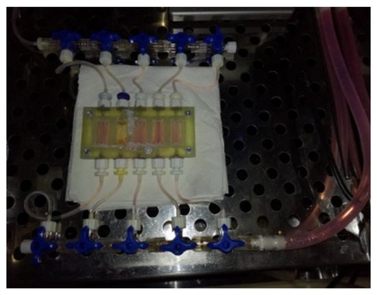
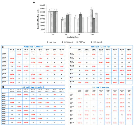
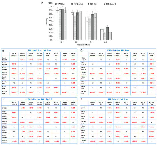
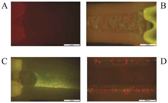
Figure 1


