Diagnostics 2022, 12(4), 959; https://doi.org/10.3390/diagnostics12040959 - 12 Apr 2022
Cited by 7 | Viewed by 3185
Abstract
Depression is characterized by feelings of sadness, loss, or anger that may interfere with everyday activities. Such a neuropsychiatric condition is commonly reported in multiple neurodegenerative disorders, which are quite different from each other. This study aimed at investigating the brain networks involved
[...] Read more.
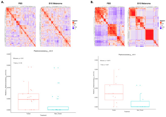
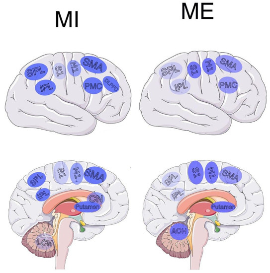
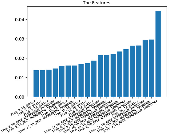
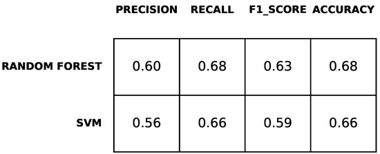
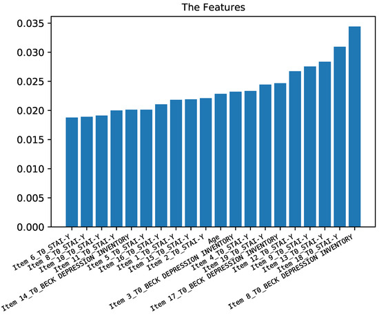
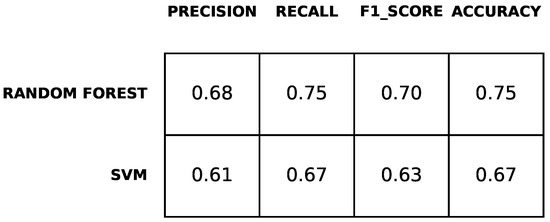
Depression is characterized by feelings of sadness, loss, or anger that may interfere with everyday activities. Such a neuropsychiatric condition is commonly reported in multiple neurodegenerative disorders, which are quite different from each other. This study aimed at investigating the brain networks involved in depression in patients with frontotemporal dementia (FTD) and Parkinson’s disease (PD) as compared to healthy controls (HC). Fifty participants were included in the study: 17 depressed FTD/PD patients; 17 non-depressed FTD/PD patients; and 16 non-depressed HCs matched for age and gender. We used the Beck depression inventory (BDI-II) to measure depression in all groups. On the same day, 3T brain magnetic resonance with structural and resting-state functional sequences were acquired. Differences in resting-state functional connectivity (FC) between depressed and non-depressed patients in all the experimental groups were assessed by using seed-to-seed and network-to-network approaches. We found a significant seed-to-seed hyperconnectivity patterns between the left thalamus and the left posterior temporal fusiform cortex, which differentiated FTD/PD depressed patients from the HCs. Network-to-network analysis revealed a significant hyperconnectivity among the default-mode network (left lateral-parietal region), the medial prefrontal cortex and the left lateral prefrontal cortex (i.e., part of the central executive network). We investigated whether such FC patterns could be related to the underlying neurodegenerative disorder by replicating the analyses with two independent samples (i.e., non-depressed PD and non-depressed FTD patients) and adding clinical parameters as covariates. We found no FC differences in these groups, thus suggesting how the FC pattern we found may signal a common depression-related neural pathway implicated in both the neurocognitive disorders.
Full article
(This article belongs to the Section Medical Imaging and Theranostics)
►
Show Figures

Figure 1


