13 pages, 805 KiB  
Article
Real-World Patterns and Decision Drivers of Radiotherapy for Lung Cancer Patients in Romania: RADIO-NET Study Results
by Mihai-Teodor Georgescu, Renata Zahu, Petronela Rusu, Gabriela Teodorescu and Gabriel Kacso
Diagnostics 2022, 12(12), 3089; https://doi.org/10.3390/diagnostics12123089 - 8 Dec 2022
Cited by 1 | Viewed by 1742
Abstract
Radiotherapy (RT) plays a crucial role in all stages of lung cancer. Data on recent real-world RT patterns and main drivers of RT decisions in lung cancer in Romania is scarce; we aimed to address these knowledge gaps through this physician-led medical chart [...] Read more.
Radiotherapy (RT) plays a crucial role in all stages of lung cancer. Data on recent real-world RT patterns and main drivers of RT decisions in lung cancer in Romania is scarce; we aimed to address these knowledge gaps through this physician-led medical chart review in 16 RT centers across the country. Consecutive patients with lung cancer receiving RT as part of their disease management between May–October 2019 (pre-COVID-19 pandemic) were included. Descriptive statistics were generated for all variables. This cohort included 422 patients: median age 63 years, males 76%, stages I–II 6%, III 43%, IV 50%, mostly adeno- and squamous cell carcinoma (76%), ECOG 0-1 50% at the time of RT. Curative intent RT was used in 36% of cases, palliative RT in 64%. Delays were reported in 13% of patients, mostly due to machine breakdown (67%). Most acute reported RT toxicity was esophagitis (19%). Multiple disease-, patient-, physician- and context-related drivers counted in the decision-making process. This is the first detailed analysis of RT use in lung cancer in Romania. Palliative RT still dominates the landscape. Earlier diagnosis, coordinated multidisciplinary strategies, and the true impact of the multimodal treatments on survival are strongly needed to improve lung cancer outcomes. Full article
(This article belongs to the Special Issue Diagnosis and Radiotherapy in Oncology)
Show Figures

Graphical abstract

21 pages, 6067 KiB  
Article
Breast Cancer Detection in Mammogram Images Using K–Means++ Clustering Based on Cuckoo Search Optimization
by Kittipol Wisaeng
Diagnostics 2022, 12(12), 3088; https://doi.org/10.3390/diagnostics12123088 - 7 Dec 2022
Cited by 16 | Viewed by 3506
Abstract
Traditional breast cancer detection algorithms require manual extraction of features from mammogram images and professional medical knowledge. Still, the quality of mammogram images hampers this and extracting high–quality features, which can result in very long processing times. Therefore, this paper proposes a new [...] Read more.
Traditional breast cancer detection algorithms require manual extraction of features from mammogram images and professional medical knowledge. Still, the quality of mammogram images hampers this and extracting high–quality features, which can result in very long processing times. Therefore, this paper proposes a new K–means++ clustering based on Cuckoo Search Optimization (KM++CSO) for breast cancer detection. The pre-processing method is used to improve the proposed KM++CSO method more segmentation efficiently. Furthermore, the interpretability is further enhanced using mathematical morphology and OTSU’s threshold. To this end, we tested the effectiveness of the KM++CSO methods on the mammogram image analysis society of the Mini–Mammographic Image Analysis Society (Mini–MIAS), the Digital Database for Screening Mammography (DDSM), and the Breast Cancer Digital Repository (BCDR) dataset through cross-validation. We maximize the accuracy and Jaccard index score, which is a measure that indicates the similarity between detected cancer and their corresponding reference cancer regions. The experimental results showed that the detection method obtained an accuracy of 96.42% (Mini–MIAS), 95.49% (DDSM), and 96.92% (BCDR). On overage, the KM++CSO method obtained 96.27% accuracy for three publicly available datasets. In addition, the detection results provided the 91.05% Jaccard index score. Full article
(This article belongs to the Section Medical Imaging and Theranostics)
Show Figures

Figure 1

13 pages, 91880 KiB  
Article
Dose Dependence Effect in Biallelic WNT10A Variant-Associated Tooth Agenesis Phenotype
by Haochen Liu, Bichen Lin, Hangbo Liu, Lanxin Su, Hailan Feng, Yang Liu, Miao Yu and Dong Han
Diagnostics 2022, 12(12), 3087; https://doi.org/10.3390/diagnostics12123087 - 7 Dec 2022
Cited by 7 | Viewed by 2153
Abstract
The goal of this study was to identify the pathogenic gene variants in patients with odonto-onycho-dermal dysplasia syndrome (OODD) or nonsyndromic tooth agenesis. Four unrelated individuals with tooth agenesis and their available family members were recruited. Peripheral blood was collected from four probands [...] Read more.
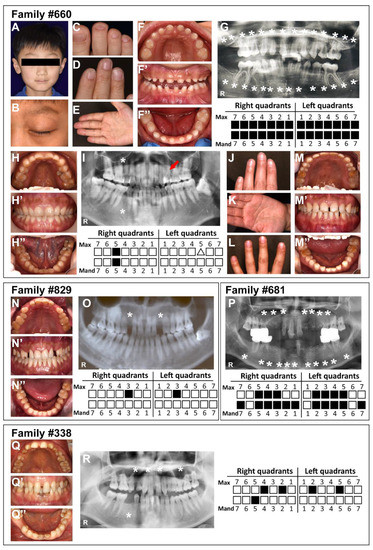
The goal of this study was to identify the pathogenic gene variants in patients with odonto-onycho-dermal dysplasia syndrome (OODD) or nonsyndromic tooth agenesis. Four unrelated individuals with tooth agenesis and their available family members were recruited. Peripheral blood was collected from four probands and five family members. Whole-exome sequencing (WES) and Sanger sequencing were used to identify the pathogenic gene variants. The harmfulness of these variations was predicted by bioinformatics. We identified four biallelic variants of the WNT10A gene in four patients, respectively: the proband#660: c.1176C > A (p.Cys392*) and c.812G > A (p.Cys271Tyr); the proband#681: c.637G > A (p.Gly213Ser) and c.985C > T (p.Arg329*); the proband#829: c.511C > T (p.Arg171Cys) and c.637G > A (p.Gly213Ser); and the proband#338: c.926A> G (p.Gln309Arg) and c.511C > T (p.Arg171Cys). Among them, two variants (c.812G > A; p.Cys271Tyr and c.985C > T; p.Arg329*) were previously unreported. Bioinformatics analysis showed that the pathogenicity of these six variants was different. Tertiary structure analysis showed that these variants were predicted to cause structural damage to the WNT10A protein. Genotype–phenotype analysis showed that the biallelic variants with more harmful effects, such as nonsense variants, caused OODD syndrome (#660 Ⅱ-1) or severe nonsyndromic tooth agenesis (NSTA) (#681 Ⅱ-1); the biallelic variants with less harmful effects, such as missense variants, caused a mild form of NSTA (#829 Ⅱ-2 and #338 Ⅱ-1). Individuals with a heterozygous variant presented a mild form of NSTA or a normal state. Our results further suggest the existence of the dose dependence of WNT10A pathogenicity on the tooth agenesis pattern, which broadens the variation spectrum and phenotype spectrum of WNT10A and could help with clinical diagnosis, treatment, and genetic counseling. Full article
(This article belongs to the Special Issue Diagnosis and Treatment of Craniofacial and Oral Disease)
Show Figures

Figure 1

21 pages, 2363 KiB  
Systematic Review
Influenza and Pneumococcal Vaccination and the Risk of COVID-19: A Systematic Review and Meta-Analysis
by Georgia V. Kapoula, Konstantina E. Vennou and Pantelis G. Bagos
Diagnostics 2022, 12(12), 3086; https://doi.org/10.3390/diagnostics12123086 - 7 Dec 2022
Cited by 12 | Viewed by 3379
Abstract
A number of studies have investigated the potential on-specific effects of some routinely administered vaccines (e.g., influenza, pneumococcal) on COVID-19 related outcomes, with contrasting results. In order to elucidate this discrepancy, we conducted a systematic review and meta-analysis to assess the association between [...] Read more.
A number of studies have investigated the potential on-specific effects of some routinely administered vaccines (e.g., influenza, pneumococcal) on COVID-19 related outcomes, with contrasting results. In order to elucidate this discrepancy, we conducted a systematic review and meta-analysis to assess the association between seasonal influenza vaccination and pneumococcal vaccination with SARS-CoV-2 infection and its clinical outcomes. PubMed and medRxiv databases were searched up to April 2022. A random effects model was used in the meta-analysis to pool odds ratio (OR) and adjusted estimates with 95% confidence intervals (CIs). Heterogeneity was quantitatively assessed using the Cochran’s Q and the I2 index. Subgroup analysis, sensitivity analysis and assessment of publication bias were performed for all outcomes. In total, 38 observational studies were included in the meta-analysis and there was substantial heterogeneity. Influenza and pneumococcal vaccination were associated with lower risk of SARS-CoV-2 infection (OR: 0.80, 95% CI: 0.75–0.86 and OR: 0.70, 95% CI: 0.57–0.88, respectively). Regarding influenza vaccination, it seems that the majority of studies did not properly adjust for all potential confounders, so when the analysis was limited to studies that adjusted for age, gender, comorbidities and socioeconomic indices, the association diminished. This is not the case regarding pneumococcal vaccination, for which even after adjustment for such factors the association persisted. Regarding harder endpoints such as ICU admission and death, current data do not support the association. Possible explanations are discussed, including trained immunity, inadequate matching for socioeconomic indices and possible coinfection. Full article
(This article belongs to the Section Clinical Laboratory Medicine)
Show Figures

Figure 1

12 pages, 876 KiB  
Article
Development of in House ELISAs to Detect Antibodies to SARS-CoV-2 in Infected and Vaccinated Humans by Using Recombinant S, S1 and RBD Proteins
by Aysun Yilmaz, Nuri Turan, Bekir Sami Kocazeybek, Harika Oyku Dinc, Hasan Emre Tali, Ozge Aydin, Hamid Besim Tali, Semaha Gul Yilmaz, Dildar Konukoglu, Sermin Borekci, Dashzeveg Bold, Gleyder Roman Sosa, Nejdiye Gungordu, Ilgim Vardaloglu, Nesrin Gareayaghi, Mine Guzel, Ebru Guner, Jean-Remy Sadeyen, Pengxiang Chang, Munir Iqbal, Juergen A. Richt and Huseyin Yilmazadd Show full author list remove Hide full author list
Diagnostics 2022, 12(12), 3085; https://doi.org/10.3390/diagnostics12123085 - 7 Dec 2022
Cited by 3 | Viewed by 2915
Abstract
(1) Background: The aim of this study was to produce in-house ELISAs which can be used to determine SARS-CoV-2-specific antibody levels directed against the spike protein (S), the S1 subunit of S and the receptor binding domain (RBD) of S in SARS-CoV-2 vaccinated [...] Read more.
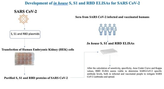
(1) Background: The aim of this study was to produce in-house ELISAs which can be used to determine SARS-CoV-2-specific antibody levels directed against the spike protein (S), the S1 subunit of S and the receptor binding domain (RBD) of S in SARS-CoV-2 vaccinated and infected humans. (2) Methods: Three in-house ELISAs were developed by using recombinant proteins of SARS-CoV-2, namely the S, S1 and RBD proteins. Specificity and sensitivity evaluations of these tests were performed using sera from SARS-CoV-2-infected (n = 70) and SARS-CoV-2-vaccinated (n = 222; CoronaVac vaccine) humans in Istanbul, Turkey. The analyses for the presence of SARS-CoV-2-specific antibodies were performed using the in-house ELISAs, a commercial ELISA (Abbott) and a commercial surrogate virus neutralization test (sVNT). We also analyzed archival human sera (n = 50) collected before the emergence of COVID-19 cases in Turkey. (3) Results: The sensitivity of the in-house S, S1 and RBD ELISAs was found to be 88.44, 90.17 and 95.38%, while the specificity was 72.27, 89.08 and 89.92%, respectively, when compared to the commercial SARS-CoV-2 antibody test kit. The area under curve (AUC) values were 0.777 for the in-house S ELISA, 0.926 for the S1 ELISA, and 0.959 for the RBD ELISA. The kappa values were 0.62, 0.79 and 0.86 for the S, S1 and RBD ELISAs, respectively. (4) Conclusions: The in-house S1 and RBD ELISAs developed in this study have acceptable performance characteristics in terms of sensitivity, specificity, AUC and kappa values. In particular, the RBD ELISA seems viable to determine SARS-CoV-2-specific antibody levels, both in infected and vaccinated people, and help mitigate SARS-CoV-2 outbreaks and spread. Full article
Show Figures

Graphical abstract

20 pages, 1550 KiB  
Article
A Deep Learning Based Approach for Grading of Diabetic Retinopathy Using Large Fundus Image Dataset
by Ayesha Mehboob, Muhammad Usman Akram, Norah Saleh Alghamdi and Anum Abdul Salam
Diagnostics 2022, 12(12), 3084; https://doi.org/10.3390/diagnostics12123084 - 7 Dec 2022
Cited by 20 | Viewed by 2976
Abstract
Diabetic Retinopathy affects one-third of all diabetic patients and may cause vision impairment. It has four stages of progression, i.e., mild non-proliferative, moderate non-proliferative, severe non-proliferative and proliferative Diabetic Retinopathy. The disease has no noticeable symptoms at early stages and may lead to [...] Read more.
Diabetic Retinopathy affects one-third of all diabetic patients and may cause vision impairment. It has four stages of progression, i.e., mild non-proliferative, moderate non-proliferative, severe non-proliferative and proliferative Diabetic Retinopathy. The disease has no noticeable symptoms at early stages and may lead to chronic destruction, thus causing permanent blindness if not detected at an early stage. The proposed research provides deep learning frameworks for autonomous detection of Diabetic Retinopathy at an early stage using fundus images. The first framework consists of cascaded neural networks, spanned in three layers where each layer classifies data into two classes, one is the desired stage and the other output is passed to another classifier until the input image is classified as one of the stages. The second framework takes normalized, HSV and RGB fundus images as input to three Convolutional Neural Networks, and the resultant probabilistic vectors are averaged together to obtain the final output of the input image. Third framework used the Long Short Term Memory Module in CNN to emphasize the network in remembering information over a long time span. Proposed frameworks were tested and compared on the large-scale Kaggle fundus image dataset EYEPAC. The evaluations have shown that the second framework outperformed others and achieved an accuracy of 78.06% and 83.78% without and with augmentation, respectively. Full article
(This article belongs to the Special Issue Data Analysis in Ophthalmic Diagnostics)
Show Figures

Figure 1

16 pages, 311 KiB  
Review
Fetal Neurology: From Prenatal Counseling to Postnatal Follow-Up
by Barbara Scelsa
Diagnostics 2022, 12(12), 3083; https://doi.org/10.3390/diagnostics12123083 - 7 Dec 2022
Cited by 11 | Viewed by 3302
Abstract
Brain abnormalities detected in fetal life are being increasingly recognized. Child neurologists are often involved in fetal consultations, and specific fetal neurology training has been implemented in many countries. Pediatric neurologists are asked to examine the data available and to contribute to the [...] Read more.
Brain abnormalities detected in fetal life are being increasingly recognized. Child neurologists are often involved in fetal consultations, and specific fetal neurology training has been implemented in many countries. Pediatric neurologists are asked to examine the data available and to contribute to the definition of the long-term outcomes. Ventriculomegaly, posterior fossa malformations, and agenesis/dysgenesis of corpus callosum are among the most common reasons for antenatal neurological consultations. Fetuses with central nervous system and extra-CNS anomalies should ideally be managed in secondary/tertiary hospitals where obstetricians who are experts in fetal medicine and pediatric specialists are available. Obstetricians play a critical role in screening, performing detailed neurosonography, and referring to other specialists for additional investigations. Clinical geneticists are frequently asked to propose diagnostic tests and counsel complex fetal malformations whose phenotypes may differ from those during postnatal life. Advances in fetal MRI and genetic investigations can support the specialists involved in counseling. Nevertheless, data interpretation can be challenging, and it requires a high level of expertise in a multidisciplinary setting. Postnatally, child neurologists should be part of an integrated multidisciplinary follow-up, together with neonatologists and pediatricians. The neurodevelopmental outcomes should be assessed at least up to school age. Children should be evaluated with formal tests of their gross motor, cognitive, language, fine motor/visuo-perceptual skills, and their behavior. In this perspective, fetal neurology can be regarded as the beginning of a long journey which continues with a prolonged, structured follow-up, support to the families, and transition to adult life. A review of the most common conditions is presented, along with the long-term outcomes and a proposal of the neurodevelopmental follow-up of children with CNS malformation which are diagnosed in uterus. Full article
(This article belongs to the Special Issue Diagnosis and Management in Prenatal Medicine—2nd Edition)
10 pages, 1599 KiB  
Article
Lung Ultrasound in Coronary Care Unit, an Important Diagnostic Tool for Concomitant Pneumonia
by Costantino Mancusi, Ilaria Fucile, Paola Gargiulo, Mariangela Mosca, Biagio Migliaccio, Christian Basile, Giuseppe Gargiulo, Ciro Santoro, Carmine Morisco, Nicola De Luca and Giovanni Esposito
Diagnostics 2022, 12(12), 3082; https://doi.org/10.3390/diagnostics12123082 - 7 Dec 2022
Cited by 6 | Viewed by 2244
Abstract
Background: In the setting of a coronary care unit (CCU), the early detection of pneumonia is of paramount important to prevent severe complications. This study was designed aiming to evaluate the diagnostic accuracy of lung ultrasound (LUS) in the detection of pneumonia and [...] Read more.
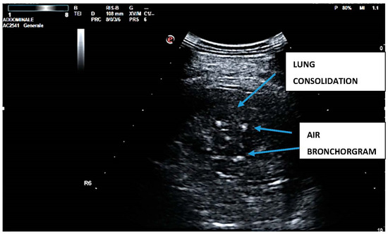
Background: In the setting of a coronary care unit (CCU), the early detection of pneumonia is of paramount important to prevent severe complications. This study was designed aiming to evaluate the diagnostic accuracy of lung ultrasound (LUS) in the detection of pneumonia and compared with chest X-ray (CXR). Method: We enrolled 110 consecutive patients admitted to the CCU of Federico II University Hospital. Each patient underwent CXR and bedside LUS on admission. The final diagnosis (pneumonia vs. no pneumonia) was established by another clinician reviewing clinical and laboratory data independent of LUS results and possibly prescribing chest contrast-enhanced CT (n = 34). Results: The mean age was 70 ± 11 years old, and 68% were males. Pneumonia was clinically diagnosed in 26 (23%) patients. LUS was positive for pneumonia in 24 patients (sensitivity 92%, specificity 81%). Chest radiography was positive for pneumonia in nine patients (sensitivity 43%, specificity 95%). Using CT scan as a reference, LUS exhibited 92% sensitivity and a specificity of 96%. In ROC curve analysis, the diagnostic accuracy of CXR and LUS for the diagnosis of pneumonia was 0.86 (95% CI 0.77–0.94), which was higher than CXR 0.68 (95% CI 0.55–0.84), p < 0.05. Conclusion: Based on the findings of the present study, the accuracy of LUS in the detection of pneumonia was significantly higher than chest X-ray with comparable sensibility to CT scan. Full article
Show Figures

Figure 1

10 pages, 1409 KiB  
Article
Automatic Feature Segmentation in Dental Periapical Radiographs
by Tugba Ari, Hande Sağlam, Hasan Öksüzoğlu, Orhan Kazan, İbrahim Şevki Bayrakdar, Suayip Burak Duman, Özer Çelik, Rohan Jagtap, Karolina Futyma-Gąbka, Ingrid Różyło-Kalinowska and Kaan Orhan
Diagnostics 2022, 12(12), 3081; https://doi.org/10.3390/diagnostics12123081 - 7 Dec 2022
Cited by 21 | Viewed by 5186
Abstract
While a large number of archived digital images make it easy for radiology to provide data for Artificial Intelligence (AI) evaluation; AI algorithms are more and more applied in detecting diseases. The aim of the study is to perform a diagnostic evaluation on [...] Read more.
While a large number of archived digital images make it easy for radiology to provide data for Artificial Intelligence (AI) evaluation; AI algorithms are more and more applied in detecting diseases. The aim of the study is to perform a diagnostic evaluation on periapical radiographs with an AI model based on Convoluted Neural Networks (CNNs). The dataset includes 1169 adult periapical radiographs, which were labelled in CranioCatch annotation software. Deep learning was performed using the U-Net model implemented with the PyTorch library. The AI models based on deep learning models improved the success rate of carious lesion, crown, dental pulp, dental filling, periapical lesion, and root canal filling segmentation in periapical images. Sensitivity, precision and F1 scores for carious lesion were 0.82, 0.82, and 0.82, respectively; sensitivity, precision and F1 score for crown were 1, 1, and 1, respectively; sensitivity, precision and F1 score for dental pulp, were 0.97, 0.87 and 0.92, respectively; sensitivity, precision and F1 score for filling were 0.95, 0.95, and 0.95, respectively; sensitivity, precision and F1 score for the periapical lesion were 0.92, 0.85, and 0.88, respectively; sensitivity, precision and F1 score for root canal filling, were found to be 1, 0.96, and 0.98, respectively. The success of AI algorithms in evaluating periapical radiographs is encouraging and promising for their use in routine clinical processes as a clinical decision support system. Full article
(This article belongs to the Section Machine Learning and Artificial Intelligence in Diagnostics)
Show Figures

Figure 1

18 pages, 460 KiB  
Systematic Review
Role of Salivary Biomarkers in Diagnosis and Detection of Dental Caries: A Systematic Review
by Ahmed Alamoudi, Raghad Alamoudi, Yara Gazzaz and Aseel M. Alqahtani
Diagnostics 2022, 12(12), 3080; https://doi.org/10.3390/diagnostics12123080 - 7 Dec 2022
Cited by 17 | Viewed by 5898
Abstract
Saliva plays a significant role in oral health and tooth integrity. Salivary components reduce tooth surface exposure to demineralization, protect against teeth wear and aid in enamel remineralization. There is a growing attempt to use salivary markers in diagnosing or predicting caries. However, [...] Read more.
Saliva plays a significant role in oral health and tooth integrity. Salivary components reduce tooth surface exposure to demineralization, protect against teeth wear and aid in enamel remineralization. There is a growing attempt to use salivary markers in diagnosing or predicting caries. However, despite the current information, there has yet to be an agreement among scholars. This study seeks to contribute more evidence on the suitability of salivary biomarkers in dental caries diagnosis. Eligible studies were electronically searched on online databases PubMed, Elsevier’s Scopus, EMBASE and Web of Science, and all the studies that met the inclusion criteria were considered. The PECOS criteria guided the study selection process based on the study question. The risk of bias was assessed using the STROBE checklist. Eighteen articles were included in the analysis. All the studies presented relevant data concerning the study objectives. There was evidence of associations between salivary biomarkers and dental caries, and the correlations were either positive or negative. The studies presented significant heterogeneity; thus, a meta-analysis was not possible. Salivary biomarkers appeared to perform crucial and complementary functions toward tooth integrity and thus may be reliable in predicting or diagnosing dental caries in patients. Full article
(This article belongs to the Section Pathology and Molecular Diagnostics)
Show Figures

Figure 1

21 pages, 3367 KiB  
Review
Advances in Bone Joint Imaging-Metal Artifact Reduction
by Sho Kohyama, Yuichi Yoshii, Yoshikazu Okamoto and Takahito Nakajima
Diagnostics 2022, 12(12), 3079; https://doi.org/10.3390/diagnostics12123079 - 7 Dec 2022
Cited by 18 | Viewed by 4085
Abstract
Numerous types of metal implants have been introduced in orthopedic surgery and are used in everyday practice. To precisely evaluate the postoperative condition of arthroplasty or trauma surgery, periprosthetic infection, and the loosening of implants, it is important to reduce artifacts induced by [...] Read more.
Numerous types of metal implants have been introduced in orthopedic surgery and are used in everyday practice. To precisely evaluate the postoperative condition of arthroplasty or trauma surgery, periprosthetic infection, and the loosening of implants, it is important to reduce artifacts induced by metal implants. In this review, we focused on technical advances in metal artifact reduction using digital tomosynthesis, computed tomography, and magnetic resonance imaging. We discussed new developments in diagnostic imaging methods and the continuous introduction of novel technologies to reduce metal artifacts; however, these innovations have not yet completely removed metal artifacts. Different algorithms need to be selected depending on the size, shape, material and implanted body parts of an implant. Future advances in metal artifact reduction algorithms and techniques and the development of new sequences may enable further reductions in metal artifacts even on original images taken previously. Moreover, the combination of different imaging modalities may contribute to further reductions in metal artifacts. Clinicians must constantly update their knowledge and work closely with radiologists to select the best diagnostic imaging method for each metal implant. Full article
(This article belongs to the Section Medical Imaging and Theranostics)
Show Figures

Figure 1

12 pages, 4170 KiB  
Article
Evaluation of Macular Thickness Changes after Uncomplicated Phacoemulsification Surgery in Healthy Subjects and Diabetic Patients without Retinopathy by Spectral Domain OCT
by Mikel García Gómez de Segura, Ana Martín-Arroyuelos, Isabel Pinilla and Javier Araiz
Diagnostics 2022, 12(12), 3078; https://doi.org/10.3390/diagnostics12123078 - 7 Dec 2022
Cited by 3 | Viewed by 2794
Abstract
Purpose: To assess differences in the evolution of macular thickness after uncomplicated phacoemulsification surgery between non-diabetic subjects and patients with diabetes mellitus (DM) without diabetic retinopathy (DR), using Spectral Domain OCT (SD-OCT). Methods: We performed a unicentric prospective study including one hundred and [...] Read more.
Purpose: To assess differences in the evolution of macular thickness after uncomplicated phacoemulsification surgery between non-diabetic subjects and patients with diabetes mellitus (DM) without diabetic retinopathy (DR), using Spectral Domain OCT (SD-OCT). Methods: We performed a unicentric prospective study including one hundred and thirty-one eyes of 70 patients divided into two groups—34 well-controlled DM patients without DR and 36 non-diabetic subjects—who underwent phacoemulsification for cataract surgery. Eyes that developed pseudophakic cystoid macular edema (PCME) were excluded from the study, leaving us with 64 patients. Macular thickness was analyzed using Cirrus HD-OCT (Macular Cube 512 × 128 protocol) preoperatively and on postoperative days 7, 30, 90, and 180. For cases with information available for both eyes, one eye was randomly selected for analysis. Results: A total of 64 eyes from 64 patients were analyzed in this study. The mean value of HbA1c in the diabetic group was 7%. After uncomplicated cataract surgery, patients showed no increase of the foveal, parafoveal, and perifoveal retinal thickness on postoperative day 7. However, thickness values increased on days 30, 90, and 180 after surgery in both groups, and peak at 90 days. There was no difference in macular thickness before or after surgery between DM and non-diabetic patients (p = 0.540). Conclusion: Macular thickness increases up to 6 months after uncomplicated cataract surgery in both DM patients without DR and non-diabetic subjects, with no differences between increases in both groups. Full article
(This article belongs to the Section Biomedical Optics)
Show Figures

Figure 1

9 pages, 967 KiB  
Article
Cloning, Expression and Evaluation of Thioredoxin Peroxidase-1 Antigen for the Serological Diagnosis of Schistosoma mekongi Human Infection
by Atcharaphan Wanlop, Jose Ma. M. Angeles, Adrian Miki C. Macalanda, Masashi Kirinoki, Yuma Ohari, Aya Yajima, Junya Yamagishi, Kevin Austin L. Ona and Shin-ichiro Kawazu
Diagnostics 2022, 12(12), 3077; https://doi.org/10.3390/diagnostics12123077 - 7 Dec 2022
Cited by 3 | Viewed by 2208
Abstract
Schistosoma mekongi, a blood fluke that causes Asian zoonotic schistosomiasis, is distributed in communities along the Mekong River in Cambodia and Lao People’s Democratic Republic. Decades of employing numerous control measures including mass drug administration using praziquantel have resulted in a decline [...] Read more.
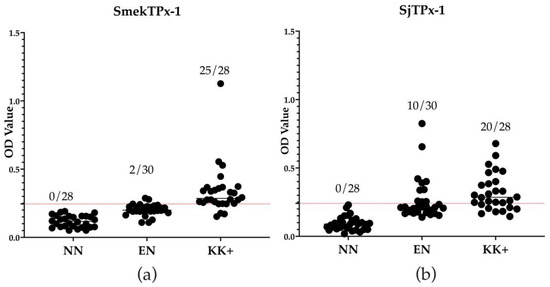
Schistosoma mekongi, a blood fluke that causes Asian zoonotic schistosomiasis, is distributed in communities along the Mekong River in Cambodia and Lao People’s Democratic Republic. Decades of employing numerous control measures including mass drug administration using praziquantel have resulted in a decline in the prevalence of schistosomiasis mekongi. This, however, led to a decrease in sensitivity of Kato–Katz stool microscopy considered as the gold standard in diagnosis. In order to develop a serological assay with high sensitivity and specificity which can replace Kato–Katz, recombinant S. mekongi thioredoxin peroxidase-1 protein (rSmekTPx-1) was expressed and produced. Diagnostic performance of the rSmekTPx-1 antigen through ELISA for detecting human schistosomiasis was compared with that of recombinant protein of S. japonicum TPx-1 (rSjTPx-1) using serum samples collected from endemic foci in Cambodia. The sensitivity and specificity of rSmekTPx-1 in ELISA were 89.3% and 93.3%, respectively, while those of rSjTPx-1 were 71.4% and 66.7%, respectively. In addition, a higher Kappa value of 0.82 calculated between rSmekTPx-1 antigen ELISA and Kato–Katz confirmed better agreement than between rSjTPx-1 antigen ELISA and Kato–Katz (Kappa value 0.38). These results suggest that ELISA with rSmekTPx-1 antigen can be a potential diagnostic method for detecting active human S. mekongi infection. Full article
(This article belongs to the Special Issue Molecular Diagnostics of Emerging Pathogens for Infectious Diseases)
Show Figures

Figure 1

16 pages, 356 KiB  
Review
Nectins and Nectin-like Molecules in Colorectal Cancer: Role in Diagnostics, Prognostic Values, and Emerging Treatment Options: A Literature Review
by Jakub Kobecki, Paweł Gajdzis, Grzegorz Mazur and Mariusz Chabowski
Diagnostics 2022, 12(12), 3076; https://doi.org/10.3390/diagnostics12123076 - 7 Dec 2022
Cited by 12 | Viewed by 2818
Abstract
In 2020, colorectal cancer was the third most common type of cancer worldwide with a clearly visible increase in the number of cases each year. With relatively high mortality rates and an uncertain prognosis, colorectal cancer is a serious health problem. There is [...] Read more.
In 2020, colorectal cancer was the third most common type of cancer worldwide with a clearly visible increase in the number of cases each year. With relatively high mortality rates and an uncertain prognosis, colorectal cancer is a serious health problem. There is an urgent need to investigate its specific mechanism of carcinogenesis and progression in order to develop new strategies of action against this cancer. Nectins and Nectin-like molecules are cell adhesion molecules that take part in a plethora of essential processes in healthy tissues as well as mediating substantial actions for tumor initiation and evolution. Our understanding of their role and a viable application of this in anti-cancer therapy has rapidly improved in recent years. This review summarizes the current data on the role nectins and Nectin-like molecules play in colorectal cancer. Full article
(This article belongs to the Special Issue Clinical Prognostic and Predictive Biomarkers)
11 pages, 1900 KiB  
Article
Evaluation of BD Onclarity™ HPV Assay on Self-Collected Vaginal and First-Void Urine Samples as Compared to Clinician-Collected Cervical Samples: A Pilot Study
by Marianna Martinelli, Chiara Giubbi, Illari Sechi, Fabio Bottari, Anna Daniela Iacobone, Rosario Musumeci, Federica Perdoni, Narcisa Muresu, Andrea Piana, Robert Fruscio, Fabio Landoni and Clementina Elvezia Cocuzza
Diagnostics 2022, 12(12), 3075; https://doi.org/10.3390/diagnostics12123075 - 7 Dec 2022
Cited by 16 | Viewed by 2483
Abstract
The accuracy of available HPV molecular assays on self-samples needs to be evaluated as compared to clinician-collected samples. This pilot study aimed to investigate the BD Onclarity™ HPV assay on vaginal and first-void urine samples. Sixty-four women referred to colposcopy for cervical dysplasia [...] Read more.
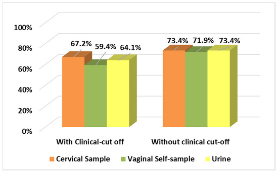
The accuracy of available HPV molecular assays on self-samples needs to be evaluated as compared to clinician-collected samples. This pilot study aimed to investigate the BD Onclarity™ HPV assay on vaginal and first-void urine samples. Sixty-four women referred to colposcopy for cervical dysplasia performed a vaginal self-collection and provided a first-void urine sample, after informed consent. A cervical specimen was collected during the clinician examination. All samples were tested using BD Onclarity™ HPV assay on the BD Viper™ LT System. Overall positive agreement (OPA) between cervical and self-sample results was evaluated using Cohen’s kappa value (κ). Using a clinical cut-off of 38.3 Ct for HPV 16 and 34.2 Ct for other HR genotypes, compared to cervical sample, the self-collected vaginal sample OPA was 85.9%, and κ = 0.699. Without a clinical cut-off, the OPA was 95.3%, and the κ = 0.890. Data obtained comparing cervical and urine samples showed an OPA of 87.5% with a κ = 0.79 using a clinical cut-off, and an OPA of 90.6% with a κ = 0.776 without a clinical cut-off. Data showed a substantial agreement between both self-collected and clinician-collected samples. A specific clinical cut-off analysis should be considered based on type of sample analysed. Full article
(This article belongs to the Special Issue Diagnosis of Lower Genital Tract Disease)
Show Figures

Figure 1