17 pages, 3410 KiB  
Article
Dissecting Gq/11-Mediated Plasma Membrane Translocation of Sphingosine Kinase-1
by Kira Vanessa Blankenbach, Ralf Frederik Claas, Natalie Judith Aster, Anna Katharina Spohner, Sandra Trautmann, Nerea Ferreirós, Justin L. Black, John J. G. Tesmer, Stefan Offermanns, Thomas Wieland and Dagmar Meyer zu Heringdorf
Cells 2020, 9(10), 2201; https://doi.org/10.3390/cells9102201 - 29 Sep 2020
Cited by 6 | Viewed by 3301
Abstract
Diverse extracellular signals induce plasma membrane translocation of sphingosine kinase-1 (SphK1), thereby enabling inside-out signaling of sphingosine-1-phosphate. We have shown before that Gq-coupled receptors and constitutively active Gαq/11 specifically induced a rapid and long-lasting SphK1 translocation, independently of canonical G [...] Read more.
Diverse extracellular signals induce plasma membrane translocation of sphingosine kinase-1 (SphK1), thereby enabling inside-out signaling of sphingosine-1-phosphate. We have shown before that Gq-coupled receptors and constitutively active Gαq/11 specifically induced a rapid and long-lasting SphK1 translocation, independently of canonical Gq/phospholipase C (PLC) signaling. Here, we further characterized Gq/11 regulation of SphK1. SphK1 translocation by the M3 receptor in HEK-293 cells was delayed by expression of catalytically inactive G-protein-coupled receptor kinase-2, p63Rho guanine nucleotide exchange factor (p63RhoGEF), and catalytically inactive PLCβ3, but accelerated by wild-type PLCβ3 and the PLCδ PH domain. Both wild-type SphK1 and catalytically inactive SphK1-G82D reduced M3 receptor-stimulated inositol phosphate production, suggesting competition at Gαq. Embryonic fibroblasts from Gαq/11 double-deficient mice were used to show that amino acids W263 and T257 of Gαq, which interact directly with PLCβ3 and p63RhoGEF, were important for bradykinin B2 receptor-induced SphK1 translocation. Finally, an AIXXPL motif was identified in vertebrate SphK1 (positions 100–105 in human SphK1a), which resembles the Gαq binding motif, ALXXPI, in PLCβ and p63RhoGEF. After M3 receptor stimulation, SphK1-A100E-I101E and SphK1-P104A-L105A translocated in only 25% and 56% of cells, respectively, and translocation efficiency was significantly reduced. The data suggest that both the AIXXPL motif and currently unknown consequences of PLCβ/PLCδ(PH) expression are important for regulation of SphK1 by Gq/11. Full article
Show Figures

Figure 1

22 pages, 4258 KiB  
Article
Interplay between Podoplanin, CD44s and CD44v in Squamous Carcinoma Cells
by Lucía Montero-Montero, Jaime Renart, Andrés Ramírez, Carmen Ramos, Mariam Shamhood, Rocío Jarcovsky, Miguel Quintanilla and Ester Martín-Villar
Cells 2020, 9(10), 2200; https://doi.org/10.3390/cells9102200 - 29 Sep 2020
Cited by 13 | Viewed by 4797
Abstract
Podoplanin and CD44 are transmembrane glycoproteins involved in inflammation and cancer. In this paper, we report that podoplanin is coordinately expressed with the CD44 standard (CD44s) and variant (CD44v) isoforms in vivo—in hyperplastic skin after a pro-inflammatory stimulus with 12-O-tetradecanoylphorbol-13-acetate (TPA)—and [...] Read more.
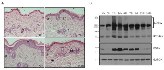
Podoplanin and CD44 are transmembrane glycoproteins involved in inflammation and cancer. In this paper, we report that podoplanin is coordinately expressed with the CD44 standard (CD44s) and variant (CD44v) isoforms in vivo—in hyperplastic skin after a pro-inflammatory stimulus with 12-O-tetradecanoylphorbol-13-acetate (TPA)—and in vitro—in cell lines representative of different stages of mouse-skin chemical carcinogenesis, as well as in human squamous carcinoma cell (SCC) lines. Moreover, we identify CD44v10 in the mouse-skin carcinogenesis model as the only CD44 variant isoform expressed in highly aggressive spindle carcinoma cell lines together with CD44s and podoplanin. We also characterized CD44v3-10, CD44v6-10 and CD44v8-10 as the major variant isoforms co-expressed with CD44s and podoplanin in human SCC cell lines. Immunofluorescence confocal microscopy experiments show that these CD44v isoforms colocalize with podoplanin at plasma membrane protrusions and cell–cell contacts of SCC cells, as previously reported for CD44s. Furthermore, CD44v isoforms colocalize with podoplanin in chemically induced mouse-skin SCCs in vivo. Co-immunoprecipitation experiments indicate that podoplanin physically binds to CD44v3-10, CD44v6-10 and CD44v8-10 isoforms, as well as to CD44s. Podoplanin–CD44 interaction is mediated by the transmembrane and cytosolic regions and is negatively modulated by glycosylation of the extracellular domain. These results point to a functional interplay of podoplanin with both CD44v and CD44s isoforms in SCCs and give insight into the regulation of the podoplanin–CD44 association. Full article
(This article belongs to the Special Issue Structure and Function of Podoplanin (PDPN) in Disease)
Show Figures

Figure 1

13 pages, 1847 KiB  
Article
HbF Levels in Sickle Cell Disease Are Associated with Proportion of Circulating Hematopoietic Stem and Progenitor Cells and CC-Chemokines
by Caterina P. Minniti, Seda S. Tolu, Kai Wang, Zi Yan, Karl Robert, Shouping Zhang, Andrew S. Crouch, Joan Uehlinger, Deepa Manwani and Eric E. Bouhassira
Cells 2020, 9(10), 2199; https://doi.org/10.3390/cells9102199 - 29 Sep 2020
Cited by 2 | Viewed by 3321
Abstract
The concentration of circulating hematopoietic stem and progenitor cells has not been studied longitudinally. Here, we report that the proportions of Lin-CD34+38- hematopoietic multipotent cells (HMCs) and of Lin-CD34+CD38+ hematopoietic progenitors cells (HPCs) are highly variable between individuals but stable over long periods [...] Read more.
The concentration of circulating hematopoietic stem and progenitor cells has not been studied longitudinally. Here, we report that the proportions of Lin-CD34+38- hematopoietic multipotent cells (HMCs) and of Lin-CD34+CD38+ hematopoietic progenitors cells (HPCs) are highly variable between individuals but stable over long periods of time, in both healthy individuals and sickle cell disease (SCD) patients. This suggests that these proportions are regulated by genetic polymorphisms or by epigenetic mechanisms. We also report that in SCD patients treated with hydroxyurea, the proportions of circulating HMCs and HPCs show a strong positive and negative correlation with fetal hemoglobin (HbF) levels, respectively. Titration of 65 cytokines revealed that the plasma concentration of chemokines CCL2, CCL11, CCL17, CCL24, CCL27, and PDGF-BB were highly correlated with the proportion of HMCs and HPCs and that a subset of these cytokines were also correlated with HbF levels. A linear model based on four of these chemokines could explain 80% of the variability in the proportion of circulating HMCs between individuals. The proportion of circulating HMCs and HPCs and the concentration of these chemokines might therefore become useful biomarkers for HbF response to HU in SCD patients. Such markers might become increasingly clinically relevant, as alternative treatment modalities for SCD are becoming available. Full article
(This article belongs to the Section Stem Cells)
Show Figures

Graphical abstract

15 pages, 1513 KiB  
Article
Conditioned Medium from Cells Overexpressing TDP-43 Alters the Metabolome of Recipient Cells
by Rudolf Hergesheimer, Débora Lanznaster, Jérôme Bourgeais, Olivier Hérault, Patrick Vourc’h, Christian R. Andres, Philippe Corcia and Hélène Blasco
Cells 2020, 9(10), 2198; https://doi.org/10.3390/cells9102198 - 29 Sep 2020
Cited by 7 | Viewed by 3613
Abstract
Amyotrophic lateral sclerosis (ALS) is a neurodegenerative disease caused by the progressive death of both upper and lower motor neurons. The disease presents a poor prognosis, and patients usually die 2–5 years after the onset of symptoms. The hallmark of this disease is [...] Read more.
Amyotrophic lateral sclerosis (ALS) is a neurodegenerative disease caused by the progressive death of both upper and lower motor neurons. The disease presents a poor prognosis, and patients usually die 2–5 years after the onset of symptoms. The hallmark of this disease is the presence of phosphorylated and ubiquitinated aggregates containing trans-active response DNA-binding protein-43 (TDP-43) in the cytoplasm of motor neurons. TDP-43 pathology has been associated with multiple pathways in ALS, such as metabolic dysfunction found in patients and in in vivo models. Recently, it has been described as a “prion-like” protein, as studies have shown its propagation in cell culture from ALS brain extract or overexpressed TDP-43 in co-culture and conditioned medium, resulting in cytotoxicity. However, the cellular alterations that are associated with this cytotoxicity require further investigation. Here, we investigated the effects of conditioned medium from HEK293T (Human Embryonic Kidney 293T) cells overexpressing TDP-43 on cellular morphology, proliferation, death, and metabolism. Although we did not find evidence of TDP-43 propagation, we observed a toxicity of TDP-43-conditioned medium and altered metabolism. These results, therefore, suggest (1) that cells overexpressing TDP-43 produce an extracellular environment that can perturb other cells and (2) that TDP-43 propagation alone may not be the only potentially cytotoxic cell-to-cell mechanism. Full article
Show Figures

Figure 1

29 pages, 3094 KiB  
Article
Hypoxia Promotes Mitochondrial Complex I Abundance via HIF-1α in Complex III and Complex IV Deficient Cells
by Amy Saldana-Caboverde, Nadee Nissanka, Sofia Garcia, Anne Lombès and Francisca Diaz
Cells 2020, 9(10), 2197; https://doi.org/10.3390/cells9102197 - 29 Sep 2020
Cited by 8 | Viewed by 3781
Abstract
Murine fibroblasts deficient in mitochondria respiratory complexes III (CIII) and IV (CIV) produced by either the ablation of Uqcrfs1 (encoding for Rieske iron sulfur protein, RISP) or Cox10 (encoding for protoheme IX farnesyltransferase, COX10) genes, respectively, showed a pleiotropic effect in complex I [...] Read more.
Murine fibroblasts deficient in mitochondria respiratory complexes III (CIII) and IV (CIV) produced by either the ablation of Uqcrfs1 (encoding for Rieske iron sulfur protein, RISP) or Cox10 (encoding for protoheme IX farnesyltransferase, COX10) genes, respectively, showed a pleiotropic effect in complex I (CI). Exposure to 1–5% oxygen increased the levels of CI in both RISP and COX10 KO fibroblasts. De novo assembly of the respiratory complexes occurred at a faster rate and to higher levels in 1% oxygen compared to normoxia in both RISP and COX10 KO fibroblasts. Hypoxia did not affect the levels of assembly of CIII in the COX10 KO fibroblasts nor abrogated the genetic defect impairing CIV assembly. Mitochondrial signaling involving reactive oxygen species (ROS) has been implicated as necessary for HIF-1α stabilization in hypoxia. We did not observe increased ROS production in hypoxia. Exposure to low oxygen levels stabilized HIF-1α and increased CI levels in RISP and COX10 KO fibroblasts. Knockdown of HIF-1α during hypoxic conditions abrogated the beneficial effect of hypoxia on the stability/assembly of CI. These findings demonstrate that oxygen and HIF-1α regulate the assembly of respiratory complexes. Full article
Show Figures

Figure 1

19 pages, 3102 KiB  
Article
Circular RNA Encoded Amyloid Beta peptides—A Novel Putative Player in Alzheimer’s Disease
by Dingding Mo, Xinping Li, Carsten A. Raabe, Timofey S. Rozhdestvensky, Boris V. Skryabin and Juergen Brosius
Cells 2020, 9(10), 2196; https://doi.org/10.3390/cells9102196 - 29 Sep 2020
Cited by 45 | Viewed by 6350
Abstract
Alzheimer’s disease (AD) is an age-related detrimental dementia. Amyloid beta peptides (Aβ) play a crucial role in the pathology of AD. In familial AD, Aβ are generated from the full-length amyloid beta precursor protein (APP) via dysregulated proteolytic processing; however, in the case [...] Read more.
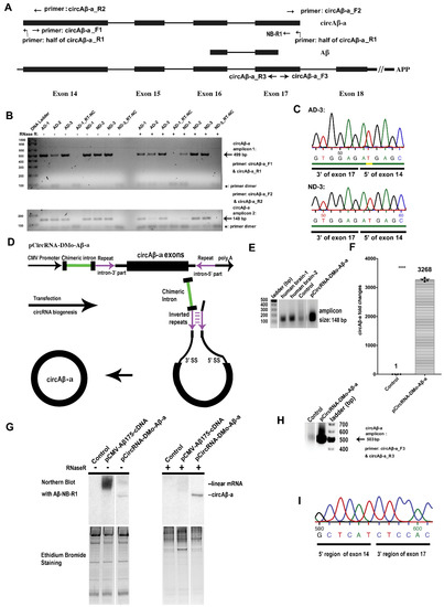
Alzheimer’s disease (AD) is an age-related detrimental dementia. Amyloid beta peptides (Aβ) play a crucial role in the pathology of AD. In familial AD, Aβ are generated from the full-length amyloid beta precursor protein (APP) via dysregulated proteolytic processing; however, in the case of sporadic AD, the mechanism of Aβ biogenesis remains elusive. circRNAs are a class of transcripts preferentially expressed in brain. We identified a circRNA harboring the Aβ-coding region of the APP gene termed circAβ-a. This circular RNA was detected in the brains of AD patients and non-dementia controls. With the aid of our recently established approach for analysis of circRNA functions, we demonstrated that circAβ-a is efficiently translated into a novel Aβ-containing Aβ175 polypeptide (19.2 KDa) in both cultured cells and human brain. Furthermore, Aβ175 was shown to be processed into Aβ peptides—a hallmark of AD. In summary, our analysis revealed an alternative pathway of Aβ biogenesis. Consequently, circAβ-a and its corresponding translation product could potentially represent novel therapeutic targets for AD treatment. Importantly, our data point to yet another evolutionary route for potentially increasing proteome complexity by generating additional polypeptide variants using back-splicing of primary transcripts that yield circular RNA templates. Full article
(This article belongs to the Special Issue Circular RNAs: Non-canonical Observations on Non-canonical RNAs)
Show Figures

Figure 1

19 pages, 2207 KiB  
Review
snR30/U17 Small Nucleolar Ribonucleoprotein: A Critical Player during Ribosome Biogenesis
by Timothy John Vos and Ute Kothe
Cells 2020, 9(10), 2195; https://doi.org/10.3390/cells9102195 - 29 Sep 2020
Cited by 11 | Viewed by 4025
Abstract
The small nucleolar RNA snR30 (U17 in humans) plays a unique role during ribosome synthesis. Unlike most members of the H/ACA class of guide RNAs, the small nucleolar ribonucleoprotein (snoRNP) complex assembled on snR30 does not direct pseudouridylation of ribosomal RNA (rRNA), but [...] Read more.
The small nucleolar RNA snR30 (U17 in humans) plays a unique role during ribosome synthesis. Unlike most members of the H/ACA class of guide RNAs, the small nucleolar ribonucleoprotein (snoRNP) complex assembled on snR30 does not direct pseudouridylation of ribosomal RNA (rRNA), but instead snR30 is critical for 18S rRNA processing during formation of the small subunit (SSU) of the ribosome. Specifically, snR30 is essential for three pre-rRNA cleavages at the A0/01, A1/1, and A2/2a sites in yeast and humans, respectively. Accordingly, snR30 is the only essential H/ACA guide RNA in yeast. Here, we summarize our current knowledge about the interactions and functions of snR30, discuss what remains to be elucidated, and present two non-exclusive hypotheses on the possible molecular function of snR30 during ribosome biogenesis. First, snR30 might be responsible for recruiting other proteins including endonucleases to the SSU processome. Second, snR30 may contribute to the refolding of pre-rRNA into a required conformation that serves as a checkpoint during ribosome biogenesis facilitating pre-rRNA cleavage. In both scenarios, the snR30 snoRNP may have scaffolding and RNA chaperoning activity. In conclusion, the snR30 snoRNP is a crucial player with an unknown molecular mechanism during ribosome synthesis, posing many interesting future research questions. Full article
(This article belongs to the Section Intracellular and Plasma Membranes)
Show Figures

Graphical abstract

19 pages, 2445 KiB  
Article
Competitive sgRNA Screen Identifies p38 MAPK as a Druggable Target to Improve HSPC Engraftment
by Denise Klatt, Teng-Cheong Ha, Maximilian Schinke, Anton Selich, Anna Lieske, Julia Dahlke, Michael Morgan, Tobias Maetzig and Axel Schambach
Cells 2020, 9(10), 2194; https://doi.org/10.3390/cells9102194 - 29 Sep 2020
Cited by 3 | Viewed by 4443
Abstract
Previous gene therapy trials for X-linked chronic granulomatous disease (X-CGD) lacked long-term engraftment of corrected hematopoietic stem and progenitor cells (HSPCs). Chronic inflammation and high levels of interleukin-1 beta (IL1B) might have caused aberrant cell cycling in X-CGD HSPCs with a concurrent loss [...] Read more.
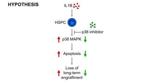
Previous gene therapy trials for X-linked chronic granulomatous disease (X-CGD) lacked long-term engraftment of corrected hematopoietic stem and progenitor cells (HSPCs). Chronic inflammation and high levels of interleukin-1 beta (IL1B) might have caused aberrant cell cycling in X-CGD HSPCs with a concurrent loss of their long-term repopulating potential. Thus, we performed a targeted CRISPR-Cas9-based sgRNA screen to identify candidate genes that counteract the decreased repopulating capacity of HSPCs during gene therapy. The candidates were validated in a competitive transplantation assay and tested in a disease context using IL1B-challenged or X-CGD HSPCs. The sgRNA screen identified Mapk14 (p38) as a potential target to increase HSPC engraftment. Knockout of p38 prior to transplantation was sufficient to induce a selective advantage. Inhibition of p38 increased expression of the HSC homing factor CXCR4 and reduced apoptosis and proliferation in HSPCs. For potential clinical translation, treatment of IL1B-challenged or X-CGD HSPCs with a p38 inhibitor led to a 1.5-fold increase of donor cell engraftment. In summary, our findings demonstrate that p38 may serve as a potential druggable target to restore engraftment of HSPCs in the context of X-CGD gene therapy. Full article
(This article belongs to the Special Issue Stem Cell-Immune Function and Cardiac Regeneration)
Show Figures

Graphical abstract

8 pages, 224 KiB  
Editorial
Stem Cell-Based Disease Modeling and Cell Therapy
by Xiaowen Bai
Cells 2020, 9(10), 2193; https://doi.org/10.3390/cells9102193 - 29 Sep 2020
Cited by 26 | Viewed by 6005
Abstract
Stem cell science is among the fastest moving fields in biology, with many highly promising directions for translatability. To centralize and contextualize some of the latest developments, this Special Issue presents state-of-the-art research of adult stem cells, induced pluripotent stem cells (iPSCs), and [...] Read more.
Stem cell science is among the fastest moving fields in biology, with many highly promising directions for translatability. To centralize and contextualize some of the latest developments, this Special Issue presents state-of-the-art research of adult stem cells, induced pluripotent stem cells (iPSCs), and embryonic stem cells as well as cancer stem cells. The studies we include describe efficient differentiation protocols of generation of chondrocytes, adipocytes, and neurons, maturation of iPSC-derived cardiomyocytes and neurons, dynamic characterization of iPSC-derived 3D cerebral organoids, CRISPR/Cas9 genome editing, and non-viral minicircle vector-based gene modification of stem cells. Different applications of stem cells in disease modeling are described as well. This volume also highlights the most recent developments and applications of stem cells in basic science research and disease treatments. Full article
(This article belongs to the Special Issue Stem Cell-based Therapy and Disease Modeling)
6 pages, 217 KiB  
Editorial
Cells in Cardiovascular Disease: Using Diversity to Confront Adversity
by José Martínez-González and Pablo García de Frutos
Cells 2020, 9(10), 2192; https://doi.org/10.3390/cells9102192 - 29 Sep 2020
Viewed by 2333
Abstract
The present Special Issue on “Cells in Cardiovascular Disease” wants to offer a general overview of current cardiovascular research and illustrate how advances in the molecular characterization at the cellular level are providing unique insights into pathologies of the circulatory system [...] Full article
(This article belongs to the Special Issue Cells in Cardiovascular Disease)
36 pages, 2689 KiB  
Review
Extracellular Vesicles as an Efficient and Versatile System for Drug Delivery
by Xuan T. T. Dang, Jayasinghe Migara Kavishka, Daniel Xin Zhang, Marco Pirisinu and Minh T. N. Le
Cells 2020, 9(10), 2191; https://doi.org/10.3390/cells9102191 - 29 Sep 2020
Cited by 89 | Viewed by 13657
Abstract
Despite the recent advances in drug development, the majority of novel therapeutics have not been successfully translated into clinical applications. One of the major factors hindering their clinical translation is the lack of a safe, non-immunogenic delivery system with high target specificity upon [...] Read more.
Despite the recent advances in drug development, the majority of novel therapeutics have not been successfully translated into clinical applications. One of the major factors hindering their clinical translation is the lack of a safe, non-immunogenic delivery system with high target specificity upon systemic administration. In this respect, extracellular vesicles (EVs), as natural carriers of bioactive cargo, have emerged as a promising solution and can be further modified to improve their therapeutic efficacy. In this review, we provide an overview of the biogenesis pathways, biochemical features, and isolation methods of EVs with an emphasis on their many intrinsic properties that make them desirable as drug carriers. We then describe in detail the current advances in EV therapeutics, focusing on how EVs can be engineered to achieve improved target specificity, better circulation kinetics, and efficient encapsulation of therapeutic payloads. We also identify the challenges and obstacles ahead for clinical translation and provide an outlook on the future perspective of EV-based therapeutics. Full article
(This article belongs to the Section Intracellular and Plasma Membranes)
Show Figures

Figure 1

22 pages, 1860 KiB  
Review
JNK3 as Therapeutic Target and Biomarker in Neurodegenerative and Neurodevelopmental Brain Diseases
by Clara Alice Musi, Graziella Agrò, Francesco Santarella, Erika Iervasi and Tiziana Borsello
Cells 2020, 9(10), 2190; https://doi.org/10.3390/cells9102190 - 28 Sep 2020
Cited by 53 | Viewed by 6196
Abstract
The c-Jun N-terminal kinase 3 (JNK3) is the JNK isoform mainly expressed in the brain. It is the most responsive to many stress stimuli in the central nervous system from ischemia to Aβ oligomers toxicity. JNK3 activity is spatial and temporal organized [...] Read more.
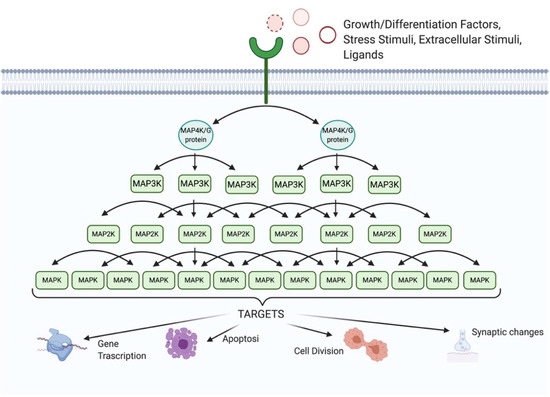
The c-Jun N-terminal kinase 3 (JNK3) is the JNK isoform mainly expressed in the brain. It is the most responsive to many stress stimuli in the central nervous system from ischemia to Aβ oligomers toxicity. JNK3 activity is spatial and temporal organized by its scaffold protein, in particular JIP-1 and β-arrestin-2, which play a crucial role in regulating different cellular functions in different cellular districts. Extensive evidence has highlighted the possibility of exploiting these adaptors to interfere with JNK3 signaling in order to block its action. JNK plays a key role in the first neurodegenerative event, the perturbation of physiological synapse structure and function, known as synaptic dysfunction. Importantly, this is a common mechanism in many different brain pathologies. Synaptic dysfunction and spine loss have been reported to be pharmacologically reversible, opening new therapeutic directions in brain diseases. Being JNK3-detectable at the peripheral level, it could be used as a disease biomarker with the ultimate aim of allowing an early diagnosis of neurodegenerative and neurodevelopment diseases in a still prodromal phase. Full article
Show Figures

Figure 1

13 pages, 2595 KiB  
Article
Neurodegeneration Caused by S1P-Lyase Deficiency Involves Calcium-Dependent Tau Pathology and Abnormal Histone Acetylation
by Shah Alam, Antonia Piazzesi, Mariam Abd El Fatah, Maren Raucamp and Gerhild van Echten-Deckert
Cells 2020, 9(10), 2189; https://doi.org/10.3390/cells9102189 - 28 Sep 2020
Cited by 24 | Viewed by 3929
Abstract
We have shown that sphingosine 1-phosphate (S1P) generated by sphingosine kinase 2 (SK2) is toxic in neurons lacking S1P-lyase (SGPL1), the enzyme that catalyzes its irreversible cleavage. Interestingly, patients harboring mutations in the gene encoding this enzyme (SGPL1) often present with [...] Read more.
We have shown that sphingosine 1-phosphate (S1P) generated by sphingosine kinase 2 (SK2) is toxic in neurons lacking S1P-lyase (SGPL1), the enzyme that catalyzes its irreversible cleavage. Interestingly, patients harboring mutations in the gene encoding this enzyme (SGPL1) often present with neurological pathologies. Studies in a mouse model with a developmental neural-specific ablation of SGPL1 (SGPL1fl/fl/Nes) confirmed the importance of S1P metabolism for the presynaptic architecture and neuronal autophagy, known to be essential for brain health. We now investigated in SGPL1-deficient murine brains two other factors involved in neurodegenerative processes, namely tau phosphorylation and histone acetylation. In hippocampal and cortical slices SGPL1 deficiency and hence S1P accumulation are accompanied by hyperphosphorylation of tau and an elevated acetylation of histone3 (H3) and histone4 (H4). Calcium chelation with BAPTA-AM rescued both tau hyperphosphorylation and histone acetylation, designating calcium as an essential mediator of these (patho)physiological functions of S1P in the brain. Studies in primary cultured neurons and astrocytes derived from SGPL1fl/fl/Nes mice revealed hyperphosphorylated tau only in SGPL1-deficient neurons and increased histone acetylation only in SGPL1-deficient astrocytes. Both could be reversed to control values with BAPTA-AM, indicating the close interdependence of S1P metabolism, calcium homeostasis, and brain health. Full article
Show Figures

Graphical abstract

9 pages, 3580 KiB  
Perspective
When the Search for Stemness Genes Meets the Skin Substitute Bioengineering Field: KLF4 Transcription Factor under the Light
by Nicolas O. Fortunel and Michèle T. Martin
Cells 2020, 9(10), 2188; https://doi.org/10.3390/cells9102188 - 28 Sep 2020
Cited by 5 | Viewed by 3829
Abstract
The transcription factor “Kruppel-like factor 4” (KLF4) is a central player in the field of pluripotent stem cell biology. In particular, it was put under the spotlight as one of the four factors of the cocktail originally described for reprogramming into induced pluripotent [...] Read more.
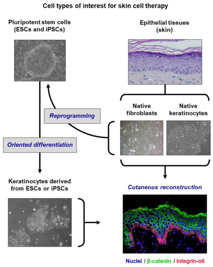
The transcription factor “Kruppel-like factor 4” (KLF4) is a central player in the field of pluripotent stem cell biology. In particular, it was put under the spotlight as one of the four factors of the cocktail originally described for reprogramming into induced pluripotent stem cells (iPSCs). In contrast, its possible functions in native tissue stem cells remain largely unexplored. We recently published that KLF4 is a regulator of “stemness” in human keratinocytes. We show that reducing the level of expression of this transcription factor by RNA interference or pharmacological repression promotes the ex vivo amplification and regenerative capacity of two types of cells of interest for cutaneous cell therapy: native keratinocyte stem and progenitor cells from adult epidermis, which have been used for more than three decades in skin graft bioengineering, and keratinocytes generated by the lineage-oriented differentiation of embryonic stem cells (ESCs), which have potential for the development of skin bio-bandages. At the mechanistic level, KLF4 repression alters the expression of a large set of genes involved in TGF-β1 and WNT signaling pathways. Major regulators of TGF-β bioavailability and different TGF-β receptors were targeted, notably modulating the ALK1/Smad1/5/9 axis. At a functional level, KLF4 repression produced an antagonist effect on TGF-β1-induced keratinocyte differentiation. Full article
(This article belongs to the Special Issue Feature Papers in Stem Cells)
Show Figures

Figure 1

19 pages, 3068 KiB  
Article
Aquaporin-4 Expression during Toxic and Autoimmune Demyelination
by Sven Olaf Rohr, Theresa Greiner, Sarah Joost, Sandra Amor, Paul van der Valk, Christoph Schmitz and Markus Kipp
Cells 2020, 9(10), 2187; https://doi.org/10.3390/cells9102187 - 28 Sep 2020
Cited by 19 | Viewed by 4041
Abstract
The water channel protein aquaporin-4 (AQP4) is required for a normal rate of water exchange across the blood–brain interface. Following the discovery that AQP4 is a possible autoantigen in neuromyelitis optica, the function of AQP4 in health and disease has become a research [...] Read more.
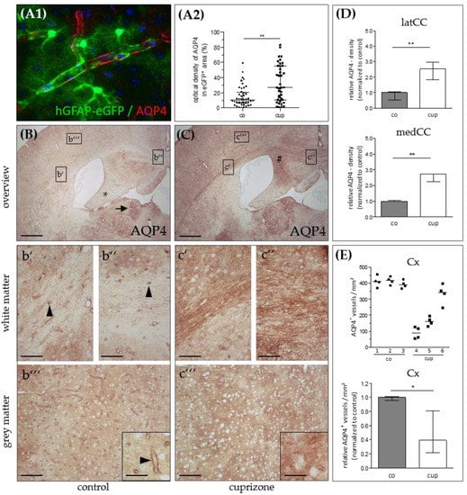
The water channel protein aquaporin-4 (AQP4) is required for a normal rate of water exchange across the blood–brain interface. Following the discovery that AQP4 is a possible autoantigen in neuromyelitis optica, the function of AQP4 in health and disease has become a research focus. While several studies have addressed the expression and function of AQP4 during inflammatory demyelination, relatively little is known about its expression during non-autoimmune-mediated myelin damage. In this study, we used the toxin-induced demyelination model cuprizone as well as a combination of metabolic and autoimmune myelin injury (i.e., Cup/EAE) to investigate AQP4 pathology. We show that during toxin-induced demyelination, diffuse AQP4 expression increases, while polarized AQP4 expression at the astrocyte endfeet decreases. The diffuse increased expression of AQP4 was verified in chronic-active multiple sclerosis lesions. Around inflammatory brain lesions, AQP4 expression dramatically decreased, especially at sites where peripheral immune cells penetrate the brain parenchyma. Humoral immune responses appear not to be involved in this process since no anti-AQP4 antibodies were detected in the serum of the experimental mice. We provide strong evidence that the diffuse increase in anti-AQP4 staining intensity is due to a metabolic injury to the brain, whereas the focal, perivascular loss of anti-AQP4 immunoreactivity is mediated by peripheral immune cells. Full article
(This article belongs to the Special Issue Glia Cells in Inflammation)
Show Figures

Figure 1