14 pages, 2686 KiB  
Article
Sex- and Female Age-Dependent Differences in Gene Expression in Diffuse Large B-Cell Lymphoma—Possible Estrogen Effects
by Dan Huang, Mattias Berglund, Anastasios Damdimopoulos, Per Antonson, Cecilia Lindskog, Gunilla Enblad, Rose-Marie Amini and Sam Okret
Cancers 2023, 15(4), 1298; https://doi.org/10.3390/cancers15041298 - 17 Feb 2023
Cited by 4 | Viewed by 2768
Abstract
For most lymphomas, including diffuse large B-cell lymphoma (DLBCL), the male incidence is higher, and the prognosis is worse compared to females. The reasons are unclear; however, epidemiological and experimental data suggest that estrogens are involved. With this in mind, we analyzed gene [...] Read more.
For most lymphomas, including diffuse large B-cell lymphoma (DLBCL), the male incidence is higher, and the prognosis is worse compared to females. The reasons are unclear; however, epidemiological and experimental data suggest that estrogens are involved. With this in mind, we analyzed gene expression data from a publicly available cohort (EGAD00001003600) of 746 DLBCL samples based on RNA sequencing. We found 1293 genes to be differentially expressed between males and females (adj. p-value < 0.05). Few autosomal genes and pathways showed common sex-regulated expression between germinal center B-cell (GCB) and activated B-cell lymphoma (ABC) DLBCL. Analysis of differentially expressed genes between pre- vs. postmenopausal females identified 208 GCB and 345 ABC genes, with only 5 being shared. When combining the differentially expressed genes between females vs. males and pre- vs. postmenopausal females, nine putative estrogen-regulated genes were identified in ABC DLBCL. Two of them, NR4A2 and MUC5B, showed induced and repressed expression, respectively. Interestingly, NR4A2 has been reported as a tumor suppressor in lymphoma. We show that ABC DLBCL females with a high NR4A2 expression showed better survival. Inversely, MUC5B expression causes a more malignant phenotype in several cancers. NR4A2 and MUC5B were confirmed to be estrogen-regulated when the ABC cell line U2932 was grafted to mice. The results demonstrate sex- and female reproductive age-dependent differences in gene expression between DLBCL subtypes, likely due to estrogens. This may contribute to the sex differences in incidence and prognosis. Full article
Show Figures

Figure 1

17 pages, 899 KiB  
Review
Practical Application of Circulating Tumor-Related DNA of Human Papillomavirus in Liquid Biopsy to Evaluate the Molecular Response in Patients with Oropharyngeal Cancer
by Agnieszka M. Mazurek and Tomasz W. Rutkowski
Cancers 2023, 15(4), 1047; https://doi.org/10.3390/cancers15041047 - 7 Feb 2023
Cited by 6 | Viewed by 2768
Abstract
Recent findings have shown that human papillomavirus (HPV) DNA is present in the blood as a tumor-specific biomarker (circulating tumor-related HPV; ctHPV) in patients with HPV-related oropharyngeal cancer (HPV-related OPC). The molecular response (MR) in patients with HPV-related OPC can be defined as [...] Read more.
Recent findings have shown that human papillomavirus (HPV) DNA is present in the blood as a tumor-specific biomarker (circulating tumor-related HPV; ctHPV) in patients with HPV-related oropharyngeal cancer (HPV-related OPC). The molecular response (MR) in patients with HPV-related OPC can be defined as the change in the number of ctHPV copies in relation to its initial quantity. The optimal model for assessing the MR using a liquid biopsy (LB) should be based on the E6/E7 sequences of the viral genome. MR assessment can help to evaluate the intensity of ongoing treatments in relation to the tumor response. The evaluation of the residual disease at the end of therapy may also be performed by MR assessment. If a partial MR (pMR) is found, caution is indicated and a subsequent LB should be considered, due to the likelihood of disease progression. Complete radiological and clinical responses together with a complete MR (cMR) convincingly indicate a low risk of treatment failure. Moreover, molecular recurrence (Mrec) during a follow-up, confirmed in two consecutive assays, even despite the lack of any other clinical or radiological symptoms of progression, indicates patients at high risk of disease recurrence. In conclusion, MR by ctHPV assessment may hasten the early detection of disease progression, at any stage of the management of the patient with HPV-related OPC. Full article
(This article belongs to the Special Issue 2nd Edition: Targeting Head and Neck Cancer)
Show Figures

Figure 1

12 pages, 2610 KiB  
Article
CD8+ Cell Density Gradient across the Tumor Epithelium–Stromal Interface of Non-Muscle Invasive Papillary Urothelial Carcinoma Predicts Recurrence-Free Survival after BCG Immunotherapy
by Julius Drachneris, Allan Rasmusson, Mindaugas Morkunas, Mantas Fabijonavicius, Albertas Cekauskas, Feliksas Jankevicius and Arvydas Laurinavicius
Cancers 2023, 15(4), 1205; https://doi.org/10.3390/cancers15041205 - 14 Feb 2023
Cited by 8 | Viewed by 2753
Abstract
Background: Bacille Calmette–Guerin (BCG) immunotherapy is the first-line treatment in patients with high-risk non-muscle invasive papillary urothelial carcinoma (NMIPUC), the most common type of bladder cancer. The therapy outcomes are variable and may depend on the immune response within the tumor microenvironment. In [...] Read more.
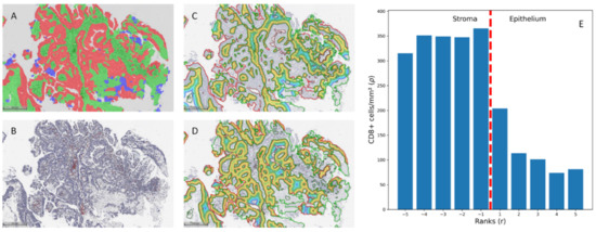
Background: Bacille Calmette–Guerin (BCG) immunotherapy is the first-line treatment in patients with high-risk non-muscle invasive papillary urothelial carcinoma (NMIPUC), the most common type of bladder cancer. The therapy outcomes are variable and may depend on the immune response within the tumor microenvironment. In our study, we explored the prognostic value of CD8+ cell density gradient indicators across the tumor epithelium–stroma interface of NMIPUC. Methods: Clinical and pathologic data were retrospectively collected from 157 NMIPUC patients treated with BCG immunotherapy after transurethral resection. Whole-slide digital image analysis of CD8 immunohistochemistry slides was used for tissue segmentation, CD8+ cell quantification, and the assessment of CD8+ cell densities within the epithelium–stroma interface. Subsequently, the gradient indicators (center of mass and immunodrop) were computed to represent the density gradient across the interface. Results: By univariable analysis of the clinicopathologic factors, including the history of previous NMIPUC, poor tumor differentiation, and pT1 stage, were associated with shorter RFS (p < 0.05). In CD8+ analyses, only the gradient indicators but not the absolute CD8+ densities were predictive for RFS (p < 0.05). The best-performing cross-validated model included previous episodes of NMIPUC (HR = 4.4492, p = 0.0063), poor differentiation (HR = 2.3672, p = 0.0457), and immunodrop (HR = 5.5072, p = 0.0455). Conclusions: We found that gradient indicators of CD8+ cell densities across the tumor epithelium–stroma interface, along with routine clinical and pathology data, improve the prediction of RFS in NMIPUC. Full article
(This article belongs to the Special Issue Tumor Immunology and Immunotherapy Resistance)
Show Figures

Figure 1

11 pages, 2865 KiB  
Article
MUC1 Expressions and Its Prognostic Values in US Gastric Cancer Patients
by Young-Il Kim, Robert Luke Pecha, Tara Keihanian, Michael Mercado, S. Valeria Pena-Munoz, Kailash Lang, George Van Buren, Sadhna Dhingra and Mohamed O. Othman
Cancers 2023, 15(4), 998; https://doi.org/10.3390/cancers15040998 - 4 Feb 2023
Cited by 15 | Viewed by 2745
Abstract
This study aims to evaluate the prognostic value of MUC expression in US GC patients. A total of 70 tumor specimens were collected from GC patients who underwent surgery or endoscopic resection between 2013 and 2019 at a tertiary referral center in the [...] Read more.
This study aims to evaluate the prognostic value of MUC expression in US GC patients. A total of 70 tumor specimens were collected from GC patients who underwent surgery or endoscopic resection between 2013 and 2019 at a tertiary referral center in the US. MUC expression status including MUC1, MUC2, MUC5AC, and MUC6 was evaluated by immunohistochemical staining. The positive rates of MUC1, MUC2, MUC5AC, and MUC6 were 71.4%, 78.6%, 74.3%, and 33.3%, respectively. Patients with positive MUC1 expression had a significantly higher rate of aggressive pathologic features including diffuse-type cancer (42.0% vs. 0%; p < 0.001), advanced GC (80.0% vs. 30.0%, p < 0.001), lymph node metastasis (62.0% vs. 20.0%; p = 0.001), and distant metastasis (32.0% vs. 5.0%; p = 0.017) compared with those with negative MUC1 expression. However, the differences in the pathologic features were not observed according to MUC2, MUC5AC, and MUC6 expression status. In early gastric cancer (EGC), patients with a high level of MUC1 expression showed a higher rate of lymphovascular invasion (71.4% vs. 21.4%; p = 0.026) and EGC meeting non-curative resection (85.7% vs. 42.9%; p = 0.061) than those with negative MUC1. In US GC patients, MUC1 expression is associated with aggressive pathological features, and might be a useful prognostic marker. Full article
(This article belongs to the Special Issue Management of Gastric Cancer)
Show Figures

Figure 1

19 pages, 6797 KiB  
Article
Ferroptosis Inducers Kill Mesenchymal Stem Cells Affected by Neuroblastoma
by Xiangze Li, Qi Wang, Chencheng Xu, Lei Zhang, Jiquan Zhou, Jingchun Lv, Min Xu and Dapeng Jiang
Cancers 2023, 15(4), 1301; https://doi.org/10.3390/cancers15041301 - 18 Feb 2023
Cited by 8 | Viewed by 2745
Abstract
Bone marrow (BM) is the most common site of neuroblastoma (NB) metastasis, and its involvement represents poor patient prognosis. In accordance with the “seed and soil” theory of tumor metastasis, BM provides a favorable environment for NB metastasis while bone marrow mesenchymal stem [...] Read more.
Bone marrow (BM) is the most common site of neuroblastoma (NB) metastasis, and its involvement represents poor patient prognosis. In accordance with the “seed and soil” theory of tumor metastasis, BM provides a favorable environment for NB metastasis while bone marrow mesenchymal stem cells (BMSCs) have been recognized as a central part of tumor stroma formation. Yet, there is currently no effective method for intervening these BMSCs. We found that BMSCs affected by NB (NB-BMSCs) could significantly promote NB growth and migration. Additionally, tumor cell-endowed BMSCs showed stronger resistance to several chemotherapeutic agents. Surprisingly, NB-BMSCs were more sensitive to ferroptosis than normal BMSCs. NB-BMSCs had lower levels of intracellular free iron while synthesizing more iron-sulfur clusters and heme. Moreover, the Xc/glutathione/glutathione peroxidase 4 (Xc/GSH/GPX4) pathway of the anti-ferroptosis system was significantly downregulated. Accordingly, ferroptosis inducers erastin and RAS-selective lethal 3 (RSL3) could significantly kill NB-BMSCs with limited effects on normal BMSCs. BMSCs from NB patients with BM metastasis also showed poor anti-ferroptosis ability compared with those from NB patients without BM metastasis. In vivo studies suggested that co-injection of mice with BMSCs and NB cells could significantly promote the growth of tumor tissues compared with injecting NB cells alone. However, treatment with erastin or RSL3 resulted in the opposite effect to some extent. Our results revealed that NB-BMSCs were vulnerable to ferroptosis from downregulation of the Xc/GSH/GPX4 pathway. Ferroptosis inducers could effectively kill NB-BMSCs, but not normal BMSCs. This study provides possible new ideas for the treatment of tumor-associated BMSCs in NB patients. Full article
(This article belongs to the Section Pediatric Oncology)
Show Figures

Graphical abstract

29 pages, 8922 KiB  
Article
Polyphenolic Boronates Inhibit Tumor Cell Proliferation: Potential Mitigators of Oxidants in the Tumor Microenvironment
by Gang Cheng, Hakim Karoui, Micael Hardy and Balaraman Kalyanaraman
Cancers 2023, 15(4), 1089; https://doi.org/10.3390/cancers15041089 - 8 Feb 2023
Cited by 2 | Viewed by 2745
Abstract
Boronate-based compounds have been used in brain cancer therapy, either as prodrugs or in combination with other modalities. Boronates containing pro-luminescent and fluorescent probes have been used in mouse models of cancer. In this study, we synthesized and developed polyphenolic boronates and mitochondria-targeted [...] Read more.
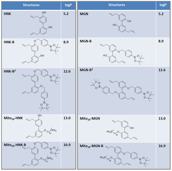
Boronate-based compounds have been used in brain cancer therapy, either as prodrugs or in combination with other modalities. Boronates containing pro-luminescent and fluorescent probes have been used in mouse models of cancer. In this study, we synthesized and developed polyphenolic boronates and mitochondria-targeted polyphenolic phytochemicals (e.g., magnolol [MGN] and honokiol [HNK]) and tested their antiproliferative effects in brain cancer cells. Results show that mitochondria-targeted (Mito) polyphenolic boronates (Mito-MGN-B and Mito-HNK-B) were slightly more potent than Mito-MGN and Mito-HNK in inhibiting proliferation of the U87MG cell line. Similar proliferation results also were observed in other cancer cell lines, such as MiaPaCa-2, A549 and UACC-62. Independent in vitro experiments indicated that reactive nitrogen species (e.g., peroxynitrite) and reactive oxygen species (e.g., hydrogen peroxide) stoichiometrically react with polyphenolic boronates and Mito-polphenolic boronates, forming polyphenols and Mito-polyphenols as major products. Previous reports suggest that both Mito-MGN and Mito-HNK activate cytotoxic T cells and inhibit immunosuppressive immune cells. We propose that Mito-polyphenolic boronate-based prodrugs may be used to inhibit tumor proliferation and mitigate oxidant formation in the tumor microenvironment, thereby generating Mito-polyphenols in situ, as well as showing activity in the tumor microenvironment. Full article
(This article belongs to the Collection Targeting Solid Tumors)
Show Figures

Figure 1

18 pages, 7836 KiB  
Article
CXCR3 Expression Is Associated with Advanced Tumor Stage and Grade Influencing Survival after Surgery of Localised Renal Cell Carcinoma
by Andrea Katharina Lindner, Agnieszka Martowicz, Gerold Untergasser, Johannes Haybaeck, Eva Compérat, Florian Kocher, Andreas Seeber, Martin Thurnher and Renate Pichler
Cancers 2023, 15(4), 1001; https://doi.org/10.3390/cancers15041001 - 4 Feb 2023
Cited by 4 | Viewed by 2742
Abstract
Background: Surgery is the standard treatment in localized renal cell carcinoma (RCC). Pembrolizumab is now approved for adjuvant therapy in high-risk patients. However, inhomogeneity of studies gives ambiguity which patient benefit most from adjuvant therapy. A high infiltration of CD8+ T cells [...] Read more.
Background: Surgery is the standard treatment in localized renal cell carcinoma (RCC). Pembrolizumab is now approved for adjuvant therapy in high-risk patients. However, inhomogeneity of studies gives ambiguity which patient benefit most from adjuvant therapy. A high infiltration of CD8+ T cells is known to be linked with poor prognosis in RCC. CXCR3 is a key player of CD8+ T cell differentiation and infiltration. We aimed to evaluate CXCR3 as a potential marker for predicting recurrence. Methods: CXCR3 and immune cell subsets (CD4, CD8, CD68 and FoXP3) were measured on RCC samples by multiplex immunofluorescence (mIF) staining. Cellular localization of CXCR3 was evaluated using single-cell RNA analysis on a publicly available dataset. Results: Tumor samples of 42 RCC patients were analyzed, from which 59.5% were classified as clear-cell RCC and of which 20 had recurrence. Single-cell RNA analysis revealed that CXCR3 was predominantly expressed in intratumoral T cells and dendritic cells. CXCR3 expression was higher in advanced tumors stages (p = 0.0044) and grade (p = 0.0518), correlating significantly with a higher CD8+ T cell expression (p < 0.001). Patients with CXCR3high RCCs had also a significant shorter RFS compared to CXCR3low (median: 78 vs. 147 months, p = 0.0213). In addition, also tumor stage pT3/4 (p < 0.0001) as well as grade G3/4 (p = 0.0008) negatively influenced RFS. Conclusion: CXCR3high cell density was associated with high T cell infiltration and advanced tumor stage, worsening RFS in surgically resected RCC patients. Beside its prognostic value, CXCR3 might be a predictive biomarker to guide therapy decision for adjuvant therapy in localized RCC. Full article
(This article belongs to the Special Issue Pathology of Urogenital Cancers)
Show Figures

Figure 1

15 pages, 1810 KiB  
Article
Ki67 as a Predictor of Response to PARP Inhibitors in Platinum Sensitive BRCA Wild Type Ovarian Cancer: The MITO 37 Retrospective Study
by Valentina Tuninetti, Eleonora Ghisoni, Sandro Pignata, Elisa Picardo, Francesco Raspagliesi, Claudia Andreetta, Elena Maldi, Grazia Artioli, Serafina Mammoliti, Lucia Zanchi, Angelica Sikokis, Nicoletta Biglia, Alessandro Parisi, Vincenzo Dario Mandato, Claudia Carella, Gennaro Cormio, Marco Marinaccio, Andrea Puppo, Biagio Paolini, Lucia Borsotti, Giulia Scotto, Margherita Turinetto, Dario Sangiolo, Massimo Di Maio and Giorgio Valabregaadd Show full author list remove Hide full author list
Cancers 2023, 15(4), 1032; https://doi.org/10.3390/cancers15041032 - 6 Feb 2023
Cited by 2 | Viewed by 2740
Abstract
Background: There is compelling need for novel biomarkers to predict response to PARP inhibitors (PARPi) in BRCA wild-type (WT) ovarian cancer (OC). Methods: MITO 37 is a multicenter retrospective study aiming at correlating Ki67 expression at diagnosis with a clinical outcome following platinum [...] Read more.
Background: There is compelling need for novel biomarkers to predict response to PARP inhibitors (PARPi) in BRCA wild-type (WT) ovarian cancer (OC). Methods: MITO 37 is a multicenter retrospective study aiming at correlating Ki67 expression at diagnosis with a clinical outcome following platinum treatment and PARPi maintenance. Clinical data were collected from high grade serous or endometroid BRCAWT OC treated with niraparib or rucaparib maintenance between 2010–2021 in 15 centers. Ki67 expression was assessed locally by certified pathologists on formalin-fixed paraffin embedded (FFPE) tissues. Median Ki67 was used as a cut-off. Results: A total of 136 patients were eligible and included in the analysis. Median Ki67 was 45.7% (range 1.0–99.9). The best response to platinum according to median Ki67 was 26.5% vs. 39.7% complete response (CR), 69.1% vs. 58.8% partial response (PR), 4.4% vs. 1.5% stable disease (SD). The best response to PARPi according to median Ki67 was 19.1% vs. 36.8% CR, 26.5% vs. 26.5% PR, 26.5 vs. 25% SD, 27.9% vs. 16.2% progressive disease (PD). No statistically significant differences in progression free survival (PFS) and overall survival (OS) were identified between low and high Ki67. PFS and OS are in line with registration trials. Conclusions: Ki67 at diagnosis did not discriminate responders to PARPi. Full article
Show Figures

Figure 1

24 pages, 3408 KiB  
Article
Molecular Insight into Drug Resistance Mechanism Conferred by Aberrant PIK3CD Splice Variant in African American Prostate Cancer
by Siyoung Ha and Bi-Dar Wang
Cancers 2023, 15(4), 1337; https://doi.org/10.3390/cancers15041337 - 20 Feb 2023
Cited by 7 | Viewed by 2739
Abstract
Targeting PI3Kδ has emerged as a promising therapy for hematologic and non-hematologic malignancies. Previously, we identified an oncogenic splice variant, PIK3CD-S, conferring Idelalisib resistance in African American (AA) prostate cancer (PCa). In the current study, we employed a comprehensive analysis combining molecular [...] Read more.
Targeting PI3Kδ has emerged as a promising therapy for hematologic and non-hematologic malignancies. Previously, we identified an oncogenic splice variant, PIK3CD-S, conferring Idelalisib resistance in African American (AA) prostate cancer (PCa). In the current study, we employed a comprehensive analysis combining molecular biology, biochemistry, histology, in silico simulation, and in vitro functional assays to investigate the PIK3CD-S expression profiles in PCa samples and to elucidate the drug resistance mechanism mediated by PI3Kδ-S (encoded by PIK3CD-S). The immunohistochemistry, RT-PCR, and Western blot assays first confirmed that PI3Kδ-S is highly expressed in AA PCa. Compared with PCa expressing the full-length PI3Kδ-L, PCa expressing PI3Kδ-S exhibits enhanced drug resistance properties, including a higher cell viability, more antiapoptotic and invasive capacities, and constitutively activated PI3K/AKT signaling, in the presence of PI3Kδ/PI3K inhibitors (Idelalisib, Seletalisib, Wortmannin, and Dactolisib). Molecular docking, ATP-competitive assays, and PI3 kinase assays have further indicated a drastically reduced affinity of PI3Kδ inhibitors with PI3Kδ-S vs. PI3Kδ-L, attributed to the lack of core binding residues in the PI3Kδ-S catalytic domain. Additionally, SRSF2 has been identified as a critical splicing factor mediating exon 20 skipping in PIK3CD pre-mRNA. The inhibition of the SRSF2 activity by SRPIN340 successfully sensitizes AA PCa cells to PI3Kδ inhibitors, suggesting a novel therapeutic option for Idelalisib-resistant tumors. Full article
(This article belongs to the Special Issue The Role of Alternative Splicing in Cancer)
Show Figures

Figure 1

19 pages, 10564 KiB  
Article
Serum Response Factor-Regulated IDO1/Kyn-Ahr Pathway Promotes Tumorigenesis of Oral Squamous Cell Carcinoma
by Mingyan Xu, Feixiang Zhu, Qi Yin, Hao Yin, Shaobin Fang, Gongwei Luo, Jie Huang, Wenxia Huang, Fan Liu, Ming Zhong and Xiaoling Deng
Cancers 2023, 15(4), 1319; https://doi.org/10.3390/cancers15041319 - 19 Feb 2023
Cited by 12 | Viewed by 2732
Abstract
Serum response factor (SRF) regulates pro-carcinogenic genes in various cancers, but its role in oral squamous cell carcinoma (OSCC) remains unclear. SRF expression in 70 OSCC samples was detected via immunohistochemistry. Abundant SRF expressed in OSCC tissues was closely associated with tumor metastasis. [...] Read more.
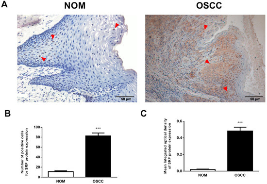
Serum response factor (SRF) regulates pro-carcinogenic genes in various cancers, but its role in oral squamous cell carcinoma (OSCC) remains unclear. SRF expression in 70 OSCC samples was detected via immunohistochemistry. Abundant SRF expressed in OSCC tissues was closely associated with tumor metastasis. SRF-overexpressing OSCC cells were constructed to evaluate how SRF affects OSCC cell tumorigenesis and epithelial-to-mesenchymal transition (EMT) in vitro and in vivo. Overexpressed SRF increased OSCC cell migration and invasion in vitro and tumor growth and invasion in vivo. This promoted EMT, characterized by decreased and increased expression of E- and N-cadherin, respectively. Furthermore, an analysis of RNA sequences of transcriptional targets of SRF showed that SRF transactivated the indoleamine 2, 3-dioxygenase 1 (IDO1)/kynurenine-aryl hydrocarbon receptor (Kyn-AhR) signaling pathway in OSCC cell lines. Direct SRF binding to the IDO1 gene promoter upregulated transcription, which was detected through chromatin immunoprecipitation and dual luciferase reporter assays. Inhibiting IDO1 or AhR impaired SRF-induced migration and invasion and prevented EMT in OSCC cells. Our results demonstrated that SRF is a critical regulator of the IDO1/Kyn-AhR signaling pathway. This in turn increases OSCC cell migration and invasion by modulating EMT, which, consequently, favors OSCC cell growth and metastasis. We revealed a novel molecular mechanism through which SRF modulates OSCC metastasis. This should provide potential targets or biomarkers for OSCC diagnosis and treatment. Full article
Show Figures

Figure 1

20 pages, 5385 KiB  
Article
miR-34a and IRE1A/XBP-1(S) Form a Double-Negative Feedback Loop to Regulate Hypoxia-Induced EMT, Metastasis, Chemo-Resistance and Autophagy
by Nassim Bouznad, Matjaz Rokavec, Meryem Gülfem Öner and Heiko Hermeking
Cancers 2023, 15(4), 1143; https://doi.org/10.3390/cancers15041143 - 10 Feb 2023
Cited by 11 | Viewed by 2732
Abstract
Tumor-associated hypoxia, i.e., decreased availability of oxygen, results in a poor clinical outcome since it promotes EMT, metastasis, and chemotherapy-resistance. We have previously identified p53 and its target miR-34a, as critical determinants of the effect of hypoxia on colorectal cancer (CRC). Here, we [...] Read more.
Tumor-associated hypoxia, i.e., decreased availability of oxygen, results in a poor clinical outcome since it promotes EMT, metastasis, and chemotherapy-resistance. We have previously identified p53 and its target miR-34a, as critical determinants of the effect of hypoxia on colorectal cancer (CRC). Here, we aimed to characterize mechanisms that contribute to the selective advantage of cells with loss of p53/miR-34a function in a hypoxic environment. Using in silico prediction, we identified XBP-1 and IRE1A as potential miR-34a targets. IRE1A and XBP-1 are central components of the unfolded protein response that is activated by ER stress, which is also induced in tumor cells as a response to harsh conditions surrounding tumors such as hypoxia and a limited supply of nutrients. Here we characterized the XBP-1(S) transcription factor and its regulator IRE1A as direct, conserved miR-34a targets in CRC cells. After hypoxia and DNA damage, IRE1A and XBP-1 were repressed by p53 in a miR-34a-dependent manner, whereas p53-deficient cells showed induction of IRE1A and XBP-1(S). Furthermore, miR-34a expression was directly suppressed by XBP-1(S). In p53-deficient CRC cells, hypoxia-induced EMT, migration, invasion, metastases formation, and resistance to 5-FU were dependent on IRE1A/XBP-1(S) activation. Hypoxia-induced autophagy was identified as an XBP-1(S)-dependent mediator of 5-FU resistance and was reversed by ectopic miR-34a expression. The HIF1A/IRE1A/XBP-1(S)/p53/miR-34a feedback loop described here represents a central regulator of the response to hypoxia and ER stress that maintains cellular homeostasis. In tumors, the inactivation of p53 and miR-34a may result in IRE1A/XPB-1(S)-mediated EMT and autophagy, which ultimately promotes metastasis and chemoresistance. Full article
(This article belongs to the Section Molecular Cancer Biology)
Show Figures

Figure 1

11 pages, 672 KiB  
Article
Bevacizumab as Single Agent in Children and Teenagers with Optic Pathway Glioma
by Pierluigi Calò, Nicolas Pianton, Alexandre Basle, Alexandre Vasiljevic, Marc Barritault, Pierre Aurélien Beuriat, Cécile Faure-Conter and Pierre Leblond
Cancers 2023, 15(4), 1036; https://doi.org/10.3390/cancers15041036 - 7 Feb 2023
Cited by 12 | Viewed by 2732
Abstract
This is a retrospective study conducted on patients with OPG, aged less than 19 years, treated with bevacizumab as a single agent, since 2010 at IHOPe (Institute of Pediatric Hematology and Oncology). Efficacy of the treatment was evaluated on the tumor response rate [...] Read more.
This is a retrospective study conducted on patients with OPG, aged less than 19 years, treated with bevacizumab as a single agent, since 2010 at IHOPe (Institute of Pediatric Hematology and Oncology). Efficacy of the treatment was evaluated on the tumor response rate on MRI with a centralized review basing upon RAPNO criteria and with visual assessment basing upon a 0.2 log change in the logMAR scale. Thirty-one patients with OPG have been included. From a radiological point of view, best anytime responses were: 1 major response, 6 partial responses, 7 minor responses and 14 stable diseases; achieving disease control in 28 (96%) out of 29 patients. Ophthalmological response was evaluated in 25 patients and disease control was achieved in 22 (88%) out of 25, with 14 steady states and 8 significant improvements. Among patients treated with chemotherapy after the bevacizumab course, nine relapsed and have been retreated with objective responses. Bevacizumab used as single agent seems effective in children and adolescents with OPG. Our work paves the way for a phase II study in which bevacizumab alone could be used as frontline therapy. Full article
(This article belongs to the Section Pediatric Oncology)
Show Figures

Figure 1

19 pages, 344 KiB  
Review
Management of Hepatocellular Carcinoma in Decompensated Cirrhotic Patients: A Comprehensive Overview
by Maria Tampaki, George V. Papatheodoridis and Evangelos Cholongitas
Cancers 2023, 15(4), 1310; https://doi.org/10.3390/cancers15041310 - 18 Feb 2023
Cited by 8 | Viewed by 2728
Abstract
Primary liver cancer is the sixth most common cancer and the fourth leading cause of cancer-related death. Hepatocellular carcinoma (HCC) accounts for 75% of primary liver cancer cases, mostly on the basis of cirrhosis. However, the data and therapeutic options for the treatment [...] Read more.
Primary liver cancer is the sixth most common cancer and the fourth leading cause of cancer-related death. Hepatocellular carcinoma (HCC) accounts for 75% of primary liver cancer cases, mostly on the basis of cirrhosis. However, the data and therapeutic options for the treatment of HCC in patients with decompensated cirrhosis are rather limited. This patient category is often considered to be in a terminal stage without the possibility of a specific treatment except liver transplantation, which is restricted by several criteria and liver donor shortages. Systemic treatments may provide a solution for patients with Child Pugh class B or C since they are less invasive. Although most of the existing trials have excluded patients with decompensated cirrhosis, there are increasing data from real-life settings that show acceptable tolerability and satisfying efficacy in terms of response. The data on the administration of locoregional treatments in such patients are also limited, but the overall survival seems to be potentially prolonged when patients are carefully selected, and close adverse event monitoring is applied. The aim of this review is to analyze the existing data regarding the administration of treatments in decompensated patients with HCC, evaluate the effect of therapy on overall survival and highlight the potential risks in terms of tolerability. Full article
(This article belongs to the Special Issue Diagnosis and Treatment for Hepatocellular Tumors)
10 pages, 1329 KiB  
Article
Prognosis of Pancreatic Cancer Based on Resectability: A Single Center Experience
by Takahiro Einama, Yasuhiro Takihata, Suefumi Aosasa, Fukumi Konno, Kazuki Kobayashi, Naoto Yonamine, Ibuki Fujinuma, Takazumi Tsunenari, Akiko Nakazawa, Eiji Shinto, Hideki Ueno and Yoji Kishi
Cancers 2023, 15(4), 1101; https://doi.org/10.3390/cancers15041101 - 9 Feb 2023
Cited by 15 | Viewed by 2720
Abstract
Although conversion surgery has increasingly been performed for initially unresectable advanced pancreatic ductal adenocarcinoma (PDAC), the rate of conversion, including that for patients who do not undergo resection, remains unclear. Patients with PDAC who were treated between January 2013 and December 2018 were [...] Read more.
Although conversion surgery has increasingly been performed for initially unresectable advanced pancreatic ductal adenocarcinoma (PDAC), the rate of conversion, including that for patients who do not undergo resection, remains unclear. Patients with PDAC who were treated between January 2013 and December 2018 were classified into three groups: resectable (R), borderline resectable (BR), and unresectable (UR). We analyzed patient outcomes, including the rate of surgical resection and survival, in each of these groups. In total, 211 patients (R, 118; BR, 22; UR, 81) were selected. Among them, 117 (99%), 18 (82%), and 15 (19%) patients in the R, BR, and UR groups, respectively, underwent surgical resection. R0 resection rates were 88, 78, and 67%, whereas median overall survival (OS) from treatment initiation were 31, 18, and 11 months (p < 0.0001) in the R, BR, and UR groups, respectively. In patients who underwent surgical resection, relapse-free survival (RFS) and OS were similar among the three groups (R vs. BR vs. UR; median RFS (months), 17 vs. 13 vs. 11, p = 0.249; median OS (months), 31 vs. 26 vs. 32, p = 0.742). Lymph node metastases and incomplete adjuvant chemotherapy were identified as independent prognostic factors for OS. Although the surgical resection rate was low, particularly in the BR and UR groups, the prognosis of patients who underwent surgical resection was similar irrespective of the initial resectability status. Full article
(This article belongs to the Special Issue Advanced Pancreatic Cancer)
Show Figures

Figure 1

13 pages, 1226 KiB  
Article
New Horizons in Metastatic Colorectal Cancer: Prognostic Role of CD44 Expression
by Pina Ziranu, Valentina Aimola, Andrea Pretta, Marco Dubois, Raffaele Murru, Nicole Liscia, Flaviana Cau, Mara Persano, Giulia Deias, Enrico Palmas, Francesco Loi, Marco Migliari, Valeria Pusceddu, Marco Puzzoni, Eleonora Lai, Stefano Cascinu, Gavino Faa and Mario Scartozzi
Cancers 2023, 15(4), 1212; https://doi.org/10.3390/cancers15041212 - 14 Feb 2023
Cited by 13 | Viewed by 2713
Abstract
Background: The transmembrane glycoprotein CD44, the major hyaluronan (HA) receptor, has been proven to regulate cell growth, survival, differentiation, and migration. It is therefore widely considered to be involved in carcinogenesis. Its role as a new therapeutic target in solid tumors is under [...] Read more.
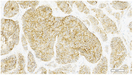
Background: The transmembrane glycoprotein CD44, the major hyaluronan (HA) receptor, has been proven to regulate cell growth, survival, differentiation, and migration. It is therefore widely considered to be involved in carcinogenesis. Its role as a new therapeutic target in solid tumors is under evaluation in clinical trials. The prognostic value remains controversial. Here, we aimed to investigate the correlation between CD44 expression and the clinicopathological features and survival in metastatic colorectal cancer (mCRC) patients. Methods: Data from 65 mCRC patients of the Medical Oncology Unit, University Hospital and University of Cagliari were retrospectively collected from 2008 to 2021. Immunohistochemical analysis was performed at the Pathology Division, University Hospital of Cagliari on 3 μm thick sections obtained from paraffin blocks. The intensity of immunohistochemical staining was subclassified into four groups: score 0 if negative or weak membrane staining in less than 10% of tumor cells; score 1+ if weak membrane staining in at least 10% of tumor cells or moderate membrane staining in less than 10% of tumor cells; score 2+ if moderate membrane staining in at least 10% of tumor cells or intensive membrane staining in less than 10% of tumor cells; score 3+ if intense membrane staining in at least 10% of tumor cells. Based on this score, we distinguished patients into low CD44 expression (score 0, 1+, 2+) and high CD44 expression (score 3+). Statistical analysis was performed with MedCalc (survival distribution: Kaplan–Meier; survival comparison: log-rank test; association between categorical variables: Fisher’s exact test). Results: Patients’ median age was 66 years (range 49–85). Regarding CD44 expression, score was 0 in 18 patients, 1+ in 15 patients, 2+ in 18 patients, and 3+ in 14 patients. Median overall survival (mOS) was 28.1 months (95%CI: 21.3–101). CD44 overexpression (3+) was correlated with poor prognosis (p = 0.0011; HR = 0.2), with a mOS of 14.5 months (95%CI 11.7 to 35.9) versus 30.7 months (95%CI 27.8 to 101) in lower CD44 expression. Higher CD44 expression was associated with clinically poor prognostic features: age ≥ 70 years (p = 0.0166); inoperable disease (p = 0.0008); stage IV at diagnosis (p = 0.0241); BRAF mutated (p = 0.0111), high-grade tumor (p = 0.0084). Conclusions: CD44 markedly correlated with aggressive tumor behavior and contributed to the earlier progression of disease, thus suggesting its role as a novel prognostic marker and potential therapeutic target for mCRC patients. Full article
(This article belongs to the Special Issue Colorectal Cancer: Recent Advances and Challenges)
Show Figures

Figure 1