14 pages, 1302 KiB  
Article
Histone Deacetylase (HDAC)-1, -2, -4, and -6 in Uveal Melanomas: Associations with Clinicopathological Parameters and Patients’ Survival
by Georgia Levidou, Pawel Gajdzis, Nathalie Cassoux, Piotr Donizy, Christos Masaoutis, Malgorzata Gajdzis, Sophie Gardrat, Alexandros Pergaris, Eougken Danas, Jerzy Klijanienko and Stamatios Theocharis
Cancers 2021, 13(19), 4763; https://doi.org/10.3390/cancers13194763 - 23 Sep 2021
Cited by 14 | Viewed by 2432
Abstract
Background: Uveal melanoma (UM) represents the most common primary intraocular malignancy in adults, exerting high metastatic potential and poor prognosis. Histone deacetylases (HDACs) play a key role in carcinogenesis, and HDAC inhibitors (HDACIs) are currently being explored as anti-cancer agents in clinical settings. [...] Read more.
Background: Uveal melanoma (UM) represents the most common primary intraocular malignancy in adults, exerting high metastatic potential and poor prognosis. Histone deacetylases (HDACs) play a key role in carcinogenesis, and HDAC inhibitors (HDACIs) are currently being explored as anti-cancer agents in clinical settings. The aim of this study was to evaluate the clinical significance of HDAC-1, -2, -4, and -6 expression in UM. Methods: HDAC-1, -2, -4, and -6 expression was examined immunohistochemically in 75 UM tissue specimens and was correlated with tumors’ clinicopathological characteristics, the presence of tumor-infiltrating lymphocytes (TILS), as well as with our patients’ overall survival (OS). Results: HDAC-2 was the most frequently expressed isoform (66%), whereas we confirmed in addition to the expected nuclear expression the presence of cytoplasmic expression of class I HDAC isoforms, namely HDAC-1 (33%) and HDAC-2 (9.5%). HDAC-4 and -6 expression was cytoplasmic. HDAC-1 nuclear expression was associated with increased tumor size (p = 0.03), HDAC-6 with higher mitotic index (p = 0.03), and nuclear HDAC-2 with epithelioid cell morphology (p = 0.03) and presence of tumor-infiltrating lymphocytes (p = 0.04). The association with the remaining parameters including Monosomy 3 was not significant. Moreover, the presence as well as the nuclear expression pattern of HDAC-2 were correlated with patients’ improved OS and remained significant in multivariate survival analysis. Conclusions: These findings provide evidence for a potential role of HDACs and especially HDAC-2 in the biological mechanisms governing UM evolution and progression. Full article
(This article belongs to the Special Issue Update in Ocular Oncology)
Show Figures

Figure 1

17 pages, 1221 KiB  
Review
Recent Advances in the Management of Patients with Non-Muscle-Invasive Bladder Cancer Using a Multidisciplinary Approach: Practical Recommendations from the Spanish Oncology Genitourinary (SOGUG) Working Group
by José Rubio-Briones, Ferran Algaba, Enrique Gallardo, José Antonio Marcos-Rodríguez, Miguel Ángel Climent and on behalf of the SOGUG Multidisciplinary Working Group
Cancers 2021, 13(19), 4762; https://doi.org/10.3390/cancers13194762 - 23 Sep 2021
Cited by 10 | Viewed by 3678
Abstract
On the basis of the discussion of the current state of research on relevant topics of non-muscle-invasive bladder cancer (NMIBC) among a group of experts of the Spanish Oncology Genitourinary (SOGUG) Working Group, recommendations were proposed to overcome the challenges posed by the [...] Read more.
On the basis of the discussion of the current state of research on relevant topics of non-muscle-invasive bladder cancer (NMIBC) among a group of experts of the Spanish Oncology Genitourinary (SOGUG) Working Group, recommendations were proposed to overcome the challenges posed by the management of NMIBC in clinical practice. A unified definition of the term ‘microhematuria’ and the profile of the patient at risk are needed. Establishing a ‘hematuria clinic’ would contribute to a centralized and more efficient evaluation of patients with this clinical sign. Second or repeated transurethral resection (re-TUR) needs to be defined, including the time window after the first procedure within which re-TUR should be performed. Complete tumor resection is mandatory when feasible, with specification of the presence or absence of muscle. Budding should be used as a classification system, and stratification of T1 tumors especially in extensive and deep tumors, is advisable. The percentage of the high-grade component should always be reported, and, in multiple tumors, grades should be reported separately. Luminal and basal subtypes can be identified because of possibly different clinical outcomes. Molecular subtypes and immunotherapy are incorporated in the management of muscle-invasive bladder cancer but data on NMIBC are still preliminary. Full article
Show Figures

Figure 1

17 pages, 4305 KiB  
Article
Fibroblasts Influence Metastatic Melanoma Cell Sensitivity to Combined BRAF and MEK Inhibition
by Delphine Morales, Pascale Vigneron, Ines Ferreira, Warda Hamitou, Mikael Magnano, Laxsika Mahenthiran, Catherine Lok and Muriel Vayssade
Cancers 2021, 13(19), 4761; https://doi.org/10.3390/cancers13194761 - 23 Sep 2021
Cited by 10 | Viewed by 2704
Abstract
The sensitivity of melanoma cells to targeted therapy compounds depends on the tumor microenvironment. Three-dimensional (3D) in vitro coculture systems better reflect the native structural architecture of tissues and are ideal for investigating cellular interactions modulating cell sensitivity to drugs. Metastatic melanoma (MM) [...] Read more.
The sensitivity of melanoma cells to targeted therapy compounds depends on the tumor microenvironment. Three-dimensional (3D) in vitro coculture systems better reflect the native structural architecture of tissues and are ideal for investigating cellular interactions modulating cell sensitivity to drugs. Metastatic melanoma (MM) cells (SK-MEL-28 BRAF V600E mutant and SK-MEL-2 BRAF wt) were cultured as a monolayer (2D) or cocultured on 3D dermal equivalents (with fibroblasts) and treated with a BRAFi (vemurafenib) combined with a MEK inhibitor (MEKi, cobimetinib). The drug combination efficiently inhibited 2D and 3D MM cell proliferation and survival regardless of their BRAF status. Two-dimensional and three-dimensional cancer-associated fibroblasts (CAFs), isolated from a cutaneous MM biopsy, were also sensitive to the targeted therapy. Conditioned media obtained from healthy dermal fibroblasts or CAFs modulated the MM cell’s response differently to the treatment: while supernatants from healthy fibroblasts potentialized the efficiency of drugs on MM, those from CAFs tended to increase cell survival. Our data indicate that the secretory profiles of fibroblasts influence MM sensitivity to the combined vemurafenib and cobimetinib treatment and highlight the need for 3D in vitro cocultures representing the complex crosstalk between melanoma and CAFs during preclinical studies of drugs. Full article
(This article belongs to the Special Issue Study on the Complex Melanoma)
Show Figures

Figure 1

14 pages, 976 KiB  
Article
Obesity-Independent Association between Glycemic Status and the Risk of Hematologic Malignancy: A Nationwide Population-Based Longitudinal Cohort Study
by Jihun Kang, Sang-Man Jin, Seok Jin Kim, Dahye Kim, Kyungdo Han, Su-Min Jeong, JiWon Chang, Sang Youl Rhee, Taewoong Choi and Dong Wook Shin
Cancers 2021, 13(19), 4760; https://doi.org/10.3390/cancers13194760 - 23 Sep 2021
Cited by 1 | Viewed by 1981
Abstract
There have been conflicting results regarding the association between diabetes and the risk of hematologic malignancies, and its interaction with obesity is unknown. This study determined the risk of hematologic malignancies according to the glycemic status in a population-based study involving health screening [...] Read more.
There have been conflicting results regarding the association between diabetes and the risk of hematologic malignancies, and its interaction with obesity is unknown. This study determined the risk of hematologic malignancies according to the glycemic status in a population-based study involving health screening 9,774,625 participants. The baseline glycemic status of the participants was categorized into no diabetes, impaired fasting glucose (IFG), newly detected diabetes, diabetes duration <5 years, and diabetes duration ≥5 year groups. The risks of overall and specific hematologic malignancies were estimated using a Cox regression analysis. During a median follow up of 7.3 years, 14,733 hematologic malignancies developed. The adjusted hazard ratio (aHR) for the risk of all the hematologic malignancies was 0.99 (95% confidence interval (CI) 0.95–1.02) for IFG, 0.99 (95% CI 0.91–1.08) for newly detected diabetes, 1.03 (95% CI 0.96–1.11) for diabetes duration <5 years, and 1.11 (95% CI 1.03, 1.20) for diabetes duration ≥5 year groups. The association was independent from obesity. The risk of non-Hodgkin’s lymphoma (NHL) increased according to the progression of dysglycemia towards a longer diabetes duration, while Hodgkin’s lymphoma did not. This study in Korea demonstrated diabetes to be associated with an increased risk of hematologic malignancies independent of obesity. The NHL risk increased with the diabetes duration. Full article
(This article belongs to the Section Cancer Epidemiology and Prevention)
Show Figures

Figure 1

10 pages, 1450 KiB  
Article
Dynamic Interplay between Lower-Grade Glioma Instability and Brain Metaplasticity: Proposal of an Original Model to Guide the Therapeutic Strategy
by Hugues Duffau
Cancers 2021, 13(19), 4759; https://doi.org/10.3390/cancers13194759 - 23 Sep 2021
Cited by 22 | Viewed by 2946
Abstract
The behavior of lower-grade glioma (LGG) is changing over time, spontaneously, and in reaction to treatments. First, due to genomic instability and clonal expansion, although LGG progresses slowly during the early period of the disease, its growth velocity will accelerate when this tumor [...] Read more.
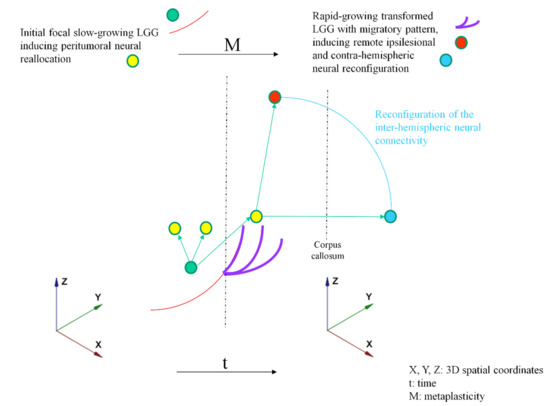
The behavior of lower-grade glioma (LGG) is changing over time, spontaneously, and in reaction to treatments. First, due to genomic instability and clonal expansion, although LGG progresses slowly during the early period of the disease, its growth velocity will accelerate when this tumor will transform to a higher grade of malignancy. Furthermore, its pattern of progression may change following therapy, e.g., by switching from a proliferative towards a more diffuse profile, in particular after surgical resection. In parallel to this plasticity of the neoplasm, the brain itself is constantly adapting to the tumor and possible treatment(s) thanks to reconfiguration within and between neural networks. Furthermore, the pattern of reallocation can also change, especially by switching from a perilesional to a contrahemispheric functional reorganization. Such a reorientation of mechanisms of cerebral reshaping, related to metaplasticity, consists of optimizing the efficiency of neural delocalization in order to allow functional compensation by adapting over time the profile of circuits redistribution to the behavioral modifications of the glioma. This interplay between LGG mutations and reactional connectomal instability leads to perpetual modulations in the glioma–neural equilibrium, both at ultrastructural and macroscopic levels, explaining the possible preservation of quality of life despite tumor progression. Here, an original model of these dynamic interactions across LGG plasticity and the brain metanetwork is proposed to guide a tailored step-by-step individualized therapeutic strategy over years. Integration of these new parameters, not yet considered in the current guidelines, might improve management of LGG patients. Full article
(This article belongs to the Special Issue Meningiomas and Low Grade Gliomas)
Show Figures

Figure 1

22 pages, 2373 KiB  
Article
Analysis of Telomere Maintenance Related Genes Reveals NOP10 as a New Metastatic-Risk Marker in Pheochromocytoma/Paraganglioma
by María Monteagudo, Paula Martínez, Luis J. Leandro-García, Ángel M. Martínez-Montes, Bruna Calsina, Marta Pulgarín-Alfaro, Alberto Díaz-Talavera, Sara Mellid, Rocío Letón, Eduardo Gil, Manuel Pérez-Martínez, Diego Megías, Raúl Torres-Ruiz, Sandra Rodriguez-Perales, Patricia González, Eduardo Caleiras, Scherezade Jiménez-Villa, Giovanna Roncador, Cristina Álvarez-Escolá, Rita M. Regojo, María Calatayud, Sonsoles Guadalix, Maria Currás-Freixes, Elena Rapizzi, Letizia Canu, Svenja Nölting, Hanna Remde, Martin Fassnacht, Nicole Bechmann, Graeme Eisenhofer, Massimo Mannelli, Felix Beuschlein, Marcus Quinkler, Cristina Rodríguez-Antona, Alberto Cascón, María A. Blasco, Cristina Montero-Conde and Mercedes Robledoadd Show full author list remove Hide full author list
Cancers 2021, 13(19), 4758; https://doi.org/10.3390/cancers13194758 - 23 Sep 2021
Cited by 19 | Viewed by 4768
Abstract
One of the main problems we face with PPGL is the lack of molecular markers capable of predicting the development of metastases in patients. Telomere-related genes, such as TERT and ATRX, have been recently described in PPGL, supporting the association between the activation [...] Read more.
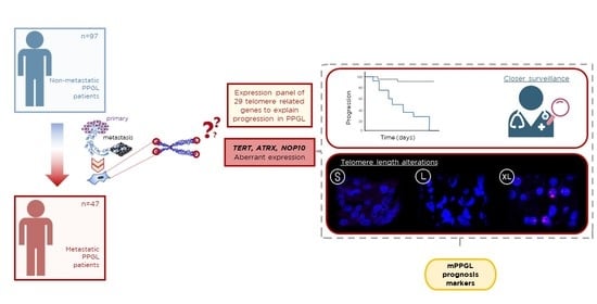
One of the main problems we face with PPGL is the lack of molecular markers capable of predicting the development of metastases in patients. Telomere-related genes, such as TERT and ATRX, have been recently described in PPGL, supporting the association between the activation of immortalization mechanisms and disease progression. However, the contribution of other genes involving telomere preservation machinery has not been previously investigated. In this work, we aimed to analyze the prognostic value of a comprehensive set of genes involved in telomere maintenance. For this study, we collected 165 PPGL samples (97 non-metastatic/63 metastatic), genetically characterized, in which the expression of 29 genes of interest was studied by NGS. Three of the 29 genes studied, TERT, ATRX and NOP10, showed differential expression between metastatic and non-metastatic cases, and alterations in these genes were associated with a shorter time to progression, independent of SDHB-status. We studied telomere length by Q-FISH in patient samples and in an in vitro model. NOP10 overexpressing tumors displayed an intermediate-length telomere phenotype without ALT, and in vitro results suggest that NOP10 has a role in telomerase-dependent telomere maintenance. We also propose the implementation of NOP10 IHC to better stratify PPGL patients. Full article
Show Figures

Graphical abstract

16 pages, 1504 KiB  
Review
Overcoming Immune Resistance in Prostate Cancer: Challenges and Advances
by Miyad Movassaghi, Rainjade Chung, Christopher B. Anderson, Mark Stein, Yvonne Saenger and Izak Faiena
Cancers 2021, 13(19), 4757; https://doi.org/10.3390/cancers13194757 - 23 Sep 2021
Cited by 20 | Viewed by 4339
Abstract
The use of immunotherapy has become a critical treatment modality in many advanced cancers. However, immunotherapy in prostate cancer has not been met with similar success. Multiple interrelated mechanisms, such as low tumor mutational burden, immunosuppressive cells, and impaired cellular immunity, appear to [...] Read more.
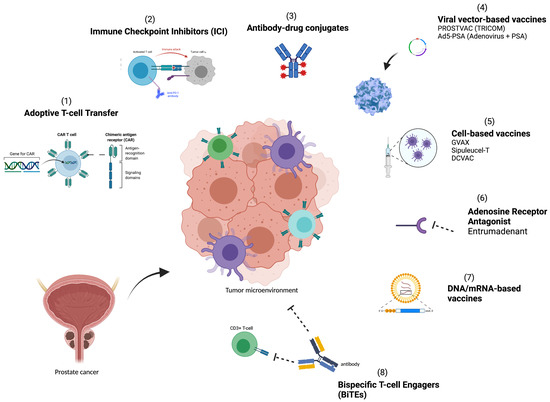
The use of immunotherapy has become a critical treatment modality in many advanced cancers. However, immunotherapy in prostate cancer has not been met with similar success. Multiple interrelated mechanisms, such as low tumor mutational burden, immunosuppressive cells, and impaired cellular immunity, appear to subvert the immune system, creating an immunosuppressive tumor microenvironment and leading to lower treatment efficacy in advanced prostate cancer. The lethality of metastatic castrate-resistant prostate cancer is driven by the lack of therapeutic regimens capable of generating durable responses. Multiple strategies are currently being tested to overcome immune resistance including combining various classes of treatment modalities. Several completed and ongoing trials have shown that combining vaccines or checkpoint inhibitors with hormonal therapy, radiotherapy, antibody–drug conjugates, chimeric antigen receptor T cell therapy, or chemotherapy may enhance immune responses and induce long-lasting clinical responses without significant toxicity. Here, we review the current state of immunotherapy for prostate cancer, as well as tumor-specific mechanisms underlying therapeutic resistance, with a comprehensive look at the current preclinical and clinical immunotherapeutic strategies aimed at overcoming the immunosuppressive tumor microenvironment and impaired cellular immunity that have largely limited the utility of immunotherapy in advanced prostate cancer. Full article
(This article belongs to the Special Issue Advances in Immunotherapy for Genitourinary Malignancies)
Show Figures

Figure 1

25 pages, 2678 KiB  
Review
Current and Future Therapies for Immunogenic Cell Death and Related Molecules to Potentially Cure Primary Breast Cancer
by Ryungsa Kim and Takanori Kin
Cancers 2021, 13(19), 4756; https://doi.org/10.3390/cancers13194756 - 23 Sep 2021
Cited by 18 | Viewed by 3803
Abstract
How primary breast cancer can be cured after (neo)adjuvant therapy remains unclear at the molecular level. Immune activation by anticancer agents may contribute to residual tumor cell eradication with postsurgical (neo)adjuvant chemotherapy. Chemotherapy-induced immunogenic cell death (ICD) may result in long-term immune activation [...] Read more.
How primary breast cancer can be cured after (neo)adjuvant therapy remains unclear at the molecular level. Immune activation by anticancer agents may contribute to residual tumor cell eradication with postsurgical (neo)adjuvant chemotherapy. Chemotherapy-induced immunogenic cell death (ICD) may result in long-term immune activation with memory effector T cells, leading to a primary breast cancer cure. Anthracycline and taxane treatments cause ICD and immunogenic modulations, resulting in the activation of antitumor immunity through damage-associated molecular patterns (DAMPs), such as adenosine triphosphate, calreticulin, high mobility group box 1, heat shock proteins 70/90, and annexin A1. This response may eradicate residual tumor cells after surgical treatment. Although DAMP release is also implicated in tumor progression, metastasis, and drug resistance, thereby representing a double-edged sword, robust immune activation by anticancer agents and the subsequent acquisition of long-term antitumor immune memory can be essential components of the primary breast cancer cure. This review discusses the molecular mechanisms by which anticancer drugs induce ICD and immunogenic modifications for antitumor immunity and targeted anti-DAMP therapy. Our aim was to improve the understanding of how to eradicate residual tumor cells treated with anticancer drugs and cure primary breast cancer by enhancing antitumor immunity with immune checkpoint inhibitors and vaccines. Full article
Show Figures

Figure 1

18 pages, 1806 KiB  
Review
Lost but Not Least—Novel Insights into Progesterone Receptor Loss in Estrogen Receptor-Positive Breast Cancer
by Michał Kunc, Marta Popęda, Wojciech Biernat and Elżbieta Senkus
Cancers 2021, 13(19), 4755; https://doi.org/10.3390/cancers13194755 - 23 Sep 2021
Cited by 11 | Viewed by 4965
Abstract
Estrogen receptor α (ERα) and progesterone receptor (PgR) are crucial prognostic and predictive biomarkers that are usually co-expressed in breast cancer (BC). However, 12–24% of BCs present ERα(+)/PgR(−) phenotype at immunohistochemical evaluation. In fact, BC may either show primary PgR(−) status (in chemonaïve [...] Read more.
Estrogen receptor α (ERα) and progesterone receptor (PgR) are crucial prognostic and predictive biomarkers that are usually co-expressed in breast cancer (BC). However, 12–24% of BCs present ERα(+)/PgR(−) phenotype at immunohistochemical evaluation. In fact, BC may either show primary PgR(−) status (in chemonaïve tumor sample), lose PgR expression during neoadjuvant treatment, or acquire PgR(−) phenotype in local relapse or metastasis. The loss of PgR expression in ERα(+) breast cancer may signify resistance to endocrine therapy and poorer outcomes. On the other hand, ERα(+)/PgR(−) BCs may have a better response to neoadjuvant chemotherapy than double-positive tumors. Loss of PgR expression may be a result of pre-transcriptional alterations (copy number loss, mutation, epigenetic modifications), decreased transcription of the PGR gene (e.g., by microRNAs), and post-translational modifications (e.g., phosphorylation, sumoylation). Various processes involved in the down-regulation of PgR have distinct consequences on the biology of cancer cells. Occasionally, negative PgR status detected by immunohistochemical analysis is paradoxically associated with enhanced transcriptional activity of PgR that might be inhibited by antiprogestin treatment. Identification of the mechanism of PgR loss in each patient seems challenging, yet it may provide important information on the biology of the tumor and predict its responsiveness to the therapy. Full article
(This article belongs to the Special Issue The Endocrine Basis of Breast Cancer)
Show Figures

Figure 1

20 pages, 1155 KiB  
Review
The ‘Yin and Yang’ of Cancer Cell Growth and Mechanosensing
by Malak Amer, Lidan Shi and Haguy Wolfenson
Cancers 2021, 13(19), 4754; https://doi.org/10.3390/cancers13194754 - 23 Sep 2021
Cited by 12 | Viewed by 5396
Abstract
In cancer, two unique and seemingly contradictory behaviors are evident: on the one hand, tumors are typically stiffer than the tissues in which they grow, and this high stiffness promotes their malignant progression; on the other hand, cancer cells are anchorage-independent—namely, they can [...] Read more.
In cancer, two unique and seemingly contradictory behaviors are evident: on the one hand, tumors are typically stiffer than the tissues in which they grow, and this high stiffness promotes their malignant progression; on the other hand, cancer cells are anchorage-independent—namely, they can survive and grow in soft environments that do not support cell attachment. How can these two features be consolidated? Recent findings on the mechanisms by which cells test the mechanical properties of their environment provide insight into the role of aberrant mechanosensing in cancer progression. In this review article, we focus on the role of high stiffness on cancer progression, with particular emphasis on tumor growth; we discuss the mechanisms of mechanosensing and mechanotransduction, and their dysregulation in cancerous cells; and we propose that a ‘yin and yang’ type phenomenon exists in the mechanobiology of cancer, whereby a switch in the type of interaction with the extracellular matrix dictates the outcome of the cancer cells. Full article
(This article belongs to the Special Issue Mechanobiology in Cancer)
Show Figures

Graphical abstract

14 pages, 1030 KiB  
Review
Overcoming Immunotherapy Resistance by Targeting the Tumor-Intrinsic NLRP3-HSP70 Signaling Axis
by Balamayooran Theivanthiran, Tarek Haykal, Linda Cao, Alisha Holtzhausen, Michael Plebanek, Nicholas C. DeVito and Brent A. Hanks
Cancers 2021, 13(19), 4753; https://doi.org/10.3390/cancers13194753 - 23 Sep 2021
Cited by 12 | Viewed by 3896
Abstract
The tumor-intrinsic NOD-like receptor family, pyrin-domain-containing-3 (NLRP3) inflammasome, plays an important role in regulating immunosuppressive myeloid cell populations in the tumor microenvironment (TME). While prior studies have described the activation of this inflammasome in driving pro-tumorigenic mechanisms, emerging data is now revealing the [...] Read more.
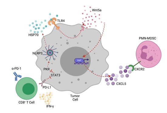
The tumor-intrinsic NOD-like receptor family, pyrin-domain-containing-3 (NLRP3) inflammasome, plays an important role in regulating immunosuppressive myeloid cell populations in the tumor microenvironment (TME). While prior studies have described the activation of this inflammasome in driving pro-tumorigenic mechanisms, emerging data is now revealing the tumor NLRP3 inflammasome and the downstream release of heat shock protein-70 (HSP70) to regulate anti-tumor immunity and contribute to the development of adaptive resistance to anti-PD-1 immunotherapy. Genetic alterations that influence the activity of the NLRP3 signaling axis are likely to impact T cell-mediated tumor cell killing and may indicate which tumors rely on this pathway for immune escape. These studies suggest that the NLRP3 inflammasome and its secreted product, HSP70, represent promising pharmacologic targets for manipulating innate immune cell populations in the TME while enhancing responses to anti-PD-1 immunotherapy. Additional studies are needed to better understand tumor-specific regulatory mechanisms of NLRP3 to enable the development of tumor-selective pharmacologic strategies capable of augmenting responses to checkpoint inhibitor immunotherapy while minimizing unwanted off-target effects. The execution of upcoming clinical trials investigating this strategy to overcome anti-PD-1 resistance promises to provide novel insight into the role of this pathway in immuno-oncology. Full article
(This article belongs to the Special Issue Targeting Innate Immunity to Treat Cancer)
Show Figures

Figure 1

16 pages, 786 KiB  
Review
Epstein–Barr Virus in Inborn Immunodeficiency—More Than Infection
by Ciro Novaes Rosa Lino and Sujal Ghosh
Cancers 2021, 13(19), 4752; https://doi.org/10.3390/cancers13194752 - 23 Sep 2021
Cited by 19 | Viewed by 5175
Abstract
Epstein–Barr Virus (EBV) is a ubiquitous virus affecting more than 90% of the world’s population. Upon infection, it establishes latency in B cells. It is a rather benign virus for immune-competent individuals, in whom infections usually go unnoticed. Nevertheless, EBV has been extensively [...] Read more.
Epstein–Barr Virus (EBV) is a ubiquitous virus affecting more than 90% of the world’s population. Upon infection, it establishes latency in B cells. It is a rather benign virus for immune-competent individuals, in whom infections usually go unnoticed. Nevertheless, EBV has been extensively associated with tumorigenesis. Patients suffering from certain inborn errors of immunity are at high risk of developing malignancies, while infection in the majority of immune-competent individuals does not seem to lead to immune dysregulation. Herein, we discuss how inborn mutations in TNFRSF9, CD27, CD70, CORO1A, CTPS1, ITK, MAGT1, RASGRP1, STK4, CARMIL2, SH2D1A, and XIAP affect the development, differentiation, and function of key factors involved in the immunity against EBV, leading to increased susceptibility to lymphoproliferative disease and lymphoma. Full article
(This article belongs to the Special Issue Epstein-Barr Virus Infection in Cancer)
Show Figures

Figure 1

17 pages, 2322 KiB  
Article
Machine-Learning Assisted Discrimination of Precancerous and Cancerous from Healthy Oral Tissue Based on Multispectral Autofluorescence Lifetime Imaging Endoscopy
by Elvis Duran-Sierra, Shuna Cheng, Rodrigo Cuenca, Beena Ahmed, Jim Ji, Vladislav V. Yakovlev, Mathias Martinez, Moustafa Al-Khalil, Hussain Al-Enazi, Yi-Shing Lisa Cheng, John Wright, Carlos Busso and Javier A. Jo
Cancers 2021, 13(19), 4751; https://doi.org/10.3390/cancers13194751 - 23 Sep 2021
Cited by 29 | Viewed by 4611
Abstract
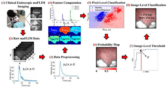
Multispectral autofluorescence lifetime imaging (maFLIM) can be used to clinically image a plurality of metabolic and biochemical autofluorescence biomarkers of oral epithelial dysplasia and cancer. This study tested the hypothesis that maFLIM-derived autofluorescence biomarkers can be used in machine-learning (ML) models to discriminate [...] Read more.
Multispectral autofluorescence lifetime imaging (maFLIM) can be used to clinically image a plurality of metabolic and biochemical autofluorescence biomarkers of oral epithelial dysplasia and cancer. This study tested the hypothesis that maFLIM-derived autofluorescence biomarkers can be used in machine-learning (ML) models to discriminate dysplastic and cancerous from healthy oral tissue. Clinical widefield maFLIM endoscopy imaging of cancerous and dysplastic oral lesions was performed at two clinical centers. Endoscopic maFLIM images from 34 patients acquired at one of the clinical centers were used to optimize ML models for automated discrimination of dysplastic and cancerous from healthy oral tissue. A computer-aided detection system was developed and applied to a set of endoscopic maFLIM images from 23 patients acquired at the other clinical center, and its performance was quantified in terms of the area under the receiver operating characteristic curve (ROC-AUC). Discrimination of dysplastic and cancerous from healthy oral tissue was achieved with an ROC-AUC of 0.81. This study demonstrates the capabilities of widefield maFLIM endoscopy to clinically image autofluorescence biomarkers that can be used in ML models to discriminate dysplastic and cancerous from healthy oral tissue. Widefield maFLIM endoscopy thus holds potential for automated in situ detection of oral dysplasia and cancer. Full article
(This article belongs to the Collection Artificial Intelligence in Oncology)
Show Figures

Figure 1

22 pages, 845 KiB  
Review
Philadelphia-Negative Chronic Myeloproliferative Neoplasms during the COVID-19 Pandemic: Challenges and Future Scenarios
by Francesca Palandri, Massimo Breccia, Valerio De Stefano and Francesco Passamonti
Cancers 2021, 13(19), 4750; https://doi.org/10.3390/cancers13194750 - 23 Sep 2021
Cited by 10 | Viewed by 3379
Abstract
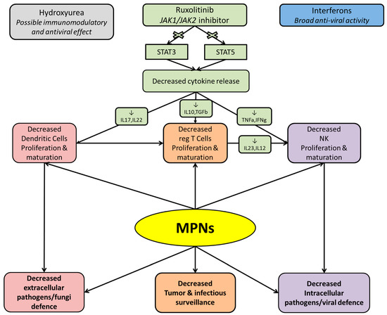
An outbreak of severe acute respiratory syndrome coronavirus 2 (SARS-CoV2) started in December 2019 in China and then become pandemic in February 2020. Several publications investigated the possible increased rate of COVID-19 infection in hematological malignancies. Based on the published data, strategies for [...] Read more.
An outbreak of severe acute respiratory syndrome coronavirus 2 (SARS-CoV2) started in December 2019 in China and then become pandemic in February 2020. Several publications investigated the possible increased rate of COVID-19 infection in hematological malignancies. Based on the published data, strategies for the management of chronic Philadelphia-negative chronic myeloproliferative neoplasms (MPNs) are provided. The risk of severe COVID-19 seems high in MPN, particularly in patients with essential thrombocythemia, but not negligible in myelofibrosis. MPN patients are at high risk of both thrombotic and hemorrhagic complications and this must be accounted in the case of COVID-19 deciding on a case-by-case basis. There are currently no data to suggest that hydroxyurea or interferon may influence the risk or severity of COVID-19 infection. Conversely, while the immunosuppressive activity of ruxolitinib might pose increased risk of infection, its abrupt discontinuation during COVID-19 syndrome is associated with worse outcome. All MPN patients should receive vaccine against COVID-19; reassuring data are available on efficacy of mRNA vaccines in MPNs. Full article
(This article belongs to the Special Issue Pathogenesis and Natural History of Myeloproliferative Neoplasms)
Show Figures

Figure 1

11 pages, 1095 KiB  
Article
Clinical Characteristics, Care Trajectories and Mortality Rate of SARS-CoV-2 Infected Cancer Patients: A Multicenter Cohort Study
by Marc-Antoine Benderra, Ainhoa Aparicio, Judith Leblanc, Demian Wassermann, Emmanuelle Kempf, Gilles Galula, Mélodie Bernaux, Anthony Canellas, Thomas Moreau, Ali Bellamine, Jean-Philippe Spano, Christel Daniel, Julien Champ, Florence Canouï-Poitrine, Joseph Gligorov, on Behalf of the AP-HP/Universities/INSERM COVID-19 Research Collaboration, Cancer AP-HP Group COVID-19 Task Force and AP-HP Covid CDW Initiative (ACCI)
Cancers 2021, 13(19), 4749; https://doi.org/10.3390/cancers13194749 - 23 Sep 2021
Cited by 15 | Viewed by 3152
Abstract
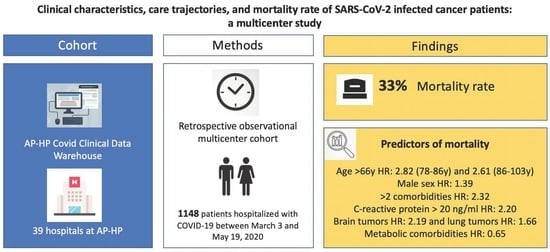
Background: COVID-19 may be more frequent and more severe in cancer patients than in other individuals. Our aims were to assess the rate of COVID-19 in hospitalized cancer patients, to describe their demographic characteristics, clinical features and care trajectories, and to assess the [...] Read more.
Background: COVID-19 may be more frequent and more severe in cancer patients than in other individuals. Our aims were to assess the rate of COVID-19 in hospitalized cancer patients, to describe their demographic characteristics, clinical features and care trajectories, and to assess the mortality rate. Methods: This multicenter cohort study was based on the Electronic Health Records of the Assistance Publique-Hôpitaux de Paris (AP-HP). Cancer patients with a diagnosis of COVID-19 between 3 March and 19 May 2020 were included. Main outcome was all-cause mortality within 30 days of COVID-19 diagnosis. Results: A total of 29,141 cancer patients were identified and 7791 (27%) were tested for SARS-CoV-2. Of these, 1359 (17%) were COVID-19-positive and 1148 (84%) were hospitalized; 217 (19%) were admitted to an intensive care unit. The mortality rate was 33% (383 deaths). In multivariate analysis, mortality-related factors were male sex (aHR = 1.39 [95% CI: 1.07–1.81]), advanced age (78–86 y: aHR = 2.83 [95% CI: 1.78–4.51] vs. <66 y; 86–103 y: aHR = 2.61 [95% CI: 1.56–4.35] vs. <66 y), more than two comorbidities (aHR = 2.32 [95% CI: 1.41–3.83]) and C-reactive protein >20 ng/mL (aHR = 2.20 [95% CI: 1.70–2.86]). Primary brains tumors (aHR = 2.19 [95% CI: 1.08–4.44]) and lung cancer (aHR = 1.66 [95% CI: 1.02–2.70]) were associated with higher mortality. Risk of dying was lower among patients with metabolic comorbidities (aHR = 0.65 [95% CI: 0.50–0.84]). Conclusions: In a hospital-based setting, cancer patients with COVID-19 had a high mortality rate. This mortality was mainly driven by age, sex, number of comorbidities and presence of inflammation. This is the first cohort of cancer patients in which metabolic comorbidities were associated with a better outcome. Full article
(This article belongs to the Collection The Impact of COVID-19 Infection in Cancer)
Show Figures

Graphical abstract