Nutrients 2018, 10(3), 279; https://doi.org/10.3390/nu10030279 - 28 Feb 2018
Cited by 28 | Viewed by 5276
Abstract
►
Show Figures
Thyroid cancer (TC) is the most common endocrine malignancy without reliable preventive agent. Resveratrol possesses in vitro anti-TC activities; while its effect(s) on thyroid tumorigenesis remains unknown. This study aims to address this issue using DEN/MNU/DHPN-induced rat carcinogenesis model. 50 male Sprague-Dawley rats
[...] Read more.
Thyroid cancer (TC) is the most common endocrine malignancy without reliable preventive agent. Resveratrol possesses in vitro anti-TC activities; while its effect(s) on thyroid tumorigenesis remains unknown. This study aims to address this issue using DEN/MNU/DHPN-induced rat carcinogenesis model. 50 male Sprague-Dawley rats were separated into four groups as Group-1 (5 rats); normally fed; Group-2 (15 rats); DEN/MNU/DHPN treatment only; Group-3 (15 rats) and -4 (15 rats); DEN/MNU/DHPN treatment; followed by resveratrol intragastric (IG) injection and intraperitoneal (IP) injection; respectively; in two-day intervals for 30 weeks. The results revealed that the average resveratrol concentration in thyroid tissues was 1.278 ± 0.419 nmol/g in IG group and 1.752 ± 0.398 nmol/g in IP group. The final body weights of Group-3 and Group-4 were lighter than that (p > 0.05) of Group-1; but heavier than Group-2 (p < 0.05). TC-related lesions (hyperplasia and adenomas) were found in 53.3% of Group-2; 33.3% Group-3 and 26.7% Group-4. Lower serum carcino-embryonic antigen (CEA) and thyroglobulin (Tg) levels; down-regulated expression of IL-6 and cyclooxygenase-2 (COX-2); reduction of NF-κB/p65 nuclear translocation; and elevated IkBα expression were found in the thyroid tissues of Group-3 and Group-4 in comparison with that of Group-2. These results demonstrate that IG and IP administered resveratrol efficiently reduces the frequency and severity of DEN/MNU/DHPN-caused TC-related lesions and would be of values in thyroid tumor prevention.
Full article

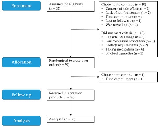
Figure 1


