by
Marco Orlando 1,† , Gianluca Molla 1,*, Pietro Castellani 2, Valentina Pirillo 1, Vincenzo Torretta 2 and Navarro Ferronato 2
, Gianluca Molla 1,*, Pietro Castellani 2, Valentina Pirillo 1, Vincenzo Torretta 2 and Navarro Ferronato 2
1
Department of Biotechnology and Life Sciences, University of Insubria, Via Dunant, 21100 Varese, Italy
2
Department of Theoretical and Applied Sciences (DiSTA), University of Insubria, Via G.B. Vico 46, 21100 Varese, Italy
†
Current address: Department of Biotechnology and Biosciences, University of Milano-Bicocca, Piazza della Scienza 2, 20126 Milano, Italy.
Int. J. Mol. Sci. 2023, 24(4), 3877; https://doi.org/10.3390/ijms24043877 - 15 Feb 2023
Cited by 90 | Viewed by 15222
Abstract
The accumulation of synthetic plastic waste in the environment has become a global concern. Microbial enzymes (purified or as whole-cell biocatalysts) represent emerging biotechnological tools for waste circularity; they can depolymerize materials into reusable building blocks, but their contribution must be considered within
[...] Read more.
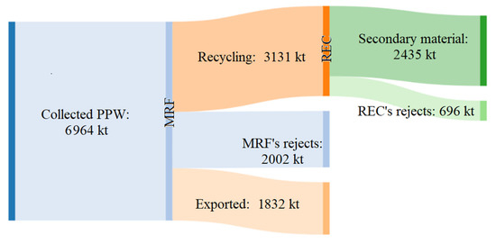
The accumulation of synthetic plastic waste in the environment has become a global concern. Microbial enzymes (purified or as whole-cell biocatalysts) represent emerging biotechnological tools for waste circularity; they can depolymerize materials into reusable building blocks, but their contribution must be considered within the context of present waste management practices. This review reports on the prospective of biotechnological tools for plastic bio-recycling within the framework of plastic waste management in Europe. Available biotechnology tools can support polyethylene terephthalate (PET) recycling. However, PET represents only ≈7% of unrecycled plastic waste. Polyurethanes, the principal unrecycled waste fraction, together with other thermosets and more recalcitrant thermoplastics (e.g., polyolefins) are the next plausible target for enzyme-based depolymerization, even if this process is currently effective only on ideal polyester-based polymers. To extend the contribution of biotechnology to plastic circularity, optimization of collection and sorting systems should be considered to feed chemoenzymatic technologies for the treatment of more recalcitrant and mixed polymers. In addition, new bio-based technologies with a lower environmental impact in comparison with the present approaches should be developed to depolymerize (available or new) plastic materials, that should be designed for the required durability and for being susceptible to the action of enzymes.
Full article
(This article belongs to the Special Issue Microbial Enzymes for Biotechnological Applications)
▼
Show Figures
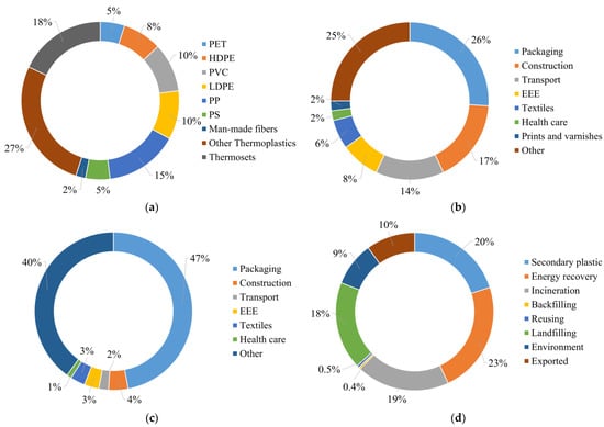
Figure 1


