22 pages, 3837 KiB  
Article
Effects of Fractionated Radiation Exposure on Vimentin Expression in Cervical Cancers: Analysis of Association with Cancer Stem Cell Response and Short-Term Prognosis
by Irina Zamulaeva 1,*, Olga Matchuk 1, Elena Selivanova 1, Liana Mkrtchian 2, Anna Yakimova 1, Victoria Gusarova 1, Nikita Lipunov 1, Liudmila Krikunova 2, Sergey Ivanov 3,4 and Andrey Kaprin 4,5
1 Department of Radiation Biochemistry, A. Tsyb Medical Radiological Research Center—Branch of the National Medical Research Radiological Center of the Ministry of Health of the Russian Federation, Korolev Str.-4, Obninsk 249036, Russia
2 Department of Radiation and Combined Treatment of Gynecological Diseases, A. Tsyb Medical Radiological Research Center—Branch of the National Medical Research Radiological Center of the Ministry of Health of the Russian Federation, Korolev Str.-4, Obninsk 249036, Russia
3 A. Tsyb Medical Radiological Research Center—Branch of the National Medical Research Radiological Center of the Ministry of Health of the Russian Federation, Korolev Str.-4, Obninsk 249036, Russia
4 Department of Urology and Operative Nephrology, RUDN University, Miklukho-Maklaya Str.-6, Moscow 117198, Russia
5 National Medical Research Radiological Center of the Ministry of Health of the Russian Federation, Korolev Str.-4, Obninsk 249036, Russia
Int. J. Mol. Sci. 2023, 24(4), 3271; https://doi.org/10.3390/ijms24043271 - 7 Feb 2023
Cited by 5 | Viewed by 2096
Abstract
Elucidation of the mechanisms for the response of cancer stem cells (CSCs) to radiation exposure is of considerable interest for further improvement of radio- and chemoradiotherapy of cervical cancer (CC). The aim of this work is to evaluate the effects of fractionated radiation [...] Read more.
Elucidation of the mechanisms for the response of cancer stem cells (CSCs) to radiation exposure is of considerable interest for further improvement of radio- and chemoradiotherapy of cervical cancer (CC). The aim of this work is to evaluate the effects of fractionated radiation exposure on the expression of vimentin, which is one of the end-stage markers of epithelial-mesenchymal transition (EMT), and analyze its association with CSC radiation response and short-term prognosis of CC patients. The level of vimentin expression was determined in HeLa, SiHa cell lines, and scrapings from the cervix of 46 CC patients before treatment and after irradiation at a total dose of 10 Gy using real-time polymerase chain reaction (PCR) assay, flow cytometry, and fluorescence microscopy. The number of CSCs was assessed using flow cytometry. Significant correlations were shown between vimentin expression and postradiation changes in CSC numbers in both cell lines (R = 0.88, p = 0.04 for HeLa and R = 0.91, p = 0.01 for SiHa) and cervical scrapings (R = 0.45, p = 0.008). Associations were found at the level of tendency between postradiation increase in vimentin expression and unfavorable clinical outcome 3–6 months after treatment. The results clarify some of the relationships between EMT, CSCs, and therapeutic resistance that are needed to develop new strategies for cancer treatment. Full article
(This article belongs to the Special Issue Advances in Radiation Toxicity)
Show Figures

Figure 1

12 pages, 1326 KiB  
Article
Autoantibodies to PAX5, PTCH1, and GNA11 as Serological Biomarkers in the Detection of Hepatocellular Carcinoma in Hispanic Americans
by Cuipeng Qiu 1, Yangcheng Ma 1, Bofei Wang 2, Xiaojun Zhang 1, Xiao Wang 1 and Jian-Ying Zhang 1,*
1 Department of Biological Sciences & NIH-Sponsored Border Biomedical Research Center, The University of Texas at El Paso, El Paso, TX 79968, USA
2 Department of Leukemia, The University of Texas at MD Anderson Cancer Center, Houston, TX 77030, USA
Int. J. Mol. Sci. 2023, 24(4), 3721; https://doi.org/10.3390/ijms24043721 - 13 Feb 2023
Cited by 4 | Viewed by 2089
Abstract
Studies have demonstrated that autoantibodies to tumor-associated antigens (TAAs) may be used as efficient biomarkers with low-cost and highly sensitive characteristics. In this study, an enzyme-linked immunosorbent assay (ELISA) was conducted to analyze autoantibodies to paired box protein Pax-5 (PAX5), protein patched homolog [...] Read more.
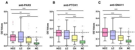
Studies have demonstrated that autoantibodies to tumor-associated antigens (TAAs) may be used as efficient biomarkers with low-cost and highly sensitive characteristics. In this study, an enzyme-linked immunosorbent assay (ELISA) was conducted to analyze autoantibodies to paired box protein Pax-5 (PAX5), protein patched homolog 1 (PTCH1), and guanine nucleotide-binding protein subunit alpha-11 (GNA11) in sera from Hispanic Americans including hepatocellular carcinoma (HCC) patients, patients with liver cirrhosis (LC), patients with chronic hepatitis (CH), as well as normal controls. Meanwhile, 33 serial sera from eight HCC patients before and after diagnosis were used to explore the potential of these three autoantibodies as early biomarkers. In addition, an independent non-Hispanic cohort was used to evaluate the specificity of these three autoantibodies. In the Hispanic cohort, at the 95.0% specificity for healthy controls, 52.0%, 44.0%, and 44.0% of HCC patients showed significantly elevated levels of autoantibodies to PAX5, PTCH1, and GNA11, respectively. Among patients with LC, the frequencies for autoantibodies to PAX5, PTCH1, and GNA11 were 32.1%, 35.7%, and 25.0%, respectively. The area under the ROC curves (AUCs) of autoantibodies to PAX5, PTCH1, and GNA11 for identifying HCC from healthy controls were 0.908, 0.924, and 0.913, respectively. When these three autoantibodies were combined as a panel, the sensitivity could be improved to 68%. The prevalence of PAX5, PTCH1, and GNA11 autoantibodies has already occurred in 62.5%, 62.5%, or 75.0% of patients before clinical diagnosis, respectively. In the non-Hispanic cohort, autoantibodies to PTCH1 showed no significant difference; however, autoantibodies to PAX5, PTCH1, and GNA11 showed potential value as biomarkers for early detection of HCC in the Hispanic population and they may monitor the transition of patients with high-risk (LC, CH) to HCC. Using a panel of the three anti-TAA autoantibodies may enhance the detection of HCC. Full article
(This article belongs to the Special Issue Molecular Mechanism of Chronic Viral and Non-viral Liver Diseases)
Show Figures

Figure 1

16 pages, 2048 KiB  
Article
Lethal Caspase-1/4-Dependent Injury Occurs in the First Minutes of Coronary Reperfusion and Requires Calpain Activity
by Xi-Ming Yang 1, Michael V. Cohen 1,2,*, Sarah Sayner 1, Jonathon P. Audia 3 and James M. Downey 1
1 Department of Physiology and Cell Biology, College of Medicine, University of South Alabama, Mobile, AL 36688, USA
2 Department of Medicine, College of Medicine, University of South Alabama, Mobile, AL 36688, USA
3 Department of Microbiology, College of Medicine, University of South Alabama, Mobile, AL 36688, USA
Int. J. Mol. Sci. 2023, 24(4), 3801; https://doi.org/10.3390/ijms24043801 - 14 Feb 2023
Cited by 4 | Viewed by 2085
Abstract
To study the relationship between caspase-1/4 and reperfusion injury, we measured infarct size (IS) in isolated mouse hearts undergoing 50 min global ischemia/2 h reperfusion. Starting VRT-043198 (VRT) at reperfusion halved IS. The pan-caspase inhibitor emricasan duplicated VRT’s protection. IS in caspase-1/4-knockout hearts [...] Read more.
To study the relationship between caspase-1/4 and reperfusion injury, we measured infarct size (IS) in isolated mouse hearts undergoing 50 min global ischemia/2 h reperfusion. Starting VRT-043198 (VRT) at reperfusion halved IS. The pan-caspase inhibitor emricasan duplicated VRT’s protection. IS in caspase-1/4-knockout hearts was similarly reduced, supporting the hypothesis that caspase-1/4 was VRT’s only protective target. NLRC4 inflammasomes activate caspase-1. NLRC4 knockout hearts were not protected, eliminating NLRC4 as caspase-1/4’s activator. The amount of protection that could be achieved by only suppressing caspase-1/4 activity was limited. In wild-type (WT) hearts, ischemic preconditioning (IPC) was as protective as caspase-1/4 inhibitors. Combining IPC and emricasan in these hearts or preconditioning caspase-1/4-knockout hearts produced an additive IS reduction, indicating that more protection could be achieved by combining treatments. We determined when caspase-1/4 exerted its lethal injury. Starting VRT after 10 min of reperfusion in WT hearts was no longer protective, revealing that caspase-1/4 inflicted its injury within the first 10 min of reperfusion. Ca++ influx at reperfusion might activate caspase-1/4. We tested whether Ca++-dependent soluble adenylyl cyclase (AC10) could be responsible. However, IS in AC10−/− hearts was not different from that in WT control hearts. Ca++-activated calpain has been implicated in reperfusion injury. Calpain could be releasing actin-bound procaspase-1 in cardiomyocytes, which would explain why caspase-1/4-related injury is confined to early reperfusion. The calpain inhibitor calpeptin duplicated emricasan’s protection. Unlike IPC, adding calpain to emricasan offered no additional protection, suggesting that caspase-1/4 and calpain may share the same protective target. Full article
(This article belongs to the Special Issue Cell Death in Cardiovascular Disease)
Show Figures

Figure 1

15 pages, 17136 KiB  
Article
Secondary Degeneration of Oligodendrocyte Precursor Cells Occurs as Early as 24 h after Optic Nerve Injury in Rats
by Lillian M. Toomey 1,2, Melissa G. Papini 1, Thomas O. Clarke 3, Alexander J. Wright 1, Eleanor Denham 1, Andrew Warnock 1, Terry McGonigle 1, Carole A. Bartlett 3, Melinda Fitzgerald 1,2 and Chidozie C. Anyaegbu 1,2,*
1 Curtin Health Innovation Research Institute, Curtin University, Bentley, WA 6102, Australia
2 Perron Institute for Neurological and Translational Science, Sarich Neuroscience Research Institute Building, 8 Verdun St., Nedlands, WA 6009, Australia
3 Experimental and Regenerative Neurosciences, School of Biological Sciences, The University of Western Australia, Perth, WA 6009, Australia
Int. J. Mol. Sci. 2023, 24(4), 3463; https://doi.org/10.3390/ijms24043463 - 9 Feb 2023
Cited by 2 | Viewed by 2080
Abstract
Optic nerve injury causes secondary degeneration, a sequela that spreads damage from the primary injury to adjacent tissue, through mechanisms such as oxidative stress, apoptosis, and blood-brain barrier (BBB) dysfunction. Oligodendrocyte precursor cells (OPCs), a key component of the BBB and oligodendrogenesis, are [...] Read more.
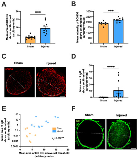
Optic nerve injury causes secondary degeneration, a sequela that spreads damage from the primary injury to adjacent tissue, through mechanisms such as oxidative stress, apoptosis, and blood-brain barrier (BBB) dysfunction. Oligodendrocyte precursor cells (OPCs), a key component of the BBB and oligodendrogenesis, are vulnerable to oxidative deoxyribonucleic acid (DNA) damage by 3 days post-injury. However, it is unclear whether oxidative damage in OPCs occurs earlier at 1 day post-injury, or whether a critical ‘window-of-opportunity’ exists for therapeutic intervention. Here, a partial optic nerve transection rat model of secondary degeneration was used with immunohistochemistry to assess BBB dysfunction, oxidative stress, and proliferation in OPCs vulnerable to secondary degeneration. At 1 day post-injury, BBB breach and oxidative DNA damage were observed, alongside increased density of DNA-damaged proliferating cells. DNA-damaged cells underwent apoptosis (cleaved caspase3+), and apoptosis was associated with BBB breach. OPCs experienced DNA damage and apoptosis and were the major proliferating cell type with DNA damage. However, the majority of caspase3+ cells were not OPCs. These results provide novel insights into acute secondary degeneration mechanisms in the optic nerve, highlighting the need to consider early oxidative damage to OPCs in therapeutic efforts to limit degeneration following optic nerve injury. Full article
Show Figures

Figure 1

18 pages, 4841 KiB  
Article
Approaches in Hydroxytyrosol Supplementation on Epithelial—Mesenchymal Transition in TGFβ1-Induced Human Respiratory Epithelial Cells
by Rabiatul Adawiyah Razali 1, Muhammad Dain Yazid 1, Aminuddin Saim 2, Ruszymah Bt Hj Idrus 1,3 and Yogeswaran Lokanathan 1,*
1 Centre for Tissue Engineering and Regenerative Medicine, Faculty of Medicine, Universiti Kebangsaan Malaysia, Cheras, Kuala Lumpur 56000, Malaysia
2 Graduate School of Medicine, KPJ Healthcare University College, Kota Seriemas, Nilai 71800, Malaysia
3 Department of Physiology, Faculty of Medicine, Universiti Kebangsaan Malaysia, Cheras, Kuala Lumpur 56000, Malaysia
Int. J. Mol. Sci. 2023, 24(4), 3974; https://doi.org/10.3390/ijms24043974 - 16 Feb 2023
Cited by 7 | Viewed by 2079
Abstract
Hydroxytyrosol (HT) is an olive polyphenol with anti-inflammatory and antioxidant properties. This study aimed to investigate the effect of HT treatment on epithelial–mesenchymal transition (EMT) in primary human respiratory epithelial cells (RECs) isolated from human nasal turbinate. HT dose–response study and growth kinetic [...] Read more.
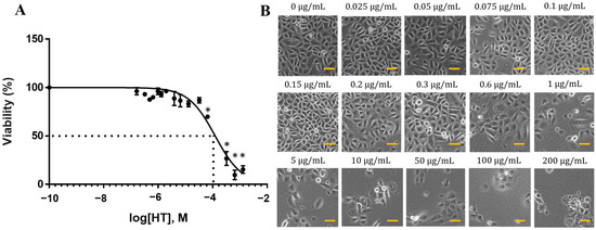
Hydroxytyrosol (HT) is an olive polyphenol with anti-inflammatory and antioxidant properties. This study aimed to investigate the effect of HT treatment on epithelial–mesenchymal transition (EMT) in primary human respiratory epithelial cells (RECs) isolated from human nasal turbinate. HT dose–response study and growth kinetic study on RECs was performed. Several approaches on HT treatment and TGFβ1 induction with varying durations and methods was studied. RECs morphology and migration ability were evaluated. Vimentin and E-cadherin immunofluorescence staining and Western blotting [E-cadherin, vimentin, SNAIL/SLUG, AKT, phosphorylated (p)AKT, SMAD2/3 and pSMAD2/3] were performed after 72-h treatment. In silico analysis (molecular docking) of HT was performed to evaluate the potential of HT to bind with the TGFβ receptor. The viability of the HT-treated RECs was concentration-dependent, where the median effective concentration (EC50) was 19.04 μg/mL. Testing of the effects of 1 and 10 µg/mL HT revealed that HT suppressed expression of the protein markers vimentin and SNAIL/SLUG while preserving E-cadherin protein expression. Supplementation with HT protected against SMAD and AKT pathway activation in the TGFβ1-induced RECs. Furthermore, HT demonstrated the potential to bind with ALK5 (a TGFβ receptor component) in comparison to oleuropein. TGFβ1-induced EMT in RECs and HT exerted a positive effect in modulating the effects of EMT. Full article
(This article belongs to the Special Issue Novel Natural Compound for Wound and Tissue Repair and Regeneration)
Show Figures

Figure 1

13 pages, 631 KiB  
Article
Toxicity and Environmental Risk Assessment of Polycarbamate and Its Main Components to Marine Algae and Crustaceans
by Toshimitsu Onduka * and Kazuhiko Mochida
Hatsukaichi Field Station, Fisheries Technology Institute, Japan Fisheries Research and Education Agency, 2-17-5 Maruishi, Hatsukaichi 739-0452, Hiroshima, Japan
Int. J. Mol. Sci. 2023, 24(4), 4183; https://doi.org/10.3390/ijms24044183 - 20 Feb 2023
Viewed by 2078
Abstract
Polycarbamate is commonly used as an antifoulant coating on fishing nets in Japan. Although its toxicity to freshwater organisms has been reported, its toxicity to marine organisms is currently unknown. We conducted algal growth inhibition and crustacean immobilization tests to assess the effects [...] Read more.
Polycarbamate is commonly used as an antifoulant coating on fishing nets in Japan. Although its toxicity to freshwater organisms has been reported, its toxicity to marine organisms is currently unknown. We conducted algal growth inhibition and crustacean immobilization tests to assess the effects of polycarbamate on marine organisms. We also evaluated the acute toxicity of the main components of polycarbamate, namely, dimethyldithiocarbamate and ethylenebisdithiocarbamate, to algae, which are the most sensitive tested organisms to polycarbamate. The toxicities of dimethyldithiocarbamate and ethylenebisdithiocarbamate partially explain that of polycarbamate. To assess the primary risk, we derived the predicted no-effect concentration (PNEC) for polycarbamate in a probabilistic manner using species sensitivity distributions. The 72 h no observed effect concentration (NOEC) of polycarbamate to the alga Skeletonema marinoi–dohrnii complex was 0.45 μg/L. The toxicity of dimethyldithiocarbamate may have contributed up to 72% of the toxicity observed for polycarbamate. The fifth percentile of hazardous concentration (HC5) derived from the acute toxicity values was 0.48 μg/L. Comparison of previously reported environmental polycarbamate concentrations in Hiroshima Bay, Japan, to the PNEC estimated using the minimum NOEC and HC5 suggest that polycarbamate currently poses a high ecological risk. Therefore, reducing the risk by restricting polycarbamate use is necessary. Full article
(This article belongs to the Section Molecular Toxicology)
Show Figures

Figure 1

12 pages, 8825 KiB  
Article
Analysis of Experimental Data on Changes in Various Structures and Functions of the Rat Brain following Intranasal Administration of Fe2O3 Nanoparticles
by Ilzira A. Minigalieva 1,2,*, Yuliya V. Ryabova 1,2, Ivan G. Shelomencev 1, Lev A. Amromin 1, Regina F. Minigalieva 1,2, Yuliya M. Sutunkova 1,2, Larisa I. Privalova 1,2 and Marina P. Sutunkova 1
1 Yekaterinburg Medical Research Center for Prophylaxis and Health Protection in Industrial Workers, 30 Popov Street, 620014 Yekaterinburg, Russia
2 Laboratory of Stochastic Transport of Nanoparticles in Living Systems, Laboratory of Multi-Scale Mathematical Modeling, Ural Federal University, 51 Lenin Avenue, 620002 Yekaterinburg, Russia
Int. J. Mol. Sci. 2023, 24(4), 3572; https://doi.org/10.3390/ijms24043572 - 10 Feb 2023
Cited by 9 | Viewed by 2077
Abstract
Particulate matter, including iron nanoparticles, is one of the constituents of ambient air pollution. We assessed the effect of iron oxide (Fe2O3) nanoparticles on the structure and function of the brain of rats. Electron microscopy showed Fe2O [...] Read more.
Particulate matter, including iron nanoparticles, is one of the constituents of ambient air pollution. We assessed the effect of iron oxide (Fe2O3) nanoparticles on the structure and function of the brain of rats. Electron microscopy showed Fe2O3 nanoparticles in the tissues of olfactory bulbs but not in the basal ganglia of the brain after their subchronic intranasal administration. We observed an increase in the number of axons with damaged myelin sheaths and in the proportion of pathologically altered mitochondria in the brains of the exposed animals against the background of almost stable blood parameters. We conclude that the central nervous system can be a target for toxicity of low-dose exposure to Fe2O3 nanoparticles. Full article
(This article belongs to the Special Issue Toxicology, Nanotoxicology and Occupational Diseases 2.0)
Show Figures

Figure 1

10 pages, 270 KiB  
Article
Analysis of Selected Salivary Adipokines and Cytokines in Patients with Obesity—A Pilot Study
by Lucyna Ostrowska 1, Joanna Smarkusz-Zarzecka 1,*, Agnieszka Gornowicz 2, Karolina Lendzion 2, Beata Zyśk 1 and Damian Pogodziński 1
1 Department of Dietetics and Clinical Nutrition, Medical University of Bialystok, 15-054 Bialystok, Poland
2 Department of Biotechnology, Medical University of Bialystok, 15-089 Bialystok, Poland
Int. J. Mol. Sci. 2023, 24(4), 4145; https://doi.org/10.3390/ijms24044145 - 18 Feb 2023
Cited by 6 | Viewed by 2076
Abstract
Obesity is a chronic, progressive and relapsing disease that produces many adverse health, social and economic effects. The aim of the study was to analyse the concentrations of selected proinflammatory parameters in the saliva of obese and normal body weight individuals. The study [...] Read more.
Obesity is a chronic, progressive and relapsing disease that produces many adverse health, social and economic effects. The aim of the study was to analyse the concentrations of selected proinflammatory parameters in the saliva of obese and normal body weight individuals. The study included 116 people divided into two groups: the study group (n = 75, subjects with obesity) and the control group (n = 41, individuals with normal body weight). Bioelectrical impedance analysis was performed, and saliva samples were collected from all study participants to determine the concentrations of selected proinflammatory adipokines and cytokines. Statistically significantly higher concentrations of MMP-2, MMP-9 and IL-1β were found in the saliva of obese women compared to women with normal body weight. Furthermore, statistically significantly higher concentrations of MMP-9, IL-6 and resistin were observed in the saliva of obese men compared to men with normal body weight. Higher concentrations of selected proinflammatory cytokines and adipokines were found in the saliva of obese individuals compared to individuals with normal body weight. It is likely that higher concentrations of MMP-2, MMP-9 and IL-1β can be detected in the saliva of obese women compared to non-obese women, while higher concentrations of MMP-9, IL-6 and resistin can be found in the saliva of obese men compared to non-obese men, which suggests that further research to confirm our observations and determine the mechanisms of development of metabolic complications associated with obesity depending on gender is needed. Full article
13 pages, 3121 KiB  
Article
HSPs/STAT3 Interplay Sustains DDR and Promotes Cytokine Release by Primary Effusion Lymphoma Cells
by Roberta Gonnella, Andrea Arena, Roberta Zarrella, Maria Saveria Gilardini Montani, Roberta Santarelli and Mara Cirone *
1 Department of Experimental Medicine, Sapienza University of Rome, Viale Regina Elena 324, 00161 Rome, Italy
These authors contributed equally to this work.
Int. J. Mol. Sci. 2023, 24(4), 3933; https://doi.org/10.3390/ijms24043933 - 15 Feb 2023
Cited by 9 | Viewed by 2075
Abstract
Primary effusion lymphoma (PEL) is a rare and aggressive B-cell lymphoma, against which current therapies usually fail. In the present study, we show that targeting HSPs, such as HSP27, HSP70 and HSP90, could be an efficient strategy to reduce PEL cell survival, as [...] Read more.
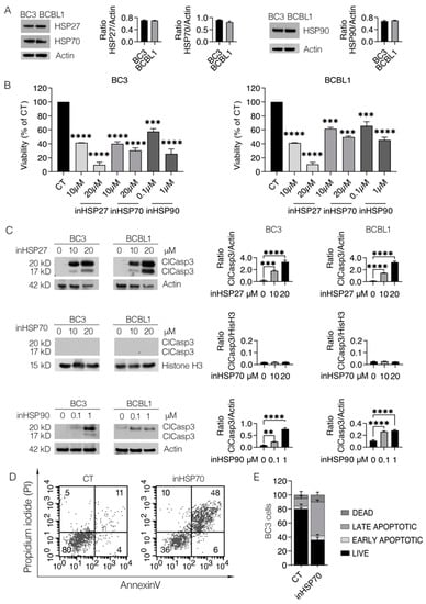
Primary effusion lymphoma (PEL) is a rare and aggressive B-cell lymphoma, against which current therapies usually fail. In the present study, we show that targeting HSPs, such as HSP27, HSP70 and HSP90, could be an efficient strategy to reduce PEL cell survival, as it induces strong DNA damage, which correlated with an impairment of DDR. Moreover, as HSP27, HSP70 and HSP90 cross talk with STAT3, their inhibition results in STAT3 de-phosphorylation and. On the other hand, the inhibition of STAT3 may downregulate these HSPs. These findings suggest that targeting HSPs has important implications in cancer therapy, as it can reduce the release of cytokines by PEL cells, which, besides affecting their own survival, could negatively influence anti-cancer immune response. Full article
Show Figures

Figure 1

13 pages, 15020 KiB  
Article
Inhibition of Ribosomal RNA Processing 15 Homolog (RRP15) Suppressed Tumor Growth, Invasion and Epithelial to Mesenchymal Transition (EMT) of Colon Cancer
by Zirong Deng, Yun Xu, Yuchen Cai, Weiling Lin, Libei Zhang, Aoqing Jiang, Yuhang Zhou, Rui Zhao, Heyan Zhao, Zhaoguo Liu * and Tingdong Yan *
1 Department of Pharmacology, School of Pharmacy, Nantong University, Nantong 226001, China
These authors contributed equally to this work.
Int. J. Mol. Sci. 2023, 24(4), 3528; https://doi.org/10.3390/ijms24043528 - 9 Feb 2023
Cited by 3 | Viewed by 2074
Abstract
Although ribosomal RNA processing 15 Homolog (RRP15) has been implicated in the occurrence of various cancers and is considered a potential target for cancer treatment, its significance in colon cancer (CC) is unclear. Thus, this present study aims to determine RRP15 expression and [...] Read more.
Although ribosomal RNA processing 15 Homolog (RRP15) has been implicated in the occurrence of various cancers and is considered a potential target for cancer treatment, its significance in colon cancer (CC) is unclear. Thus, this present study aims to determine RRP15 expression and biological function in CC. The results demonstrated a strong expression of RRP15 in CC compared to normal colon specimens, which was correlated with poorer overall survival (OS) and disease-free survival (DFS) of the patients. Among the nine investigated CC cell lines, RRP15 demonstrated the highest and lowest expression in HCT15 and HCT116 cells, respectively. In vitro assays demonstrated that the knockdown of RRP15 inhibited the growth, colony-forming ability and invasive ability of the CC cells whereas its overexpression enhanced the above oncogenic function. Moreover, subcutaneous tumors in nude mice showed that RRP15 knockdown inhibited the CC growth while its overexpression enhanced their growth. Additionally, the knockdown of RRP15 inhibited the epithelial–mesenchymal transition (EMT), whereas overexpression of RRP15 promoted the EMT process in CC. Collectively, inhibition of RRP15 suppressed tumor growth, invasion and EMT of CC, and might be considered a promising therapeutic target for treating CC. Full article
(This article belongs to the Special Issue A New Frontier on Cancer Invasion and Metastasis Research 2022)
Show Figures

Figure 1

7 pages, 404 KiB  
Communication
“Pesto” Mutation: Phenotypic and Genotypic Characteristics of Eight GCK/MODY Ligurian Patients
by Alessandro Salina 1,†, Marta Bassi 2,3,†, Concetta Aloi 1,*, Marina Francesca Strati 2,3, Renata Bocciardi 2,4, Giuseppe d’Annunzio 3, Mohamad Maghnie 2,3 and Nicola Minuto 3
1 LABSIEM (Laboratory for the Study of Inborn Errors of Metabolism), Pediatric Clinic, IRCCS Istituto Giannina Gaslini, 16147 Genoa, Italy
2 Department of Neuroscience, Rehabilitation, Ophthalmology, Genetics, Maternal and Child Health (DINOGMI), University of Genoa, 16100 Genoa, Italy
3 Department of Pediatrics, IRCCS Istituto Giannina Gaslini, 16147 Genoa, Italy
4 UOC Genetica Medica, IRCCS Istituto Giannina Gaslini, 16147 Genoa, Italy
These authors contributed equally to this work.
Int. J. Mol. Sci. 2023, 24(4), 4034; https://doi.org/10.3390/ijms24044034 - 17 Feb 2023
Cited by 2 | Viewed by 2070
Abstract
Maturity Onset Diabetes of the Young (MODY) is a monogenic form of diabetes mellitus (DM) that accounts for around 2–5% of all types of diabetes. Autosomal dominant inheritance in pathogenic variations of 14 genes related to β-cell functions can lead to monogenic types [...] Read more.
Maturity Onset Diabetes of the Young (MODY) is a monogenic form of diabetes mellitus (DM) that accounts for around 2–5% of all types of diabetes. Autosomal dominant inheritance in pathogenic variations of 14 genes related to β-cell functions can lead to monogenic types of diabetes. In Italy, GCK/MODY is the most frequent form and it is caused by mutations of the glucokinase (GCK). Patients with GCK/MODY usually have stable mild fasting hyperglycaemia with mildly elevated HbA1c levels and rarely need pharmacological treatment. Molecular analysis of the GCK coding exons was carried out by Sanger sequencing in eight Italian patients. All the probands were found to be heterozygous carriers of a pathogenic gross insertion/deletion c.1279_1358delinsTTACA; p.Ser426_Ala454delinsLeuGln. It was previously described for the first time by our group in a large cohort of Italian GCK/MODY patients. The higher levels of HbA1c (6.57% vs. 6.1%), and the higher percentage of patients requiring insulin therapy (25% vs. 2%) compared to the previously studied Italian patients with GCK/MODY, suggest that the mutation discovered could be responsible for a clinically worse form of GCK/MODY. Moreover, as all the patients carrying this variant share an origin from the same geographic area (Liguria), we postulate a possible founder effect and we propose to name it the “pesto” mutation. Full article
(This article belongs to the Special Issue Diabetes: Molecular Mechanisms)
Show Figures

Figure 1

11 pages, 2320 KiB  
Article
Do Aging and Parity Affect VEGF-A/VEGFR Content and Signaling in the Ovary?—A Mouse Model Study
by Valentina Di Nisio 1,*, Gianna Rossi 2, Alessandro Chiominto 3, Ezio Pompili 3 and Sandra Cecconi 2
1 Department of Clinical Science, Intervention and Technology, Division of Obstetrics and Gynecology, Karolinska Institutet and Karolinska University Hospital, 14186 Huddinge, Sweden
2 Department of Life, Health and Environmental Sciences, University of L’Aquila, Via Vetoio, 67100 L’Aquila, Italy
3 Department of Pathology, San Salvatore Hospital, 67100 L’Aquila, Italy
Int. J. Mol. Sci. 2023, 24(4), 3318; https://doi.org/10.3390/ijms24043318 - 7 Feb 2023
Cited by 3 | Viewed by 2068
Abstract
In this study, the effects of aging and parity on VEGF-A/VEGFR protein content and signaling in the mice ovaries were determined. The research group consisted of nulliparous (virgins, V) and multiparous (M) mice during late-reproductive (L, 9–12 months) and post-reproductive (P, 15–18 months) [...] Read more.
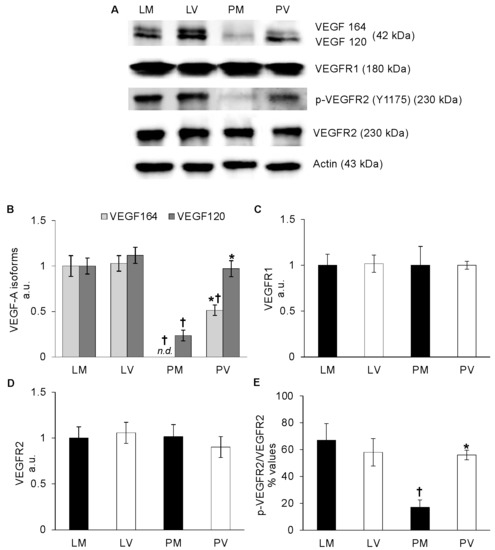
In this study, the effects of aging and parity on VEGF-A/VEGFR protein content and signaling in the mice ovaries were determined. The research group consisted of nulliparous (virgins, V) and multiparous (M) mice during late-reproductive (L, 9–12 months) and post-reproductive (P, 15–18 months) stages. Whilst ovarian VEGFR1 and VEGFR2 remained unchanged in all the experimental groups (LM, LV, PM, PV), protein content of VEGF-A and phosphorylated VEGFR2 significantly decreased only in PM ovaries. VEGF-A/VEGFR2-dependent activation of ERK1/2, p38, as well as protein content of cyclin D1, cyclin E1, and Cdc25A were then assessed. In ovaries of LV and LM, all of these downstream effectors were maintained at a comparable low/undetectable level. Conversely, the decrease recorded in PM ovaries did not occur in the PV group, in which the significant increase of kinases and cyclins, as well phosphorylation levels mirrored the trend of the pro-angiogenic markers. Altogether, the present results demonstrated that, in mice, ovarian VEGF-A/VEGFR2 protein content and downstream signaling can be modulated in an age- and parity-dependent manner. Moreover, the lowest levels of pro-angiogenic and cell cycle progression markers detected in PM mouse ovaries sustains the hypothesis that parity could exert a protective role by downregulating the protein content of key mediators of pathological angiogenesis. Full article
Show Figures

Figure 1

24 pages, 3426 KiB  
Article
Genomic Distribution of Pro-Virulent cpdB-like Genes in Eubacteria and Comparison of the Enzyme Specificity of CpdB-like Proteins from Salmonella enterica, Escherichia coli and Streptococcus suis
by João Meireles Ribeiro 1, José Canales 1, María Jesús Costas 1, Alicia Cabezas 1, Rosa María Pinto 1, Miguel García-Díaz 2, Paloma Martín-Cordero 3 and José Carlos Cameselle 1,*
1 Grupo de Enzimología, Departamento de Bioquímica y Biología Molecular y Genética, Facultad de Medicina y Ciencias de la Salud, Universidad de Extremadura, 06006 Badajoz, Spain
2 Unidad de Aparato Digestivo, Hospital de Zafra, Área de Salud Llerena-Zafra, Servicio Extremeño de Salud, 06300 Zafra, Spain
3 Servicio de Microbiología, Hospital Universitario de Badajoz, Servicio Extremeño de Salud, 06006 Badajoz, Spain
Int. J. Mol. Sci. 2023, 24(4), 4150; https://doi.org/10.3390/ijms24044150 - 19 Feb 2023
Cited by 6 | Viewed by 2066
Abstract
The cpdB gene is pro-virulent in avian pathogenic Escherichia coli and in Salmonella enterica, where it encodes a periplasmic protein named CpdB. It is structurally related to cell wall-anchored proteins, CdnP and SntA, encoded by the also pro-virulent cdnP and sntA genes [...] Read more.
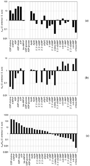
The cpdB gene is pro-virulent in avian pathogenic Escherichia coli and in Salmonella enterica, where it encodes a periplasmic protein named CpdB. It is structurally related to cell wall-anchored proteins, CdnP and SntA, encoded by the also pro-virulent cdnP and sntA genes of Streptococcus agalactiae and Streptococcus suis, respectively. CdnP and SntA effects are due to extrabacterial hydrolysis of cyclic-di-AMP, and to complement action interference. The mechanism of CpdB pro-virulence is unknown, although the protein from non-pathogenic E. coli hydrolyzes cyclic dinucleotides. Considering that the pro-virulence of streptococcal CpdB-like proteins is mediated by c-di-AMP hydrolysis, S. enterica CpdB activity was tested as a phosphohydrolase of 3′-nucleotides, 2′,3′-cyclic mononucleotides, linear and cyclic dinucleotides, and cyclic tetra- and hexanucleotides. The results help to understand cpdB pro-virulence in S. enterica and are compared with E. coli CpdB and S. suis SntA, including the activity of the latter on cyclic-tetra- and hexanucleotides reported here for the first time. On the other hand, since CpdB-like proteins are relevant to host-pathogen interactions, the presence of cpdB-like genes was probed in eubacterial taxa by TblastN analysis. The non-homogeneous genomic distribution revealed taxa with cpdB-like genes present or absent, identifying eubacteria and plasmids where they can be relevant. Full article
(This article belongs to the Special Issue Genomics: Infectious Disease and Host-Pathogen Interaction 2.0)
Show Figures

Figure 1

14 pages, 1144 KiB  
Brief Report
Kinesins Modify ERR1-Dependent Transcription Using a Conserved Nuclear Receptor Box Motif
by A. M. Pramodh Bandara Seneviratne 1,2,*, Sarah Lidagoster 1, Sofia Valbuena-Castor 1, Kareena Lashley 1, Sumit Saha 1, Aleksandra Alimova 1,2 and Geri Kreitzer 1,2,*
1 CUNY School of Medicine, City College of New York, New York, NY 10031, USA
2 Department of Molecular, Cellular and Biomedical Sciences, CUNY School of Medicine, City College of New York, New York, NY 10031, USA
Int. J. Mol. Sci. 2023, 24(4), 3795; https://doi.org/10.3390/ijms24043795 - 14 Feb 2023
Cited by 2 | Viewed by 2057
Abstract
Kinesin family motors are microtubule (MT)-stimulated ATPases known best as transporters of cellular cargoes through the cytoplasm, regulators of MT dynamics, organizers of the mitotic spindle, and for insuring equal division of DNA during mitosis. Several kinesins have also been shown to regulate [...] Read more.
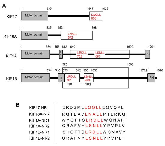
Kinesin family motors are microtubule (MT)-stimulated ATPases known best as transporters of cellular cargoes through the cytoplasm, regulators of MT dynamics, organizers of the mitotic spindle, and for insuring equal division of DNA during mitosis. Several kinesins have also been shown to regulate transcription by interacting with transcriptional cofactors and regulators, nuclear receptors, or with specific promotor elements on DNA. We previously showed that an LxxLL nuclear receptor box motif in the kinesin-2 family motor KIF17 mediates binding to the orphan nuclear receptor estrogen related receptor alpha (ERR1) and is responsible for the suppression of ERR1-dependent transcription by KIF17. Analysis of all kinesin family proteins revealed that multiple kinesins contain this LxxLL motif, raising the question as to whether additional kinesin motors contribute to the regulation of ERR1. In this study, we interrogate the effects of multiple kinesins with LxxLL motifs on ERR1-mediated transcription. We demonstrate that the kinesin-3 family motor KIF1B contains two LxxLL motifs, one of which binds to ERR1. In addition, we show that expression of a KIF1B fragment containing this LxxLL motif inhibits ERR1-dependent transcription by regulating nuclear entry of ERR1. We also provide evidence that the effects of expressing the KIF1B-LxxLL fragment on ERR1 activity are mediated by a mechanism distinct from that of KIF17. Since LxxLL domains are found in many kinesins, our data suggest an expanded role for kinesins in nuclear receptor mediated transcriptional regulation. Full article
Show Figures

Figure 1

15 pages, 3371 KiB  
Article
Group V Chitin Deacetylases Influence the Structure and Composition of the Midgut of Beet Armyworm, Spodoptera exigua
by Han Wu 1,2,†, Dan Zhao 1,†, Xiao-Chang Guo 1, Zhao-Rui Liu 1, Rui-Jun Li 1, Xiu-Jun Lu 1 and Wei Guo 1,2,*
1 College of Plant Protection, Hebei Agricultural University, Baoding 071001, China
2 Graduate School of Chinese Academy of Agricultural Sciences, Beijing 100081, China
These authors contributed equally to this work.
Int. J. Mol. Sci. 2023, 24(4), 3076; https://doi.org/10.3390/ijms24043076 - 4 Feb 2023
Cited by 4 | Viewed by 2054
Abstract
Chitin deacetylase (CDA) can accelerate the conversion of chitin to chitosan, influencing the mechanical properties and permeability of the cuticle structures and the peritrophic membrane (PM) in insects. Putative Group V CDAs SeCDA6/7/8/9 (SeCDAs) were identified and characterized from beet armyworm Spodoptera exigua [...] Read more.
Chitin deacetylase (CDA) can accelerate the conversion of chitin to chitosan, influencing the mechanical properties and permeability of the cuticle structures and the peritrophic membrane (PM) in insects. Putative Group V CDAs SeCDA6/7/8/9 (SeCDAs) were identified and characterized from beet armyworm Spodoptera exigua larvae. The cDNAs of SeCDAs contained open reading frames of 1164 bp, 1137 bp, 1158 bp and 1152 bp, respectively. The deduced protein sequences showed that SeCDAs are synthesized as preproteins of 387, 378, 385 and 383 amino acid residues, respectively. It was revealed via spatiotemporal expression analysis that SeCDAs were more abundant in the anterior region of the midgut. The SeCDAs were down-regulated after treatment with 20-hydroxyecdysone (20E). After treatment with a juvenile hormone analog (JHA), the expression of SeCDA6 and SeCDA8 was down-regulated; in contrast, the expression of SeCDA7 and SeCDA9 was up-regulated. After silencing SeCDAV (the conserved sequences of Group V CDAs) via RNA interference (RNAi), the layer of intestinal wall cells in the midgut became more compact and more evenly distributed. The vesicles in the midgut were small and more fragmented or disappeared after SeCDAs were silenced. Additionally, the PM structure was scarce, and the chitin microfilament structure was loose and chaotic. It was indicated in all of the above results that Group V CDAs are essential for the growth and structuring of the intestinal wall cell layer in the midgut of S. exigua. Additionally, the midgut tissue and the PM structure and composition were affected by Group V CDAs. Full article
(This article belongs to the Section Molecular Biology)
Show Figures

Figure 1