Int. J. Mol. Sci. 2022, 23(20), 12516; https://doi.org/10.3390/ijms232012516 - 19 Oct 2022
Cited by 12 | Viewed by 3976
Abstract
There has been an immense effort by global pharmaceutical companies to develop anti-COVID-19 drugs, including small molecule-based RNA replication inhibitors via drug repositioning and antibody-based spike protein blockers related to cell entry by SARS-CoV-2. However, several limitations to their clinical use have emerged
[...] Read more.
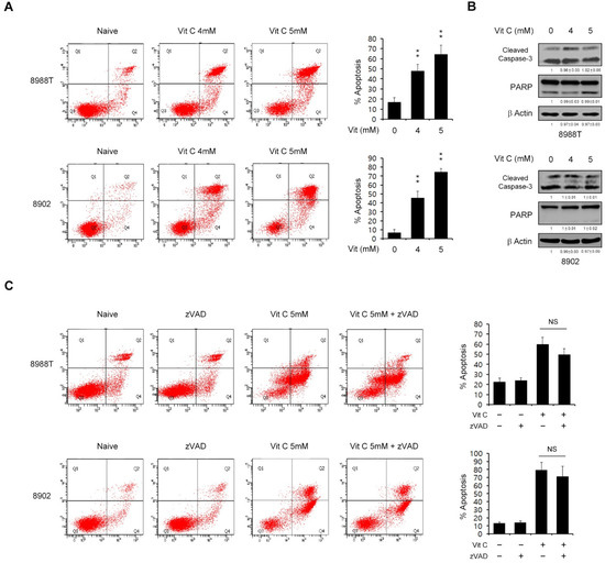
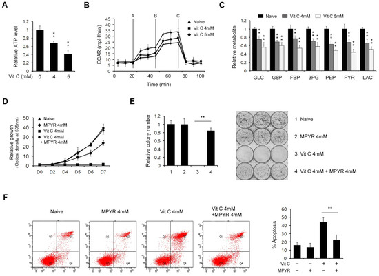
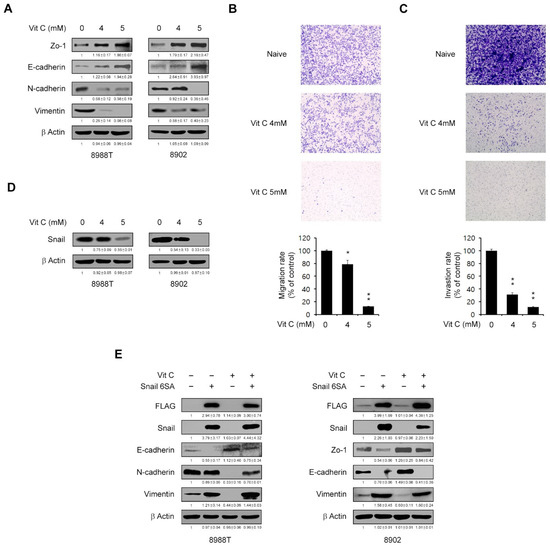
There has been an immense effort by global pharmaceutical companies to develop anti-COVID-19 drugs, including small molecule-based RNA replication inhibitors via drug repositioning and antibody-based spike protein blockers related to cell entry by SARS-CoV-2. However, several limitations to their clinical use have emerged in addition to a lack of progress in the development of small molecule-based cell entry inhibitors from natural products. In this study, we tested the effectiveness of kuwanon C (KC), which has mainly been researched using in silico docking simulation and can serve as an effective building block for developing anti-COVID-19 drugs, in blocking the spike S1 RBD:ACE2 receptor interaction. KC is a natural product derived from Morus alba L., commonly known as mulberry, which has known antiviral efficacy. Molecular interaction studies using competitive ELISA and the BLItz system revealed that KC targets both the spike S1 RBD and the ACE2 receptor, successfully disrupting their interaction, as supported by the in silico docking simulation. Furthermore, we established a mechanism of action by observing how KC prevents the infection of SARS-CoV-2 spike pseudotyped virus in ACE2/TPRSS2-overexpressing HEK293T cells. Finally, we demonstrated that KC inhibits clinical isolates of SARS-CoV-2 in Vero cells. Future combinations of small molecule-based cell entry inhibitors, such as KC, with the currently prescribed RNA replication inhibitors are anticipated to significantly enhance the efficacy of COVID-19 therapies.
Full article
(This article belongs to the Special Issue Natural Product-Based Drug Discovery for COVID-19 Disease)
►
Show Figures

Figure 1


