by
Daisuke Akahori 1, Naoki Inui 1,2,*, Yusuke Inoue 1, Hideki Yasui 1 , Hironao Hozumi 1, Yuzo Suzuki 1, Masato Karayama 1, Kazuki Furuhashi 1, Noriyuki Enomoto 1, Tomoyuki Fujisawa 1 and Takafumi Suda 1
, Hironao Hozumi 1, Yuzo Suzuki 1, Masato Karayama 1, Kazuki Furuhashi 1, Noriyuki Enomoto 1, Tomoyuki Fujisawa 1 and Takafumi Suda 1
1
Second Division, Department of Internal Medicine, Hamamatsu University School of Medicine, 1-20-1 Handayama, Higashi-ku, Hamamatsu 431-3192, Shizuoka, Japan
2
Department of Clinical Pharmacology and Therapeutics, Hamamatsu University School of Medicine, 1-20-1 Handayama, Higashi-ku, Hamamatsu 431-3192, Shizuoka, Japan
Int. J. Mol. Sci. 2022, 23(16), 8996; https://doi.org/10.3390/ijms23168996 - 12 Aug 2022
Cited by 17 | Viewed by 3421
Abstract
Pulmonary fibrosis is a progressive and fatal disorder characterized by dysregulated repair after recurrent injury. Destruction of the lung architecture with excess extracellular matrix deposition induces respiratory failure with hypoxia and progressive dyspnea. The impact of hypoxia on pulmonary endothelial cells during pulmonary
[...] Read more.
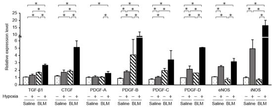
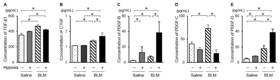
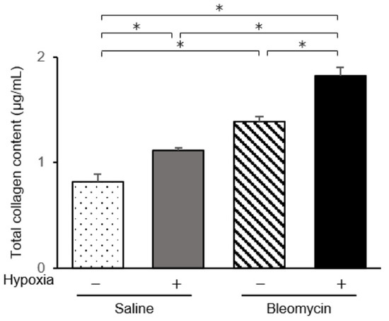
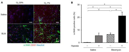
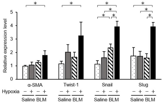
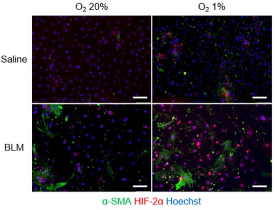
Pulmonary fibrosis is a progressive and fatal disorder characterized by dysregulated repair after recurrent injury. Destruction of the lung architecture with excess extracellular matrix deposition induces respiratory failure with hypoxia and progressive dyspnea. The impact of hypoxia on pulmonary endothelial cells during pulmonary fibrogenesis is unclear. Using a magnetic-activated cell sorting system, pulmonary endothelial cells were isolated from a mouse model of pulmonary fibrosis induced by intratracheally administered bleomycin. When endothelial cells were exposed to hypoxic conditions, a hypoxia-inducible factor (HIF)-2α protein was detected in CD31- and α-smooth muscle actin (SMA)-positive cells. Levels of plasminogen activator inhibitor 1, von Willebrand factor, and matrix metalloproteinase 12 were increased in endothelial cells isolated from bleomycin-treated mice exposed to hypoxic conditions. When endothelial cells were cultured under hypoxic conditions, levels of fibrotic mediators, transforming growth factor-β and connective tissue growth factor, were elevated only in endothelial cells from bleomycin-treated and not from saline-treated lungs. The increased expression of α-SMA and mesenchymal markers and collagen production in bleomycin- or hypoxia-stimulated endothelial cells were further elevated in endothelial cells from bleomycin-treated mouse lungs cultured under hypoxic conditions. Exposure to hypoxia damaged endothelial cells and enhanced fibrogenesis-related damage in bleomycin-treated pulmonary endothelial cells.
Full article
(This article belongs to the Special Issue Molecular and Cellular Mechanisms of Idiopathic Pulmonary Fibrosis and Interstitial Lung Diseases)
▼
Show Figures
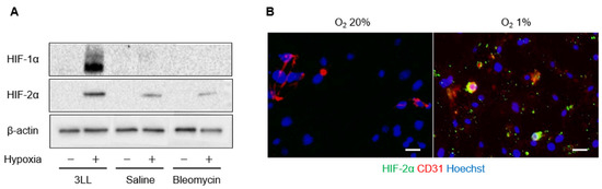
Figure 1


