Int. J. Mol. Sci. 2022, 23(15), 8229; https://doi.org/10.3390/ijms23158229 - 26 Jul 2022
Cited by 13 | Viewed by 4183
Abstract
The prevalence of non-alcoholic steatohepatitis (NASH) is rapidly increasing and associated with cardiovascular disease (CVD), the major cause of mortality in NASH patients. Although sharing common risk factors, the mechanisms by which NASH may directly contribute to the development to CVD remain poorly
[...] Read more.
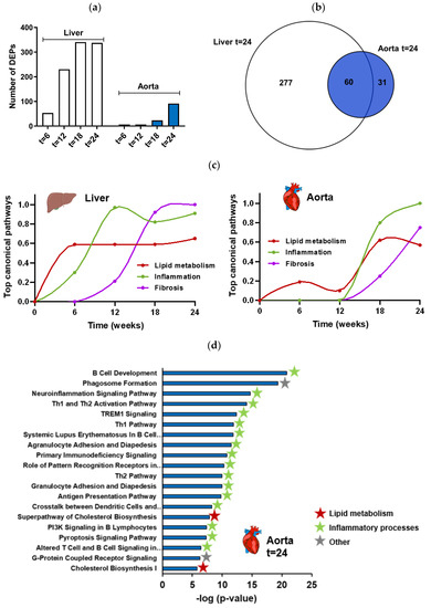
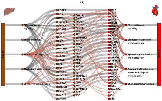
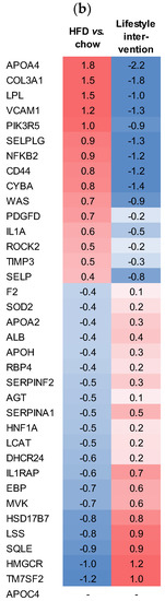
The prevalence of non-alcoholic steatohepatitis (NASH) is rapidly increasing and associated with cardiovascular disease (CVD), the major cause of mortality in NASH patients. Although sharing common risk factors, the mechanisms by which NASH may directly contribute to the development to CVD remain poorly understood. The aim of this study is to gain insight into key molecular processes of NASH that drive atherosclerosis development. Thereto, a time-course study was performed in Ldlr−/−.Leiden mice fed a high-fat diet to induce NASH and atherosclerosis. The effects on NASH and atherosclerosis were assessed and transcriptome analysis was performed. Ldlr−/−.Leiden mice developed obesity, hyperlipidemia and insulin resistance, with steatosis and hepatic inflammation preceding atherosclerosis development. Transcriptome analysis revealed a time-dependent increase in pathways related to NASH and fibrosis followed by an increase in pro-atherogenic processes in the aorta. Gene regulatory network analysis identified specific liver regulators related to lipid metabolism (SC5D, LCAT and HMGCR), inflammation (IL1A) and fibrosis (PDGF, COL3A1), linked to a set of aorta target genes related to vascular inflammation (TNFA) and atherosclerosis signaling (CCL2 and FDFT1). The present study reveals pathogenic liver processes that precede atherosclerosis development and identifies hepatic key regulators driving the atherogenic pathways and regulators in the aorta.
Full article
(This article belongs to the Special Issue Recent Advances in Molecular Research of Metabolic Disorders)
►
Show Figures
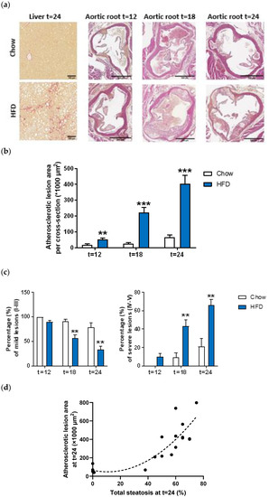
Figure 1


