Int. J. Mol. Sci. 2021, 22(24), 13528; https://doi.org/10.3390/ijms222413528 - 16 Dec 2021
Cited by 19 | Viewed by 4686
Abstract
Obesity is characterized as a complex and multifactorial excess accretion of adipose tissue accompanied with alterations in the immune and metabolic responses. Although the chemokine systems have been documented to be involved in the control of tissue inflammation and metabolism, the dual role
[...] Read more.
Obesity is characterized as a complex and multifactorial excess accretion of adipose tissue accompanied with alterations in the immune and metabolic responses. Although the chemokine systems have been documented to be involved in the control of tissue inflammation and metabolism, the dual role of chemokines and chemokine receptors in the pathogenesis of the inflammatory milieu and dysregulated energy metabolism in obesity remains elusive. The objective of this review is to present an update on the link between chemokines and obesity-related inflammation and metabolism dysregulation under the light of recent knowledge, which may present important therapeutic targets that could control obesity-associated immune and metabolic disorders and chronic complications in the near future. In addition, the cellular and molecular mechanisms of chemokines and chemokine receptors including the potential effect of post-translational modification of chemokines in the regulation of inflammation and energy metabolism will be discussed in this review.
Full article
(This article belongs to the Special Issue Recent Research on Diabetes Mellitus and Its Complications)
►
Show Figures
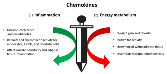
Figure 1


