Int. J. Mol. Sci. 2018, 19(6), 1641; https://doi.org/10.3390/ijms19061641 - 1 Jun 2018
Cited by 83 | Viewed by 13694
Abstract
Achieving surface design and control of biomaterial scaffolds with nanometer- or micrometer-scaled functional films is critical to mimic the unique features of native extracellular matrices, which has significant technological implications for tissue engineering including cell-seeded scaffolds, microbioreactors, cell assembly, tissue regeneration, etc. Compared
[...] Read more.
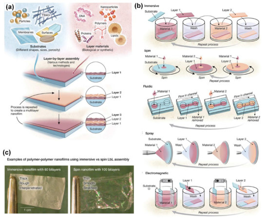
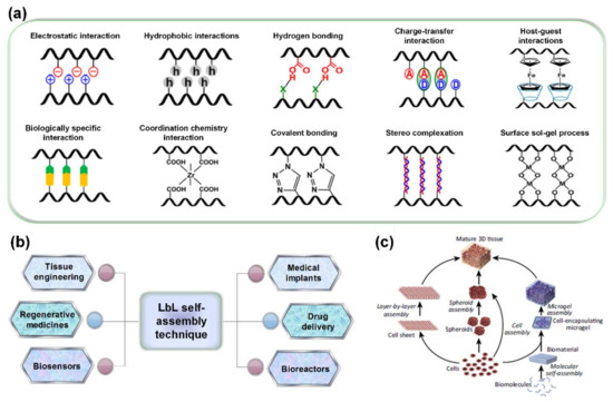
Achieving surface design and control of biomaterial scaffolds with nanometer- or micrometer-scaled functional films is critical to mimic the unique features of native extracellular matrices, which has significant technological implications for tissue engineering including cell-seeded scaffolds, microbioreactors, cell assembly, tissue regeneration, etc. Compared with other techniques available for surface design, layer-by-layer (LbL) self-assembly technology has attracted extensive attention because of its integrated features of simplicity, versatility, and nanoscale control. Here we present a brief overview of current state-of-the-art research related to the LbL self-assembly technique and its assembled biomaterials as scaffolds for tissue engineering. An overview of the LbL self-assembly technique, with a focus on issues associated with distinct routes and driving forces of self-assembly, is described briefly. Then, we highlight the controllable fabrication, properties, and applications of LbL self-assembly biomaterials in the forms of multilayer nanofilms, scaffold nanocoatings, and three-dimensional scaffolds to systematically demonstrate advances in LbL self-assembly in the field of tissue engineering. LbL self-assembly not only provides advances for molecular deposition but also opens avenues for the design and development of innovative biomaterials for tissue engineering.
Full article
(This article belongs to the Special Issue Novel Biomaterials for Tissue Engineering 2018)
►
Show Figures

Graphical abstract


