by
Ho Jun Lee 1 and Yong Ju Jang 2,*
and Yong Ju Jang 2,*
1
Department of Otorhinolaryngology-Head and Neck Surgery, Chuncheon Sacred Heart Hospital, College of Medicine, Hallym University, Chuncheon 24253, Korea
2
Department of Otolaryngology, Asan Medical Center, University of Ulsan College of Medicine, Seoul 05505, Korea
Int. J. Mol. Sci. 2018, 19(3), 711; https://doi.org/10.3390/ijms19030711 - 2 Mar 2018
Cited by 428 | Viewed by 20342
Abstract
Hypertrophic scars and keloids are fibroproliferative disorders that may arise after any deep cutaneous injury caused by trauma, burns, surgery, etc. Hypertrophic scars and keloids are cosmetically problematic, and in combination with functional problems such as contractures and subjective symptoms including pruritus, these
[...] Read more.
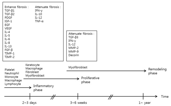
Hypertrophic scars and keloids are fibroproliferative disorders that may arise after any deep cutaneous injury caused by trauma, burns, surgery, etc. Hypertrophic scars and keloids are cosmetically problematic, and in combination with functional problems such as contractures and subjective symptoms including pruritus, these significantly affect patients’ quality of life. There have been many studies on hypertrophic scars and keloids; but the mechanisms underlying scar formation have not yet been well established, and prophylactic and treatment strategies remain unsatisfactory. In this review, the authors introduce and summarize classical concepts surrounding wound healing and review recent understandings of the biology, prevention and treatment strategies for hypertrophic scars and keloids.
Full article
(This article belongs to the Special Issue Recent Advances in Scar Biology)
▼
Show Figures
Figure 1

