1
Division of Laboratory Medicine, Department of Pathology and Laboratory Diagnostics, Istituto Nazionale Tumori “Fondazione G. Pascale”, IRCCS, Naples 80131, Italy
2
Pathology Unit, Department of Pathology and Laboratory Diagnostics, Istituto Nazionale Tumori “Fondazione G. Pascale”, IRCCS, Naples 80131, Italy
†
These authors contributed equally to this work.
Int. J. Mol. Sci. 2017, 18(4), 804; https://doi.org/10.3390/ijms18040804 - 12 Apr 2017
Cited by 5 | Viewed by 6788
Abstract
Current criteria for differential diagnosis of multiple myeloma (MM), Monoclonal gammopathy of undetermined significance (MGUS), and smoldering multiple myeloma (SMM) are included in the 2003 guidelines by the International Myeloma Working Group (IMWG). An updated version was then published in 2014, highlighting the
[...] Read more.
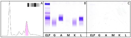
Current criteria for differential diagnosis of multiple myeloma (MM), Monoclonal gammopathy of undetermined significance (MGUS), and smoldering multiple myeloma (SMM) are included in the 2003 guidelines by the International Myeloma Working Group (IMWG). An updated version was then published in 2014, highlighting the importance of serum free light chain (sFLC) detection, as well as the κ/λ ratio as excellent indicators of clonality. At present, two commercial assays for sFLC quantification are available: the Freelite™ assay and the N-Latex assay. The first was developed by The Binding Site based on a mixture of polyclonal antibodies directed against a variety of FLC epitopes. It may be run on a wide range of nephelometers, as well as on turbidimeters. The second method was developed by Siemens and runs exclusively on Siemens instruments. It employs a probe mixture of mouse monoclonal antibodies. The aim of our study was to evaluate sFLC measurement and calculated κ/λ ratio in 85 patients with monoclonal gammopathies (MGs) in order to compare methods. We demonstrated that there is only a moderate concordance between the two FLC assays. In particular, in one case, we observed no qualitative alterations of the serum protein pattern, and in the absence of a Freelite™ assay, sFLC measurement would not have been possible to highlight the increase of λ FLC.
Full article
(This article belongs to the Section Molecular Pathology, Diagnostics, and Therapeutics)
▼
Show Figures
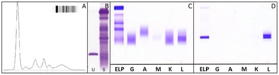
Figure 1


