Neuroglia 2024, 5(2), 89-104; https://doi.org/10.3390/neuroglia5020007 - 21 Apr 2024
Abstract
►
Show Figures
The traditional neuron-centric view of the central nervous system (CNS) is shifting toward recognizing the importance of communication between the neurons and the network of glial cells. This shift is leading to a more comprehensive understanding of how glial cells contribute to CNS
[...] Read more.
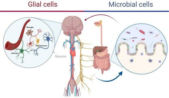
The traditional neuron-centric view of the central nervous system (CNS) is shifting toward recognizing the importance of communication between the neurons and the network of glial cells. This shift is leading to a more comprehensive understanding of how glial cells contribute to CNS function. Alongside this shift, recent discoveries have illuminated the significant role of the human microbiome, comprising trillions of microorganisms, mirroring the number of human cells in an individual. This paper delves into the multifaceted functions of neuroglia, or glial cells, which extend far beyond their traditional roles of supporting and protecting neurons. Neuroglia modulate synaptic activity, insulate axons, support neurogenesis and synaptic plasticity, respond to injury and inflammation, and engage in phagocytosis. Meanwhile, the microbiome, long overlooked, emerges as a crucial player in brain functionality akin to glial cells. This review aims to underscore the importance of the interaction between glial cells and resident microorganisms in shaping the development and function of the human brain, a concept that has been less studied. Through a comprehensive examination of existing literature, we discuss the mechanisms by which glial cells interface with the microbiome, offering insights into the contribution of this relationship to neural homeostasis and health. Furthermore, we discuss the implications of dysbiosis within this interaction, highlighting its potential contribution to neurological disorders and paving the way for novel therapeutic interventions targeting both glial cells and the microbiome.
Full article
Graphical abstract
