


cells-logo

Journal Browser

Journal Browser

Molecular Signaling, Circuit Neuroplasticity and the Cognitive Function

A Topical Collection in Cells (ISSN 2073-4409) belonging to the section "Cellular Neuroscience".

Viewed by 34150

Editor


E-Mail Website
Collection Editor
Department of Neurophysiology and Neuropharmacology, Medical University of Vienna, 1090 Vienna, Austria
Interests: synaptogenesis; synaptic transmission; synaptic plasticity; learning and memory; fear; anxiety; cognition; biopsychology

Topical Collection Information

Dear Colleagues,

What is cognition from a molecular and cellular perspective? How does the plastic rearrangement of synaptic contacts generate specific patterns of neuronal activity so that some—and not other—cognitive functions emerge? Which specific molecular signaling pathways become suppressed in the aged brain to restrain the unleashing of those neuronal skills that make youngsters such efficient learners?

No matter how enigmatic the phenomenon of cognition appears to be, it is nothing but reasonable to conclude that cognition, understood as a property of given nervous systems, is not only a cognoscible phenomenon but also one not exclusive to humans and, moreover, not exclusive to brains. Our approaches to the problem of the physical nature of cognition can thus afford perspectives free of anthropomorphized boundaries. In this regard, the use of animal models and the combination of in vivo and in vitro approaches comprise powerful experimental tools in neuroscience in the search for the structural, molecular, cellular, and functional underpinnings of the cognitive function.

The Cells team is delighted to invite you to contribute with your original research articles and reviews to this Topic Collection addressing molecular, cellular, and neural circuit functional mechanisms of the nervous system that generate and regulate cognitive function (including—but not limited to—attention, emotion, social cognition, and learning and memory) in health and disease.

We look forward to learning about your findings.

Dr. Francisco Monje
Collection Editor

Manuscript Submission Information

Manuscripts should be submitted online at www.mdpi.com by registering and logging in to this website. Once you are registered, click here to go to the submission form. All submissions that pass pre-check are peer-reviewed. Accepted papers will be published continuously in the journal (as soon as accepted) and will be listed together on the collection website. Research articles, review articles as well as communications are invited. For planned papers, a title and short abstract (about 250 words) can be sent to the Editorial Office for assessment.

Submitted manuscripts should not have been published previously, nor be under consideration for publication elsewhere (except conference proceedings papers). All manuscripts are thoroughly refereed through a single-anonymized peer-review process. A guide for authors and other relevant information for submission of manuscripts is available on the Instructions for Authors page. Cells is an international peer-reviewed open access semimonthly journal published by MDPI.

Please visit the Instructions for Authors page before submitting a manuscript. The Article Processing Charge (APC) for publication in this open access journal is 2700 CHF (Swiss Francs). Submitted papers should be well formatted and use good English. Authors may use MDPI's English editing service prior to publication or during author revisions.

Keywords

  • synaptogenesis
  • synaptic transmission
  • synaptic plasticity
  • learning and memory
  • fear
  • anxiety
  • cognition

Published Papers (7 papers)

2026

Jump to: 2025, 2024, 2023, 2022

20 pages, 6545 KB  
Article
Male and Female Mice Show Similar Fear Memory Performance Despite Hippocampal Immediate Early Gene Expression Differences During Encoding and Consolidation
by Katherine O. McDonald, Temmie Yu, Aditi Prabhu and Sara J. Aton
Cells 2026, 15(17), 1523; https://doi.org/10.3390/cells15171523 - 24 Aug 2026
Viewed by 242
Abstract
Accurate and efficient memory processing is essential for survival. A body of ongoing work in both human subjects and animal models suggests that memory processing may differ substantially between males and females. In mice, contextual fear memory (CFM) encoding, consolidation, and recall have [...] Read more.
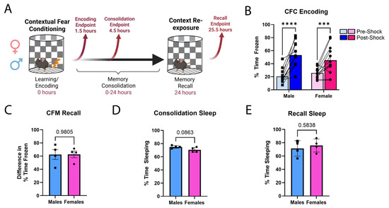
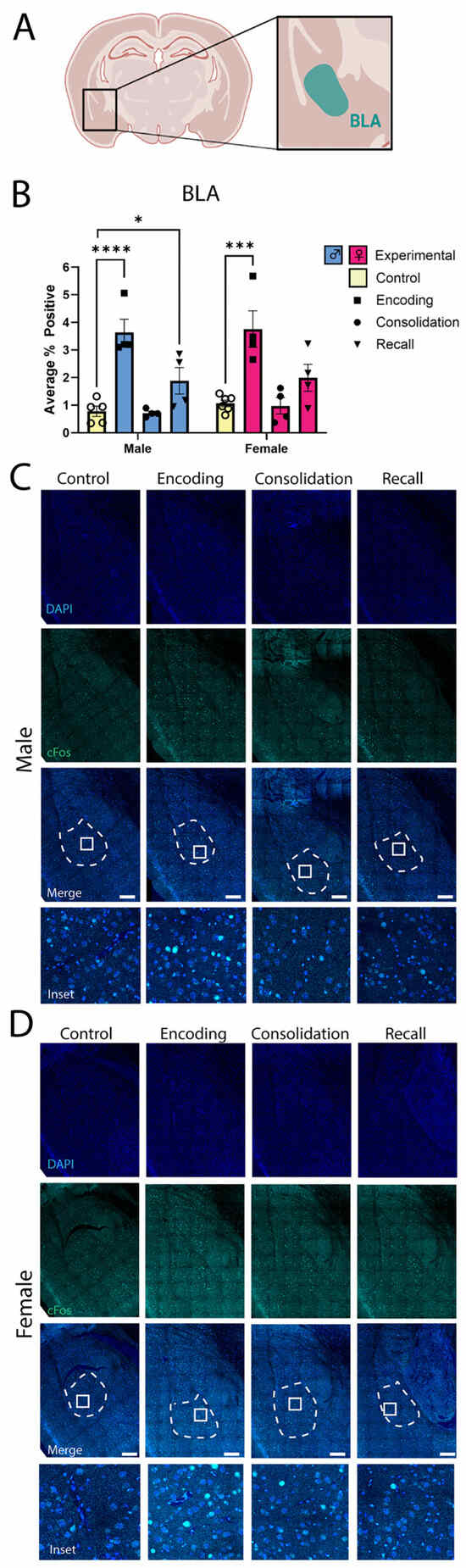
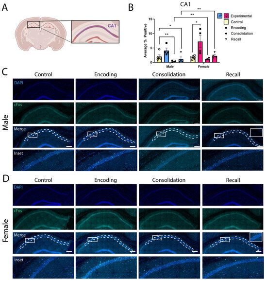
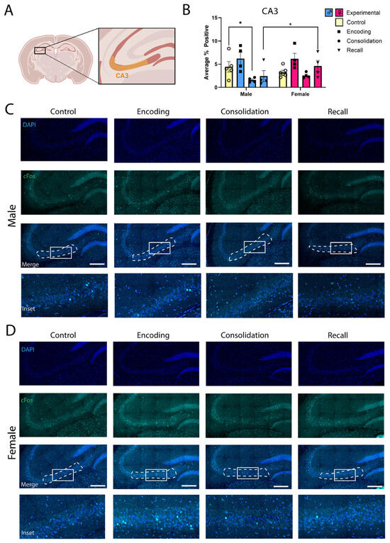
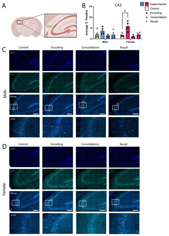
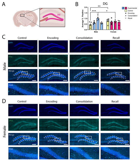
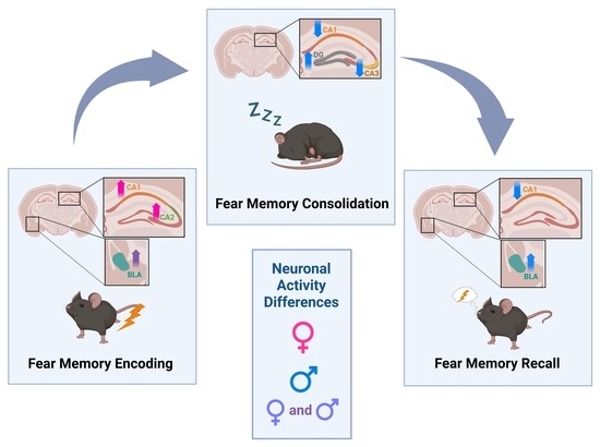
Accurate and efficient memory processing is essential for survival. A body of ongoing work in both human subjects and animal models suggests that memory processing may differ substantially between males and females. In mice, contextual fear memory (CFM) encoding, consolidation, and recall have been well studied, and the mouse hippocampus and amygdala have been implicated in these processes. The present pilot study addresses whether the activation of these brain regions differs substantially between male and female mice at each stage of CFM processing. We find that male and female mice show no differences in sleep behavior, which is essential for CFM consolidation, following single-trial contextual fear conditioning (CFC). We also find no significant differences in CFM recall performance between male and female mice. However, females show a trend for larger increases in CA1 cFos expression, relative to males, during CFM encoding. On the other hand, only males—but not females—show an apparent increase in cFos expression among dentate gyrus (DG) granule cells during CFM consolidation. Males also show a trend for a larger apparent reduction in cFos in CA1 and CA3 during CFM consolidation, relative to females. These preliminary findings highlight the idea that the neurobiological underpinnings of memory processing may differ between males and females, even when performance during recall is identical. Full article
Show Figures

Graphical abstract
54 pages, 7903 KB  
Review
Sex Does Matter! The Influence of Sex on Outcomes of Transcranial Direct Current Stimulation (tDCS)
by James Chmiel, Marta Stępień-Słodkowska, Aleksandra Kładna and Mirela Niedzielska
Cells 2026, 15(16), 1474; https://doi.org/10.3390/cells15161474 - 17 Aug 2026
Viewed by 321
Abstract
Background: Transcranial direct current stimulation (tDCS) produces heterogeneous outcomes across cognitive, behavioural, and clinical studies. Sex-related differences may contribute to this variability. Objective: This narrative review examined whether and how sex moderates tDCS outcomes across behavioural, physiological, clinical, and modelling studies and considered [...] Read more.
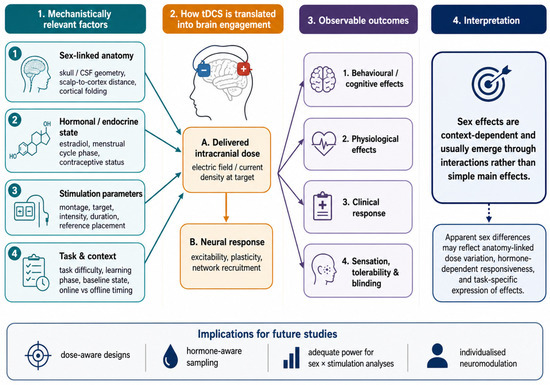
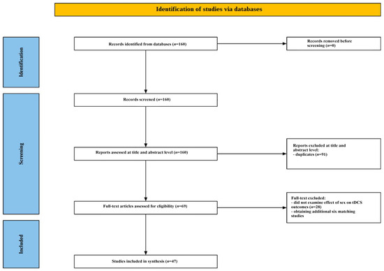
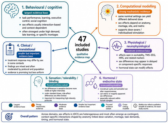
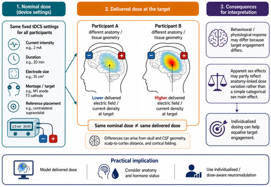
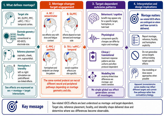
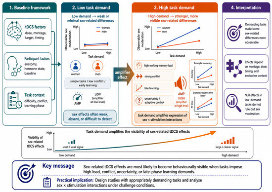
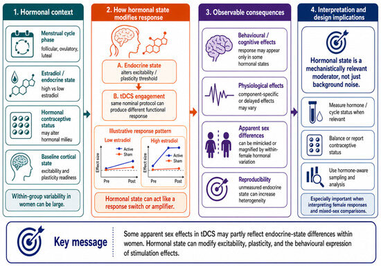
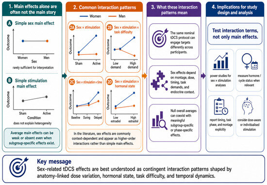
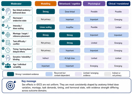
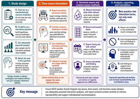
Background: Transcranial direct current stimulation (tDCS) produces heterogeneous outcomes across cognitive, behavioural, and clinical studies. Sex-related differences may contribute to this variability. Objective: This narrative review examined whether and how sex moderates tDCS outcomes across behavioural, physiological, clinical, and modelling studies and considered potential underlying mechanisms. Materials and Methods: A structured literature search identified studies examining sex, or variables reported by the original authors as gender but operationalised through female–male group comparisons, as moderators of tDCS outcomes. After screening, 41 studies met the inclusion criteria, and 6 additional studies were identified through citation searching, yielding 47 studies. Because of substantial heterogeneity in populations, protocols, and outcomes, findings were synthesised narratively. Results: Sex-related effects were common but highly context-dependent, varying by montage, cortical target, stimulation intensity, reference placement, and outcome domain. They often emerged as interaction effects rather than main effects and were most apparent during demanding tasks, later learning phases, or delayed after-effects. Computational models indicated that identical stimulation settings may produce different intracranial electric fields in women and men because of anatomical differences. Hormonal and endocrine states, particularly in females, may further modify or amplify these effects. Sensation, tolerability, and blinding may also contribute under higher-intensity protocols but do not fully explain sex-contingent outcomes. Conclusions: Sex influences tDCS outcomes through interacting anatomical, hormonal, and task-related mechanisms rather than in a uniform manner. Future studies should treat sex as a mechanistically relevant moderator, incorporate dose- and hormone-aware designs, and use adequately powered analyses to improve reproducibility and support individualised neuromodulation. Full article
Show Figures

Figure 1

2025

Jump to: 2026, 2024, 2023, 2022

35 pages, 638 KB  
Review
The Influence of Circadian Rhythms on Transcranial Direct Current Stimulation (tDCS) Effects: Theoretical and Practical Considerations
by James Chmiel and Agnieszka Malinowska
Cells 2025, 14(15), 1152; https://doi.org/10.3390/cells14151152 - 25 Jul 2025
Cited by 12 | Viewed by 6526
Abstract
Transcranial direct current stimulation (tDCS) can modulate cortical excitability in a polarity-specific manner, yet identical protocols often produce inconsistent outcomes across sessions or individuals. This narrative review proposes that much of this variability arises from the brain’s intrinsic temporal landscape. Integrating evidence from [...] Read more.
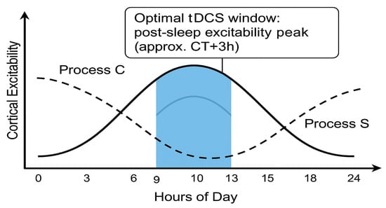
Transcranial direct current stimulation (tDCS) can modulate cortical excitability in a polarity-specific manner, yet identical protocols often produce inconsistent outcomes across sessions or individuals. This narrative review proposes that much of this variability arises from the brain’s intrinsic temporal landscape. Integrating evidence from chronobiology, sleep research, and non-invasive brain stimulation, we argue that tDCS produces reliable, polarity-specific after-effects only within a circadian–homeostatic “window of efficacy”. On the circadian (Process C) axis, intrinsic alertness, membrane depolarisation, and glutamatergic gain rise in the late biological morning and early evening, whereas pre-dawn phases are marked by reduced excitability and heightened inhibition. On the homeostatic (Process S) axis, consolidated sleep renormalises synaptic weights, widening the capacity for further potentiation, whereas prolonged wakefulness saturates plasticity and can even reverse the usual anodal/cathodal polarity rules. Human stimulation studies mirror this two-process fingerprint: sleep deprivation abolishes anodal long-term-potentiation-like effects and converts cathodal inhibition into facilitation, while stimulating at each participant’s chronotype-aligned (phase-aligned) peak time amplifies and prolongs after-effects even under equal sleep pressure. From these observations we derive practical recommendations: (i) schedule excitatory tDCS after restorative sleep and near the individual wake-maintenance zone; (ii) avoid sessions at high sleep pressure or circadian troughs; (iii) log melatonin phase, chronotype, recent sleep and, where feasible, core temperature; and (iv) consider mild pre-heating or time-restricted feeding as physiological primers. By viewing Borbély’s two-process model and allied metabolic clocks as adjustable knobs for plasticity engineering, this review provides a conceptual scaffold for personalised, time-sensitive tDCS protocols that could improve reproducibility in research and therapeutic gain in the clinic. Full article
Show Figures

Figure 1

2024

Jump to: 2026, 2025, 2023, 2022

28 pages, 4678 KB  
Article
High-Resolution Proteomics Unravel a Native Functional Complex of Cav1.3, SK3, and Hyperpolarization-Activated Cyclic Nucleotide-Gated Channels in Midbrain Dopaminergic Neurons
by Maya Belghazi, Cécile Iborra, Ophélie Toutendji, Manon Lasserre, Dominique Debanne, Jean-Marc Goaillard and Béatrice Marquèze-Pouey
Cells 2024, 13(11), 944; https://doi.org/10.3390/cells13110944 - 30 May 2024
Cited by 4 | Viewed by 2874
Abstract
Pacemaking activity in substantia nigra dopaminergic neurons is generated by the coordinated activity of a variety of distinct somatodendritic voltage- and calcium-gated ion channels. We investigated whether these functional interactions could arise from a common localization in macromolecular complexes where physical proximity would [...] Read more.
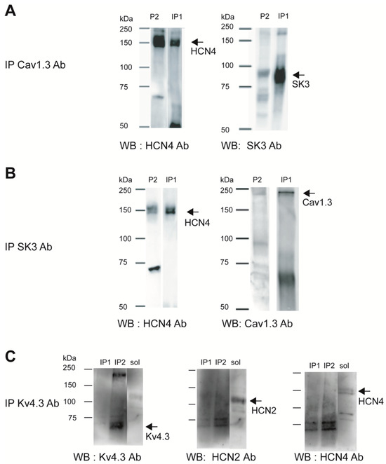
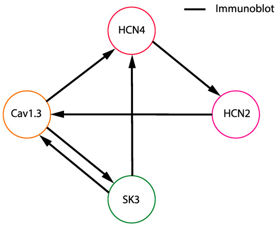
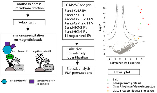
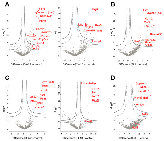
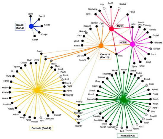
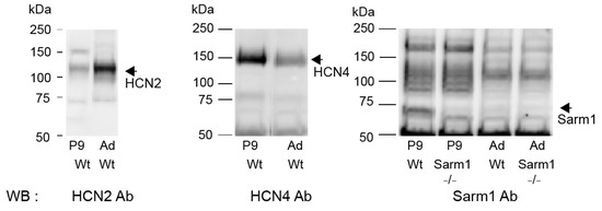
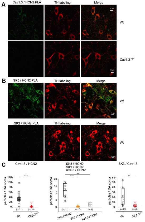
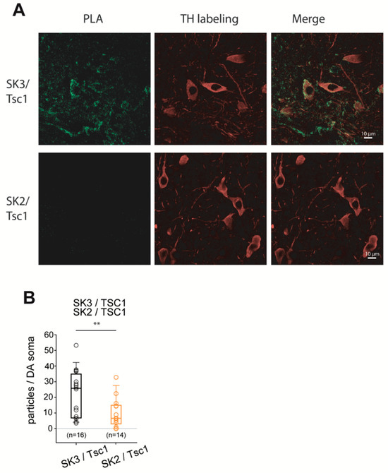
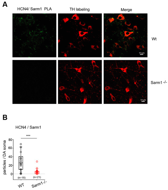
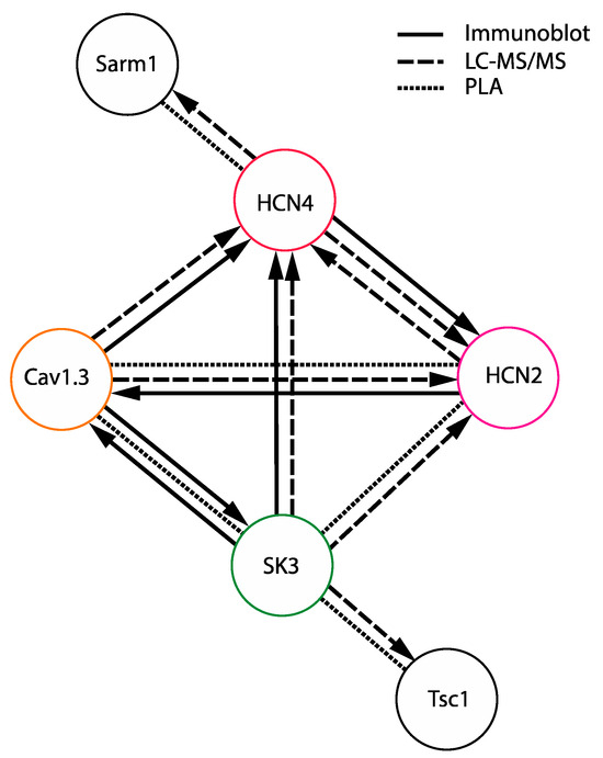
Pacemaking activity in substantia nigra dopaminergic neurons is generated by the coordinated activity of a variety of distinct somatodendritic voltage- and calcium-gated ion channels. We investigated whether these functional interactions could arise from a common localization in macromolecular complexes where physical proximity would allow for efficient interaction and co-regulations. For that purpose, we immunopurified six ion channel proteins involved in substantia nigra neuron autonomous firing to identify their molecular interactions. The ion channels chosen as bait were Cav1.2, Cav1.3, HCN2, HCN4, Kv4.3, and SK3 channel proteins, and the methods chosen to determine interactions were co-immunoprecipitation analyzed through immunoblot and mass spectrometry as well as proximity ligation assay. A macromolecular complex composed of Cav1.3, HCN, and SK3 channels was unraveled. In addition, novel potential interactions between SK3 channels and sclerosis tuberous complex (Tsc) proteins, inhibitors of mTOR, and between HCN4 channels and the pro-degenerative protein Sarm1 were uncovered. In order to demonstrate the presence of these molecular interactions in situ, we used proximity ligation assay (PLA) imaging on midbrain slices containing the substantia nigra, and we could ascertain the presence of these protein complexes specifically in substantia nigra dopaminergic neurons. Based on the complementary functional role of the ion channels in the macromolecular complex identified, these results suggest that such tight interactions could partly underly the robustness of pacemaking in dopaminergic neurons. Full article
Show Figures

Figure 1

20 pages, 1615 KB  
Review
Astrocytic GABAergic Regulation in Alcohol Use and Major Depressive Disorders
by Dina N. Ali, Hossam M. Ali, Matthew R. Lopez, Shinwoo Kang and Doo-Sup Choi
Cells 2024, 13(4), 318; https://doi.org/10.3390/cells13040318 - 9 Feb 2024
Cited by 27 | Viewed by 11662
Abstract
Gamma-aminobutyric acid (GABA) is the major inhibitory neurotransmitter in the central nervous system (CNS). Most GABAergic neurons synthesize GABA from glutamate and release it in the synaptic cleft in the CNS. However, astrocytes can also synthesize and release GABA, activating GABA receptors in [...] Read more.
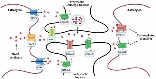
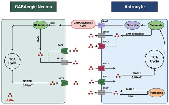
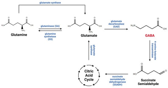
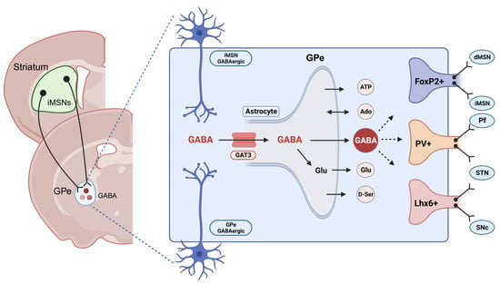
Gamma-aminobutyric acid (GABA) is the major inhibitory neurotransmitter in the central nervous system (CNS). Most GABAergic neurons synthesize GABA from glutamate and release it in the synaptic cleft in the CNS. However, astrocytes can also synthesize and release GABA, activating GABA receptors in the neighboring neurons in physiological and pathological conditions. As the primary homeostatic glial cells in the brain, astrocytes play a crucial role in regulating GABA homeostasis and synaptic neurotransmission. Accumulating evidence demonstrates that astrocytic GABA dysregulation is implicated in psychiatric disorders, including alcohol use disorder (AUD) and major depressive disorder (MDD), the most prevalent co-occurring psychiatric disorders. Several current medications and emerging pharmacological agents targeting GABA levels are in clinical trials for treating AUD and MDD. This review offers a concise summary of the role of astrocytic GABA regulation in AUD and MDD. We also provide an overview of the current understanding and areas of debate regarding the mechanisms by which astrocytes regulate GABA in the CNS and their potential significance in the molecular basis of AUD and MDD, paving the way toward future research directions and potential therapeutic target areas within this field. Full article
Show Figures

Graphical abstract
2023

Jump to: 2026, 2025, 2024, 2022

12 pages, 294 KB  
Review
The Role of Oxytocin in Alzheimer’s Disease and Its Relationship with Social Interaction
by Junpei Takahashi, Daisuke Yamada, Wakana Nagano and Akiyoshi Saitoh
Cells 2023, 12(20), 2426; https://doi.org/10.3390/cells12202426 - 10 Oct 2023
Cited by 21 | Viewed by 5587
Abstract
Alzheimer’s disease (AD)—the most common cause of dementia in the elderly—is characterized by progressive memory loss and β-amyloid protein (Aβ) accumulation in the brain. Recently, loneliness was found to be a high risk factor for AD, and social isolation has become a major [...] Read more.
Alzheimer’s disease (AD)—the most common cause of dementia in the elderly—is characterized by progressive memory loss and β-amyloid protein (Aβ) accumulation in the brain. Recently, loneliness was found to be a high risk factor for AD, and social isolation has become a major cause of AD. AD. Oxytocin (OXT), the main hormone involved in social bonding, has been implicated in social interactions, notably in building trust and relationships. Moreover, social isolation or social enrichment modulates the activation of neurons related to OXT. Recently, we reported that OXT reverses learning and memory impairment in AD animal models. Based on the limited number of studies currently available, OXT might be a therapeutic target for AD. Further studies are necessary in order to better understand the role of oxytocin in AD. In this review, we described the relationships between OXT, AD, and social interaction. Full article

2022

Jump to: 2026, 2025, 2024, 2023

16 pages, 2609 KB  
Article
Sustained Activation of the Anterior Thalamic Neurons with Low Doses of Kainic Acid Boosts Hippocampal Neurogenesis
by Farah Chamaa, Batoul Darwish, Rami Arnaout, Ziad Nahas, Elie D. Al-Chaer, Nayef E. Saadé and Wassim Abou-Kheir
Cells 2022, 11(21), 3413; https://doi.org/10.3390/cells11213413 - 28 Oct 2022
Cited by 5 | Viewed by 3788
Abstract
Adult hippocampal neurogenesis is prone to modulation by several intrinsic and extrinsic factors. The anterior nucleus (AN) of the thalamus has extensive connections with the hippocampus, and stimulation of this region may play a role in altering neurogenesis. We have previously shown that [...] Read more.
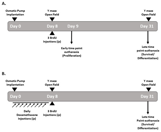
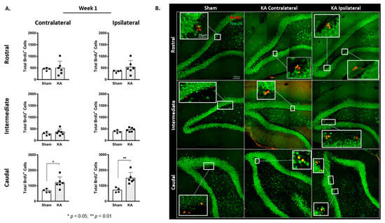
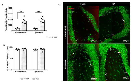
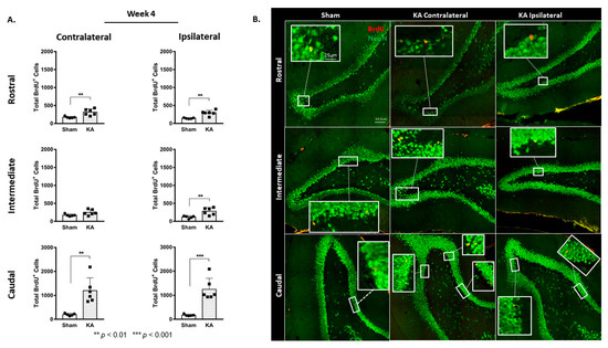
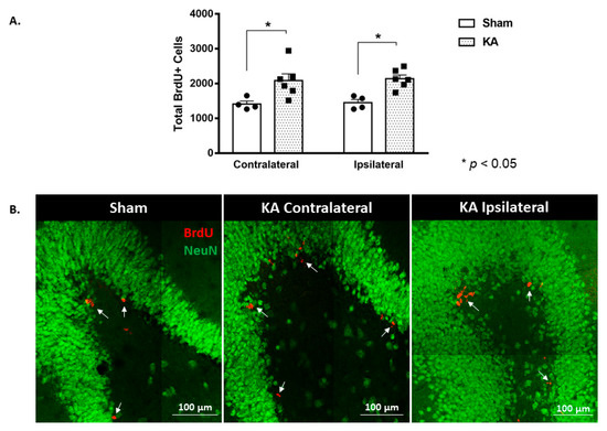
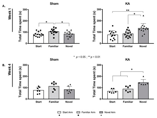
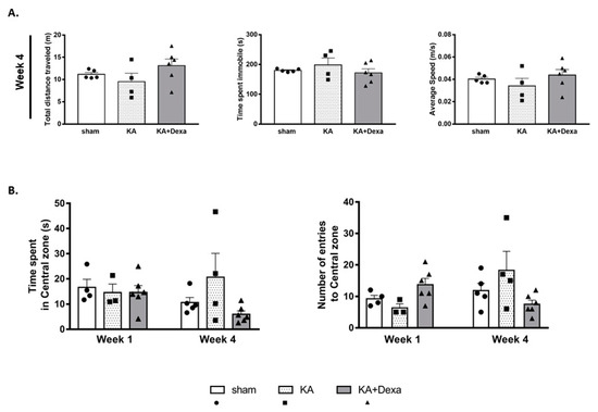
Adult hippocampal neurogenesis is prone to modulation by several intrinsic and extrinsic factors. The anterior nucleus (AN) of the thalamus has extensive connections with the hippocampus, and stimulation of this region may play a role in altering neurogenesis. We have previously shown that electrical stimulation of the AN can substantially boost hippocampal neurogenesis in adult rats. Here, we performed selective unilateral chemical excitation of the cell bodies of the AN as it offers a more specific and sustained stimulation when compared to electrical stimulation. Our aim is to investigate the long-term effects of KA stimulation of the AN on baseline hippocampal proliferation of neural stem cells and neurogenesis. Continuous micro-perfusion of very low doses of kainic acid (KA) was administered into the right AN for seven days. Afterwards, adult male rats received 5′-bromo-2′-deoxyuridine (BrdU) injections (200 mg/kg, i.p) and were euthanized at either one week or four weeks post micro-perfusion. Open field and Y-maze tests were performed before euthanasia. The KA stimulation of the AN evoked sustained hippocampal neurogenesis that was associated with improved spatial memory in the Y-maze test. Administering dexamethasone prior to and simultaneously with the KA stimulation decreased both the hippocampal neurogenesis and the improved spatial recognition memory previously seen in the Y-maze test. These results suggest that hippocampal neurogenesis may be a downstream effect of stimulation in general, and of excitation of the cell bodies of the AN in particular, and that stimulation of that area improves spatial memory in rats. Full article
Show Figures

Figure 1

Back to TopTop