
Recent Advances in Nanomaterial-Based Sensing for Food Safety Analysis
by
 ,
,
Xiujuan Qiao
1,2,3,†,
Jingyi He
1,2,†,
Ruixi Yang
1,2,
Yanhui Li
1,2,
Gengjia Chen
1,2,
Sanxiong Xiao
4,
Bo Huang
1,2,
Yahong Yuan
1,2,
Qinglin Sheng
1,2,* and
Tianli Yue
1,2,* 1
College of Food Science and Technology, Northwest University, Xi’an 710069, China
2
Laboratory of Nutritional and Healthy Food-Individuation Manufacturing Engineering/Research Center of Food Safety Risk Assessment and Control, Xi’an 710069, China
3
College of Chemistry and Molecular Engineering, Qingdao University of Science and Technology, Qingdao 266042, China
4
Xi’an Product Quality Supervision & Inspection Institute, Xi’an 710069, China
*
Authors to whom correspondence should be addressed.
†
These authors contributed equally to this work.
Processes 2022, 10(12), 2576; https://doi.org/10.3390/pr10122576
Submission received: 29 October 2022
/
Revised: 23 November 2022
/
Accepted: 1 December 2022
/
Published: 3 December 2022
(This article belongs to the Special Issue Food Safety Management and Quality Control Techniques)
Abstract
:The increasing public attention on unceasing food safety incidents prompts the requirements of analytical techniques with high sensitivity, reliability, and reproducibility to timely prevent food safety incidents occurring. Food analysis is critically important for the health of both animals and human beings. Due to their unique physical and chemical properties, nanomaterials provide more opportunities for food quality and safety control. To date, nanomaterials have been widely used in the construction of sensors and biosensors to achieve more accurate, fast, and selective food safety detection. Here, various nanomaterial-based sensors for food analysis are outlined, including optical and electrochemical sensors. The discussion mainly involves the basic sensing principles, current strategies, and novel designs. Additionally, given the trend towards portable devices, various smartphone sensor-based point-of-care (POC) devices for home care testing are discussed.
1. Introduction
With the environmental pollution caused by social and technological development, the food safety problem has become increasingly serious. In addition, illegal businesses’ blind pursuit of interests has forced food safety problems to occur frequently, which is closely related to people’s health [1]. For example, clenbuterol, a banned chemical, was used by farmers in pigs to promote their leanness [2]. In 2008, melamine illegally added to infant formula caused more than 290,000 infants to suffer from pathological urinary tract stones [3,4]. All these events have made the public aware of the importance and urgency of addressing food safety issues.
Food safety issues continue to occur, prompting us to seek more effective food analysis techniques [5,6]. However, food analysis is a complicated process and a big challenge for human beings. For example, many potentially dangerous ingredients may occur in different kinds of food and at every stage in handling food [7]. These dangerous contaminants include heavy metals, various toxins, residual drugs, pesticides, viruses, illegal additives, and bacteria [8,9,10]. In addition, the complications will be increased with food industry globalization [11]. In view of the requirement of consumers for health, food safety issues have prompted the public to construct fast, cost-effective, specific, and sensitive analytical methods and analytical techniques for food analysis.
Sensitivity and specificity are the most important features of sensor sensing applications, and their sensing performance can be enhanced by proper modification of the sensing surface [12]. One of the most important ways to increase the sensing performance is the application of nanomaterials. Nanomaterials, with a size from 1 nm to 100 nm, can be easily developed into different forms for different detection needs, such as 0 dimension (0D) of nanoparticles/nanoclusters [13,14,15,16,17], I dimension (1D) of nanowire/nanorod [18,19,20], II dimension (2D) of nanosheet [21,22], and III dimension (3D) of nanonet/nanoflower/nano-bulk [23,24,25]. Due to their unique light, electrical, and mechanical properties, their large surface area, good biocompatibility, catalytic activity, and rich bonding sites, nanomaterials have been widely used in various fields [26,27,28,29,30,31], especially for the production of sensing surface elements, that can not only increase the sensitivity but also provide lower limits of detection (LOD) [32,33,34].
There are many kinds of nanomaterial-based sensors and biosensors, which could be classified as optical and electrochemical sensors according to their transduction signals. Despite the fact that some reviews on the applications of nanomaterials for use in food safety sensing have been reported [35,36,37,38], a comprehensive overview of various nanomaterial-based sensors for food safety detection needs to be updated. Therefore, in this manuscript, we review the latest advances in novel sensors based on nanomaterials to detect food contamination over the past few years, including toxins, heavy metals, pesticides, drug residues, pathogens, and other hazardous substances (Figure 1). Various sensors in this manuscript could be divided into diverse optical methods and multiple electrochemical methods. In addition, the current smartphone-based portable devices have also been summarized here for the determination of contaminants in household food.
2. Current Approaches for Food Safety Sensing
Nowadays, nanomaterial-based sensors and biosensors are being explored to reduce the occurrence of food safety problems. Generally, a biosensor contains several parts: sensing elements, recognition elements, and transducer elements. Sensing elements, involving nanomaterials of 0D, 1D, 2D, and 3D nanomaterials. The high conductivity, large surface area and unique physicochemical properties of nanomaterials mean that they are characterized by good sensitivity in the detection of food contaminants [39,40,41].
Apart from the sensitivity, the selectivity is also one of the most prominent features for the construction of biosensors. The integration of a recognition element with sensing element can provide a useful tool to improve the selectivity for monitoring food pollutants. The recognition elements usually involve aptamers [42], antibodies [43,44], DNA [45], peptides [46], and cells [47], which can selectively and specifically respond to analysis targets [48]. The transducer element, which plays crucial roles in the performance of sensor devices [49], can convert a biological response into detectable signals [50]. According to different kinds of transducer signals, current approaches based on nanomaterials for food contaminants’ detection could be classified into optical methods or electrochemical methods. Optical methods contain colorimetry, chemiluminescence (CL), fluorescence, and so on. Electrochemical methods contain impedimetric sensors, potentiometric sensors, and amperometric sensors (Figure 2).
Non-covalent and covalent methods are used for the biofunctionalization of nanomaterials [51,52]. Non-covalent bonding forces include electrostatic interaction (Figure 3A), π-π stacking (Figure 3B), van der Waals forces, etc. The non-covalent method is to simply link biomolecules with nanomaterials. Compared with non-covalent interaction, covalent binding has advantages in terms of reproducibility and the stability of the nanomaterials’ surface functionalization and physisorption. Covalent links could be formed by biomolecular links (Figure 3C), classic amide coupling reactions (Figure 3D), cross-linking, or click chemistry. These strategies preserve all specific properties of both the biomolecule and nanomaterial.
3. Optical Sensors and Biosensors
In optical sensors, quantum dots, gold/silver nanoparticles, upconversion nanoparticles, metal oxide nanomaterials, and organic fluorescent molecular-based nanomaterials have been widely used to improve sensing performance [53,54]. Depending on the signal output format, this section focuses on colorimetric, fluorescent, and surface plasmon resonance (SPR) sensors for food inspection.
3.1. Colorimetric Sensors and Biosensors
Colorimetric assays have attracted more attention because they are the simplest and easiest sensing strategies among various optical methods. Regarding colorimetric assay, color changes can be easily detected by visual observation without any complex and expensive equipment [55]. Compared to traditional detection methods that require high-pressure input and the presence of inert gases, nanomaterials provide a feasible in-situ detection solution [56]. Among them, gold nanoparticles (Au NPs), the most preferred candidate materials, are widely applied to fabricate sensors of colorimetric assays for food contaminants due to their unique optical features, simple synthesis method, high stability, and easy modification [57,58].
The typical mechanism of the fabrication of colorimetric sensors based on Au NPs mainly relies on Au NPs’ distance changes. Au NPs dispersed in solution usually appear red with the maximum absorption wavelength at ~520 nm. However, when Au NPs polymerize, the solution color changes to dark blue or purple (surface plasmon band from visible region to the near-infrared region). Directly prepared gold nanoparticles cannot meet the needs of sensing detection, and their surfaces need to be modified [59]. As shown in Figure 4, the aggregation can be both induced by targets on unmodified Au NPs and functional Au NPs. Additionally, the aggregation of Au NPs can also be induced by salt. Conversely, when the aggregated Au NPs are redispersed, it causes the solution color to change from purple or blue to red. The redispersion of Au NPs can be mediated by specific target molecule interaction (Figure 5). Surface microenvironment and external environment changes lead to the agglomeration and redispersion of Au NPs, which promotes their application in the field of colorimetric assays.
Recently, researchers have developed a solid-phase sandwich-type colorimetric immunosensor for the rapid detection of Staphylococcus A (SEA) in food. With the help of covalent affinity with protein A, SEA antibodies are modified on the slides to form a test region. The same antibody is conjugated to the gold nanoparticles by physical adsorption as nanoimmune probes. When slides are continuously exposed to SEA and AuNP-antibody bioconjugates, distinct red spots appear in the detection area due to the aggregation of gold nanoparticles. The limit of detection of SEA in milk by biosensors is 1.5 ng mL−1 [59]. The sensor does not require any signal amplification strategy, and the detection of various targets can be achieved by changing the immune probe. The colorimetric properties of gold nanoparticles also contribute to the detection of food freshness. Li et al. [60] constructed a detection system composed of polyethylene glycol (PEG)-modified AuNPs and dopamine to achieve convenient and effective colorimetric detection of food freshness (Figure 6). The system exhibits a significant burgundy to black color change at amine concentrations of 1–100 μg mL−1 with a detection limit of 2.8 μg mL−1.
3.2. Fluorescence Sensors and Biosensors
Compared with subjective colorimetric method, fluorescence phenomena are more attractive due to their low backgrounds, high sensitivity, strong objectivity, low detection limit, and high repeatability in food detection. Thus, developing sensitive, low cost and eco-friendly fluorescence materials with desirable fluorescence properties is urgently essential for food analysis. Since carbonate quantum dots (CDs) were firstly discovered by Xu et al. in 2004 [62], the fluorescence emission property of CD makes them attractive as sensing probes for food analysis [63,64]. For example, Yue et al. [65] synthesized copper-modified fluorescent carbon dots (Cu-CDs) with stable double emission by hydrothermal methods, which were successfully applied to the detection of methyltobutazine (TM) in fruits according to the linear relationship between the degree of fluorescence quenching of Cu-CDs and the concentration of the target. Yin et al. [66] prepared a novel N,Cl co-doped carbon dots (N,Cl-CDs) method based on deep eutectic solvent (DES) to achieve the rapid and accurate quantification of morphine fluorescence methods in food.
In addition to its excellent fluorescence properties, CDs can be prepared from any starting material containing carbon, include most food by-products, such as fruits, livestock, and vegetables. Based on this property, agricultural products containing a large amount of natural carbon to form CDs have great potential in bio-environmental sensing platforms [67,68,69,70], and also provide new solutions to the serious problem of food waste in global production and consumption processes [71,72,73]. The sensors for food analysis based on CDs produced from agriculture products are summarized as follows in Table 1.
Other nanomaterials with non-fluorescent properties can achieve fluorescence effect-based food detection by combining with fluorescent molecules. The chromophore of most conventional fluorescent probes produces strong fluorescence in its separated state, but the presence of aggregation-caused quenching (ACQ) results in molecules not radiating energy at high concentrations or in a concentrated state. In fact, the presence of the ACQ phenomenon limits the application of fluorescent probes to some extent [88,89,90]. Since the discovery of aggregation-induced emission effect (AIE) by Tang Group in 2001 [91], the use of AIE materials as fluorescent probes has gradually become a research hotspot [92,93,94,95,96]. This is because the AIE effect can overcome the shortcomings of the ACQ effect mentioned above, and thus is able to be used for high concentrations of fluorescence [97,98,99,100,101]. AIE emission can be clearly explained by the key role of molecular motions [102,103].
Generally, detection strategies for AIE luminescence (AIEgens) include: (1) electrostatic interaction and hydrogen bond interaction, (2) solubility change of AIEgens, (3) disruption of AIE luminescence quenching, (4) preparation of nano-sized AIE particles, and (5) target-induced disaggregation of AIEgens. Based on these sensing mechanisms, a number of AIEgens have been exploited as sensing probes for food detection. Jia et al. [104] proposed a novel label-free fluorescent aptamer that was modified on graphene oxide (GO) to detect AFB1 in food samples by observing the fluorescence of quaternized tetraphenylethene salt and AFB1 aptamer aggregates (TPE-Z/AFB1) (Figure 7A). The fluorescence of the TPE-Z aptamer aggregates almost has non-fluorescence emission in the Tris-HCl buffer solution. In the presence of AFB1, the release of TPE-Z/AFB1 aptamers from the GO surface results in fluorescence recovery. The sensor has a detection limit of 0.25 ng mL−1. Wang et al. [105] constructed a tetraphenylethylene derivative functionalized mesoporous silica nanoparticle for the detection of furazolidone (FZD) and furacillin (NF) antibiotics in water.
In addition, the self-polymerization and disassembly of AIE nanomaterials can also be used for food inspection. As shown in Figure 7B, Mehta et al. [106] developed fluorescent peptidyl probes (2,3) with benzimidazolyl-cyanovinylidene (1) fluorophores based on their AIE effect for the detection of Ag+ and Ag NPs in water samples with detection limits of 0.64 ppb and 1.1 ppb, respectively. As for Figure 6C, Han et al. [107] reported a copper nanocluster (Cu NCs) with 2,3,5,6-tetrafluorothiophenol as a reducing agent and protective agent for the detection of histamine in food. When histamine is present, the nanoaggregates of Cu NCs are destroyed and spherical particles appear, leading to the quenching of their AIE effect.
3.3. Surface Plasmon Resonance (SPR) Sensors and Biosensors
Surface plasmon resonance (SPR) technology is a special optical method that detects changes in the refractive index caused by the interaction between molecules and ligands in a sample on the sensor surface [108]. When a molecule fixed on the surface of the sensor binds to a target in solution, it causes a change in the refractive index, resulting in a surface plasmon at the dielectric interface. Compared with conventional optical techniques, SPR technique exhibits some advantages, for example, the direct monitoring of the refractive index changes for food safety without need of specific properties (absorption, fluorescence, or scattering bands) and without the requirement of radioactive or fluorescent markers [109] of the analyte. Generally, SPR biosensors consist of a light source, optical system, sensing system, and detection system. The optical system emits the incident light; the change in resonance angle/wavelength can be converted by the sensing system; the magnitude signal of the resonance angle or wavelength is detected by system measures. SPR biosensors’ design principles always include fiber-optic surface plasmon resonance (FOSPR) [110], localized surface plasmon resonance (LSPR) [111], surface plasmon resonance imaging (SPRI), and transmission surface plasmon resonance (TSPR) [112]. The schematic illustrations with basic composition and advantages about the four types of SPR biosensors’ platforms have been provided in Figure 8.
Nanoparticle-based SPR technology has been widely used for the detection of foodborne contaminants. Vaisocherová-Lísalová et al. [113] constructed a multichannel SPR biosensor modified with an antibody-functionalized poly(carboxybetaine acrylamide) (pCBAA) coating, which enhanced the sensor response with the help of Au NPs for foodborne E. Coli O157:H7 and Salmonella sp., which were detected with limits of 57 cfu mL−1 and 17 cfu mL−1, respectively. Écija-Arenas et al. [114] modified a single layer of graphene on the transducer gold surface by chemical vapor deposition (CVD), and then modified 1-pyrenebutyric acid (PBA) to graphene by π stacking to ensure the covalent binding of subsequent aptamers. The specific capture of kanamycin by aptamers resulted in a change in SPR signaling with a detection limit of 285 nmol L−1.
4. Electrochemical Sensors and Biosensors
The electrochemical-based detection method for food safety is one of the most popular methods among detection strategies [115]. Due to their cost-effectiveness, simplicity, inherent sensitivity, high sensing speed, and compatibility with portable devices, electrochemical-based sensors have become the most rapidly growing sensor class [116]. Traditional electrodes based on mercury [117,118] have gradually been replaced by other suitable nanomaterials’ modified electrodes, such as biocompatible and excellent conductive carbon nanomaterials [119,120,121,122,123] or metal nanoparticles [124,125,126], nanozymes with high stability and tunable catalytic activity [127], and metal-organic frameworks (MOFs) with more active sites and high porosity [128].
Electrochemical sensors can be divided into potentiometric sensors, electrochemical sensors, and impedance sensors, according to the different conduction behavior of impedance, current, or potential signals caused by the interaction between the target and the recognition element on the sensor surface. This section focuses on the application of the above three types of nanomaterial-based electrochemical sensors in food safety detection.
4.1. Impedance Sensors and Biosensors
Electrochemical impedance spectroscopy (EIS) is a method based on electrical impedance detection at the electrode/electrolyte interface that can effectively detect food contaminants [129]. When the target in the electrolyte binds to the recognition element on the electrode surface, a specific concentration of the detected target is achieved by observing the impedance change at the electrode/electrolyte interface. This value is obtained by applying a small sinusoidal voltage at a specific frequency. For example, Mejri-Omrani et al. [130] reported an electrochemical aptasensor for directly detecting Ochratoxin A (OTA) by EIS technique. Polypyrrole (PPy) covalently bonded with polyamidoamine dendrimers as sensing elements, were coated on the surface of a gold electrode to reach PAMAM G4. Label-free OTA aptamers were thereafter covalently bonded to the PAMAM G4 for building an electrochemical aptasensor. When OTA and aptamers bind, the conformation of PAMAM G4 changes, leading to a difference in the electrical signal. The results showed that the method was able to detect concentrations of OTA in foods below the European regulations allowed, reaching 2 ng L−1. Miao et al. [131] constructed an electrochemical impedance sensor based on magnetic molecularly imprinted polymer for the detection of the insecticide dichlorodiphenyltrichloroethane (DDT) in food. The sensor uses a molecularly imprinted polymer synthesized from magnetic Fe3O4 and polydopamine (PDA@Fe3O4 MIP MNPs) as the electrode signal material, which is adsorbed by the identification unit of the PDA layer when DDT is present, resulting in a change in the electrochemical impedance signal of the material. The results show that the sensor has a good linear relationship with DDT concentration in the range of 1 × 10⁻¹¹ to 1 × 10⁻³ mol L−1, and the detection limit reaches 6 × 10⁻¹² mol L−1 (Figure 9A).
In addition, EIS is also used in the field of portable, miniaturized platforms for sensing foodborne bacteria of L. monocytogenes. These antibody-functionalized EIS microelectrodes are not only specific for the identification of Listeria monocytogenes, but their microfluidic devices can also be used for detection in liquid samples, such as milk. This highly efficient microfluidic device provides a platform for the detection of Listeria monocytogenes with a limit of detection (LOD) of 5.5 cfu mL−1 (Figure 9B) [132].
4.2. Voltammetry Sensors and Biosensors
Cyclic voltammetry (CV), linear sweep voltammetry (LSV), differential pulse voltammetry (DPV), and square wave voltammetry (SWV) are the most commonly used techniques in building sensors for food safety analysis. CV is one of the most widely used voltammetric techniques, which is based on the qualitative and quantitative analysis of targets by determining electrochemical information in the presence of redox intermediates or reversible reactions. In CV experiments, the potential superimposed on the working electrode is positively and linearly related to time. When the set ramp potential is reached, the applied potential returns to the initial potential, and a CV scan signal is obtained by recording the generated current. For other voltammetry sensors, DPV technique and SWV is generally applied for quantitative analysis in biosensors, especially for aptasensors, due to its merits of high sensitivity, short analysis time, and simultaneous multi-component detection. In addition, these techniques can be used to explore the kinetics, thermodynamics, and mechanisms of chemical reactions and analytical measurements. DPV differs from ESI technology in that DPV detects by applying a series of regular voltage pulses to the current immediately after a linear sweep and recording the resulting current difference as a function of the applied potential. In SWV, an excitation signal is obtained by applying a symmetrical square wave pulse of the staircase waveform to the working electrode.
Summerson and Prasco [133] report an inexpensive and convenient electrochemical sensor for the real-time monitoring of ochratoxin A (OTA) (Figure 10). When the target binds specifically to the aptamer, the conformational change of the aptamer causes MB to approach the electrode surface and enhance the electron transport ability of the electrode. SWV records the signals generated by different concentrations of OTA into the detection system. This is a simple design without any signal amplification strategy for food safety analysis.
To achieve higher detection goals, designers often introduce enzymes. For example, Ezhilan et al. [134] reported that a sensitive acetylcholinesterase cyclic voltammetric biosensor based on Pt/ZnO/acetylcholinesterase/chitosan enables the detection of melamine and urea in adulterated milk. The acetylthiocholine acted as an electrochemical active substance so that the subsequent redox signal was recorded in CV by acetylcholinesterase catalysis. Urea and melamine bind to the serine hydroxyl group of acetylcholinesterase as competitive inhibitors, thus affecting the catalytic activity of acetylcholinease. This biosensor detection mechanism is based on linear regression models for the calculation of binary mixtures of urea and melamine in adulterated cow milk. The proposed bioelectrode for the detection of urea and melamine has a limit of detection of 1 pM and 3 pM, respectively.
For signal amplification, Wang et al. [135] constructed an exonuclease III (Exo III)-driven double-amplified electrochemical aptamer sensor for the detection of chloramphenicol (CAP). First, the researchers synthesized Zr-MOF complexes of PtPd@Ni-Co hollow nanoboxes (PtPd@Ni-Co HNBs) and poly (diallyldimethylammonium chloride)-functionalized graphene (PDDA-Gr) as electrode modification materials to increase electrode surface area and conductivity. The captured DNA and assistant DNA were then modified on the surface of Zr-MOF to promote signal amplification. When CAP is present, it causes the release of a large amount of trigger DNA (Tr DNA) in Cycle I, then Tr DNA and Exo III initiating Cycle II, causing the exposed capture DNA to further bind to the signal probe (MB/HP-UiO-66/Signal DNA) and resulting in an electrochemical ‘signal on’. Under optimal conditions, the aptamer sensor has a good linear range, from 10 fM to 10 nM with a chloramphenicol detection limit of 0.985 fM. By changing the aptamer sequence, this sensing strategy can be applied to the detection of a variety of different targets (Figure 11).
Xu and Hou et al. [136] reported a highly sensitive aptamer sensor based on two electroactive species for the detection of malathion. When malathion is present, its specific binding to the aptamer causes the thiamine (Tn)-labeled aptamer to detach from the electrode surface while inducing the ferrocene (Fc)-labeled capture probe to form a hairpin structure, so detection can be achieved by signal shutdown of Tn and signal conduction of Fc. At the same time, the signal cycle amplification is achieved with the help of exonuclease I (Exo I). In this work, under the detection conditions, the proposed aptasensor exhibited excellent stability, specificity, and repeatability with a wide linear range from 0.5 to 600 ng L−1 (Figure 12).
As described above, enzymes play a vital role in food testing and have been used to detect chemical or biological contaminants such as small molecules, heavy metal ions, and proteins [137]. However, the disadvantages of natural enzymes are that they are costly, time-consuming, vulnerable to extreme pH and temperature, and so on, which brings great challenges to their application. In this regard, nanozymes, the newly emerging artificial enzyme mimics, could be accepted to surpass these obstacles [138]. Nanozymes, having both nanomaterial features and natural enzyme-like properties, are attractive substitutes to natural enzymes [139]. Currently, nanozymes are widely considered to be the next generation of artificial enzyme stars due to their attractive nanoscale advantages of low cost, long-term storage, high stability to severe environments, and ease of mass production [140,141]. The materials used to synthesize nanoenzymes mainly include metals, metal oxides, MOFs, covalent organic frameworks (COF), polymers, etc. Various enzyme-like materials include nanomaterials, polymers, compounds, micelles, metalloproteins, and coordination complexes. These nanozymes are usually applied to mimic many kinds of enzymes, for example: oxidase (OXD), catalase (CAT) [142], superoxide dismutase (SOD) [143], peroxidase (POD) [144], glucose oxidase (GOX) [145], sphosphatase, and so on (Figure 13).
Nanozymes still have excellent test performance in complex food matrix environments. Wu et al. [146] developed a two-dimensional MnO2 nanosheet-mediated electrochemical sensor with oxidase-like and peroxidase properties for the detection of organophosphate pesticides. Hu et al. [147] developed a functional 2D MOF nanoenzyme for the detection of Staphylococcus aureus. The 2D MOF material has peroxidase activity, which can effectively catalyze o-phenylenediamine to 2,2-diaminoazobenzene to detect the concentration of Staphylococcus aureus. Under optimal conditions, the sensor has a detection limit of 6 cfu mL−1 (Figure 14).
4.3. Potentiometric Sensors and Biosensors
Potentiometry (PM) is used in quantitative electroanalytical analysis of the target analyte by measuring the electrochemical potential of charged species between two electrodes (reference electrode and working electrode) in electrolytic tanks. Typically, PM testing is used when there is no or very little current in the system. This PM technique can achieve outstanding sensitivity for detection sensors. For example, Arvand et al. [148] report a potential sensor based on a mesoporous aluminosilicate-modified poly(vinyl chloride) film for the determination of Al3+ in food and drug products. However, a highly accurate and stable reference electrode is required in this system, which will affect the application of PM in analytical detection [149].
In addition to the above examples, Table 2 (below) lists other nanomaterial-based (photo)electrochemical sensing food detection schemes.
5. Mobile Sensors and Biosensors
Most of the current conventional sensing techniques for food safety are still dependent on laboratories, which are costly, time-consuming, complicated, labor-intensive, etc. Thus, the conventional sensing techniques make these sensing platforms unsuitable for point-of-care (POC) and consumer-oriented detection. These disadvantages could not timely prevent harmful substances from harming the human body and the environment. Therefore, there is an intense desire to develop portable sensing devices as homecare test devices [176]. To this end, the detection application of smartphone-based biosensors in food safety has become a hot spot for researchers [177,178,179,180].
With the rapid development of nanomaterials and the widespread use of smartphones for everyone, the combination of food safety monitoring services and smartphones will have the potential to offer portable sensing devices as homecare test devices. For example, Man et al. [181] developed a Au NPs-based microfluidic colorimetric immunoassay device for the detection of alternariol monomethyl ether (AME) with a detection limit of 200 pg mL−1 and a recovery rate of 90.63% to 93.9% for smartphone imaging (Figure 15A). Cheng et al. [182] report a two-dimensional (2D) Pt-Ni(OH)2 nanosheet-based amplified bidirectional lateral flow immunoassay device for the simultaneous detection of acetochlor and cymethrin, with detection limits of 0.63 ng/mL and 0.24 ng/mL, respectively (Figure 15B). In order to apply POC to home life, its devices need to be highly reliable, low cost, and user-friendly. Based on the above requirements, the rapid development of smartphones, which contain various embedded sensors, could enable successful application in food safety monitoring. For example, smartphones’ built-in sensors of audio jacks and cameras and external sensors (e.g., probe, microfluidic devices and circuit) have been widely used in POC devices [183,184]. Smartphone-based POC sensors aim to accomplish more timely, more efficient, and easier testing in resource-limited settings.
5.1. Smartphone-Based Optical Biosensors
Because of the advantages of smartphones, they are commonly applied in building optical POC biosensing systems, including colorimetry [185,186], surface plasmon resonance [187], fluorescence [188,189], image analysis, etc. The combination of smartphones and biosensing devices can promote precision and immediacy in food safety analysis.
Zeinhom et al. [190] have established a novel colorimetric biosensor for the detection of S. Enteritidis in cheese, milk, and water samples through visual and quantitative techniques. This testing system is based on magnetic beads labeled S. Enteritidis antibodies as a capture platform, horseradish peroxidase (HRP) as a signal amplification, where HRP enzyme and capture antibodies are both located on inorganic nanoflowers. The visual signal produced by nanotechnology at low concentrations is easily recorded by smartphones. The detection principle of the capture platform form and antibody-enzyme-inorganic nanoflower signal amplification strategy for S. Enteritidis is shown in Figure 16. This successful application of smartphones in the detection of S. Enteritidis in cheese and milk indicates the possibility of its application in various food products.
Compared with colorimetric technics, fluorescence technics show more attractive features of high precision, sensitivity, and accuracy. The same group have developed a smartphone-based fluorescence biosensors for E. coli O157:H7 detection. This POC biosensor, based on smartphones, includes a long-pass thin-film interference filter, a laser-diode-based photosource, a 3D printer, and insert lenses. A commercial blue laser (eBay) was employed as the excitation light source, powered by two batteries. This process was excited at 405 nm with output power of ∼5 mW, and the detection fluorescence emission was collected by a signal-collecting external lens. The proposed biosensor provides a low noise to background imagine system, with a detection limit of 1 cfu mL−1 and 10 cfu mL−1 for yoghurt and egg, respectively (Figure 17A) [191]. Typically, actual food samples contain a number of different substances with similar biochemical/chemical structures and properties. Therefore, the smartphone-based multiplexing detection equipment has been applied to food [192,193,194,195]. Xing et al. [196] report a microfluidic biosensor based on the principle of fluorescence resonance energy transfer (FRET) between GO and fluorescent molecules, combined with smartphones, for the simultaneous detection of multiplex foodborne bacterial ssDNA (Figure 17B).
5.2. Smartphone-Based Electrochemical Biosensors
Nowadays, different kinds of electrochemical techniques, such as impedimetric [197], potentiometric [198], and amperometric [199] techniques based on smartphones have been researched. A number of POC or miniaturized electrochemical devices have been developed. Generally, electrochemical assay based on smartphone needs to meet the requirements of an external potentiostat for electrochemical signal acquisition and processing, disposable and low-cost electrodes, data transmission mode, and extra batteries for power supply.
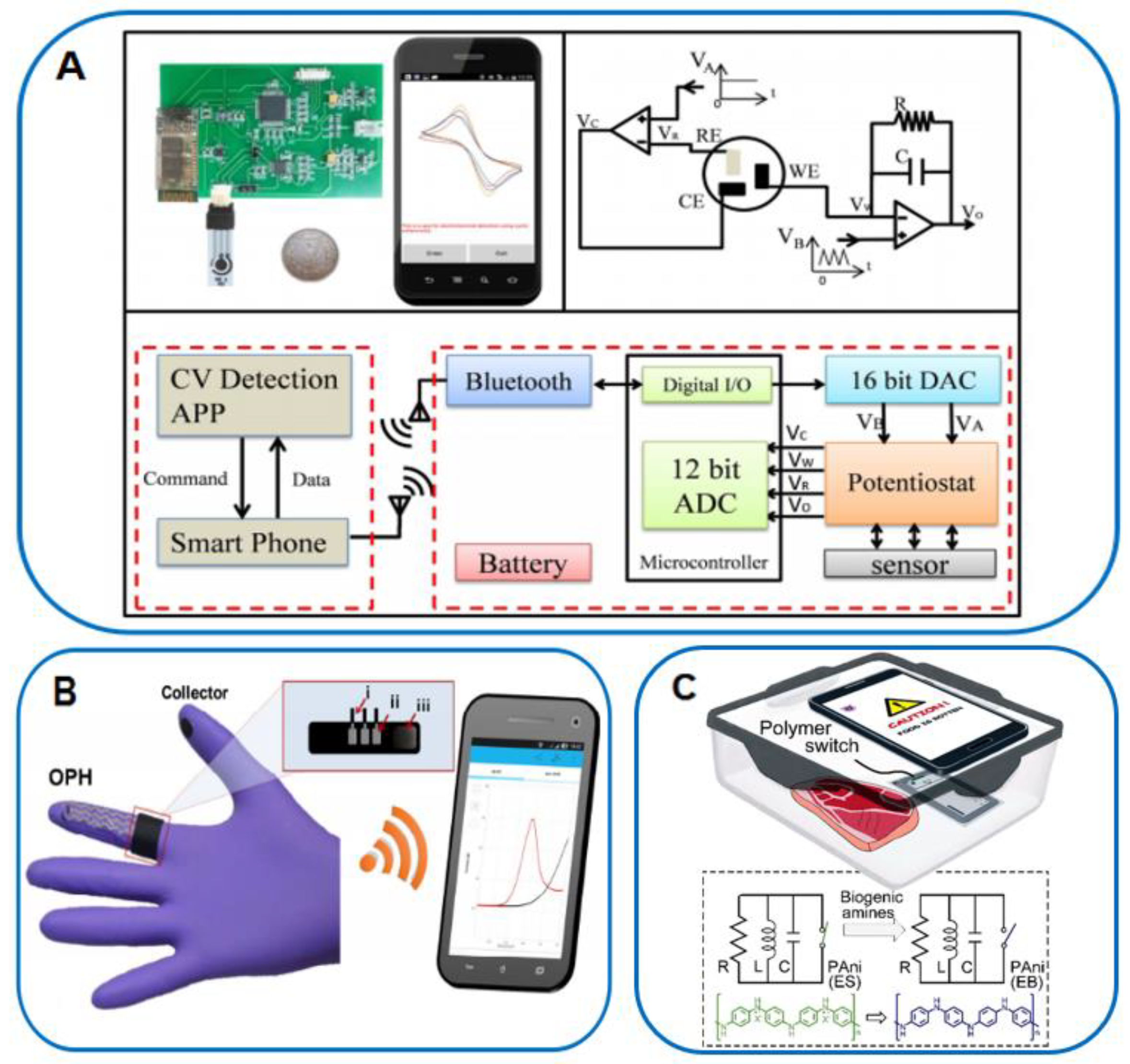
In view of the above requirements, screen printing technology can be put into consideration to develop POC and low-cost electrochemical biosensors, because it is possible to easily prepare different kinds of electrodes on paper, plastics, and other flexible substrates [200]. Ji et al. [201] developed an electrochemical detection sensor based on a combination of screen-printed electrodes and a smartphone. This system was composed of a simple GO modification electrode, an energy transformation module to provide stimuli signals, a low-cost potentiostat for CV measurements, a bluetooth module for transmitting electrochemical data, and a smartphone as the controller and displayer. The redox couples CV signal could perform on smartphone by roughly controlling different scan rates with test errors less than 3.8% when compared to detection results on commercial electrochemical workstations (Figure 18A).
In addition, Mishra et al. [202] have developed a wearable electrochemical platform with electrodes imprinted on the surface of nitrile experimental gloves, which have the advantages of low cost and being disposable. Characterization was performed at room temperature by using a Palmsens handheld potentiostat (EmStat3 Blue with 10.0 × 6.0 × 3.4 cm3 dimensions, Palmsens, Houten, Netherlands) powered by a rechargeable Li-Po battery. This enzyme-based, disposable ‘glove lab’ biosensing system transmits data to Android smartphone devices in real time with a compact electronic interface for electrochemical sensing and wireless Bluetooth. The electrode system and the wireless electronic interface in the form of a long snake are modified on the surface of the glove with three layers of stress-resistant elastic ink. The first layer is a silver layer of Ag/AgCl particles combined with elastic Ecoflex material, the second layer is an elastic styrene-isoprene copolymer-modified carbon ink, and the third layer is a transparent flexible insulator. This organophosphorus hydrolase (OPH)-based biosensor provides a new idea for detecting organophosphate (OP) in food, and the cost, convenience, and speed advantages of the sensor also open the door to more applications of flexible wearable sensors in hand multiplexed sensing (Figure 18B).
In order to further reduce external attachments, Near Field Communication (NFC) technology is gradually entering the field of view. NFC is able to provide wireless power and data transmission through inductive coupling, thus solving the problem of using additional battery accessories in smartphone electrochemical sensors. NFC technology shows great potential in the development of new POC sensing systems due to its characteristics, such as low cost and possessing no battery to develop. Currently, NFC-based smartphone-integrated POC biosensors have already been used for biochemical detection [203,204] and food safety sensing [205]. Figure 18C shows an example of switchable NFC tags to detect food spoilage with a smartphone [205]. This protocol reports a gas sensor based on iron(III) p-toluenesulfonate-doped polyaniline (PAni) nanostructures for the detection of biogenic amines in meat.
Figure 18.
(A) An electrochemical detections sensor based on combination of screen-printed electrodes and smartphone [201]; (B) a wearable and disposable electrochemical platform based on a combination of a nitrile experimental glove surface and smartphone [202]; (C) switchable NFC technology for the detection of food spoilage with smartphone [205].
6. Conclusion and Outlook
Safety issues in all aspects of food production have always been a global concern, and in order to protect people from the threat of food contaminants, more convenient detection methods are needed. Compared with traditional methods, nanomaterial-based sensors are widely used in food safety testing because of their low cost, high efficiency, and reliability. In terms of electrode modification, nanomaterials can improve detection sensitivity due to their large specific surface area, good electrical conductivity and good catalytic activity. In terms of selectivity, nanomaterials provide excellent selectivity for sensing analysis by binding to aptamers, antibodies, cells, etc. This review summarizes various nanomaterial-based optical and electrochemical sensing analysis techniques for food contaminants, mainly mentioning basic sensing principles, examples of current sensing strategies, and novel designs based on nanomaterials. Through the design of nanomaterials and the construction of sensors, the performance of sensors has been significantly improved, providing new topics for food safety testing.
In addition, given the trend of portable devices and widespread use of mobile phones, the new generation of smartphone-based point-of-care devices has significant advantages (cost-effectiveness, ease of operation, minimal equipment, and ease of data management) when assembling various sensors for home care testing, which is the direction of the future. If these sensors are used in the food industry, they will be beneficial for food quality control. In the future, mobile diagnostics will show great potential by combining technologies such as portable smartphones, cloud computing, and new sensing designs.
Author Contributions
Writing—original draft preparation, X.Q. and J.H.; investigation and discussion, R.Y., Y.L. and G.C.; revision and discussion, S.X., B.H. and Y.Y.; revision, discussion, and project administration, T.Y. and Q.S. All authors have read and agreed to the published version of the manuscript.
Funding
This work was supported by National Key R&D Program of China (2019YFC1606703) and the Natural Science Foundation of Shaanxi Province in China (2020JM-429).
Conflicts of Interest
The authors declare no conflict of interest.
References
- Boyaci, I.H.; Temiz, H.T.; Genis, H.E.; Soykut, E.A.; Nur Yazgan, N.N.; Güven, B.; Uysal, R.S.; Bozkurt, A.G.; İlaslan, K.; Torun, O.; et al. Dispersive and FT-Raman spectroscopic methods in food analysis. RSC Adv. 2015, 5, 56606–56624. [Google Scholar] [CrossRef]
- Li, G.L.; Fu, Y.H.; Han, X.S.; Li, X.Y.; Li, C.C. Metabolomic investigation of porcine muscle and fatty tissue after Clenbuterol treatment using gas chromatography/mass spectrometry. J. Chromatogr. A 2016, 1456, 242–248. [Google Scholar] [CrossRef] [PubMed]
- Chen, J.X.; Xu, F.; Jiang, H.Y.; Hou, Y.L.; Rao, Q.X.; Guo, P.J.; Ding, S.Y. A novel quantum dot-based fluoroimmunoassay method for detection of Enrofloxacin residue in chicken muscle tissue. Food Chem. 2009, 113, 1197–1201. [Google Scholar] [CrossRef]
- Wu, Y.N.; Zhao, Y.F.; Li, J.G. A Survey on Occurrence of Melamine and Its Analogues in Tainted Infant Formula in China. Biomed. Environ. Sci. 2009, 22, 95–99. [Google Scholar] [CrossRef] [PubMed]
- García-Cañas, V.; Simó, C.; Herrero, M.; Ibáñez, E.; Cifuentes, A. Present and future challenges in food analysis: Foodomics. Anal. Chem. 2012, 84, 10150–10159. [Google Scholar] [CrossRef] [PubMed] [Green Version]
- Ricci, F.; Volpe, G.; Micheli, L.; Palleschi, G. A review on novel developments and applications of immunosensors in food analysis. Anal. Chim. Acta 2007, 605, 111–129. [Google Scholar] [CrossRef] [PubMed]
- Scognamiglio, V.; Arduini, F.; Palleschi, G.; Rea, G. Biosensing technology for sustainable food safety. TrAC Trends Anal. Chem. 2014, 62, 1–10. [Google Scholar] [CrossRef]
- Amine, A.; Mohammadi, H.; Bourais, I.; Palleschi, G. Enzyme inhibition-based biosensors for food safety and environmental monitoring. Biosens. Bioelectron. 2006, 21, 1405–1423. [Google Scholar] [CrossRef] [PubMed]
- Campàs, M.; Garibo, D.; Prieto-Simon, B. Novelano biotechnological concepts in electrochemical biosensors for the analysis of toxins. Analyst 2012, 137, 1055–1067. [Google Scholar] [CrossRef]
- Bahadır, E.B.; Sezgintürk, M.K. Applications of commercial biosensors in clinical, food, environmental, and biothreat/biowarfareanalyses. Anal. Biochem. 2015, 478, 107–120. [Google Scholar] [CrossRef]
- Sharma, R.; Ragavan, K.V.; Thakur, M.S.; Raghavarao, K.S.M.S. Recent advances in nanoparticle based aptasensors for food contaminants. Biosens. Bioelectron. 2015, 74, 612–627. [Google Scholar] [CrossRef] [PubMed]
- Majdinasab, M.; Yaqub, M.; Rahim, A.; Catanante, G.; Hayat, A.; Marty, J.L. An overview on recent progress inelectrochemical biosensors for antimicrobial drug residues in animal-derived food. Sensors 2017, 17, 1947. [Google Scholar] [CrossRef] [PubMed] [Green Version]
- Zhukhovitskiy, A.V.; MacLeod, M.J.; Johnson, J.A. Carbene Ligands in Surface Chemistry: From Stabilization of Discrete Elemental Allotropes to Modification of Nanoscale and Bulk Substrates. Chem. Rev. 2015, 115, 11503–11532. [Google Scholar] [CrossRef]
- Wang, G.Q.; Rühling, A.; Amirjalayer, S.; Marek, K.; Ernst, J.B.; Richter, C.; Gao, H.J.; Timmer, A.; Gao, H.Y.; Doltsinis, N.L.; et al. Ballbot-type motion of N-heterocyclic carbenes on gold surfaces. Nat. Chem. 2017, 9, 152–156. [Google Scholar] [CrossRef] [PubMed]
- Misztalewska-Turkowicz, I.; Markiewicz, K.H.; Michalak, M.; Wilczewska, A.Z. NHC-copper complexes immobilized on magnetic nanoparticles: Synthesis and catalytic activity in the CuAAC reactions. J. Catal. 2018, 362, 46–54. [Google Scholar] [CrossRef]
- Engel, S.; Fritz, E.C.; Ravoo, B.J. New trends in the functionalization of metallic gold: From organosulfur ligands to N-heterocyclic carbenes. Chem. Soc. Rev. 2017, 46, 2057–2075. [Google Scholar] [CrossRef]
- Bernardos, M.D.D.L.; Pérez-Rodríguez, S.; Gual, A.; Claver, C.; Godard, C. Facile synthesis of NHC-stabilized Ni nanoparticles and their catalytic application in the Z-selective hydrogenation of alkynes. Chem. Commun. 2017, 53, 7894–7897. [Google Scholar] [CrossRef] [PubMed] [Green Version]
- Zhang, J.; Langille, M.R.; Mirkin, C.A. Synthesis of Silver Nanorods by Low Energy Excitation of Spherical Plasmonic Seeds. Nano Lett. 2011, 11, 2495–2498. [Google Scholar] [CrossRef]
- Yu, X.F.; Liu, J.W.; Cong, H.P.; Xue, L.; Arsad, M.N.; Albar, H.A.; Sobahi, T.R.; Gao, Q.; Yu, S.H. Template- and surfactant-free synthesis of ultrathin CeO2 nanowires in a mixed solvent and their superior adsorption capability for water treatment. Chem. Sci. 2015, 6, 2511–2515. [Google Scholar] [CrossRef] [PubMed] [Green Version]
- Khanal, B.P.; Zubarev, E.R. Chemical Transformation of Nanorods to Nanowires: Reversible Growth and Dissolution of Anisotropic Gold Nanostructures. ACS Nano 2019, 13, 2370–2378. [Google Scholar] [CrossRef] [PubMed]
- Wang, H.T.; Sun, W.S.; Liang, X.; Zou, H.Y.; Jiao, X.; Lin, K.A.; Li, T.L. Two-dimensional Fe2O3 nanosheets as adsorbent for the removal of Pb(II) from aqueous solution. J. Mol. Liq. 2021, 335, 116197. [Google Scholar] [CrossRef]
- Zhang, S.J.; Pelligra, C.I.; Feng, X.D.; Osuji, C.O. Directed assembly of hybrid nanomaterials and nanocomposites. Adv. Mater. 2018, 30, 1705794. [Google Scholar] [CrossRef] [PubMed]
- Zhao, Y.F.; Zhou, H.; Chen, W.X.; Tong, Y.J.; Zhao, C.; Lin, Y.; Jiang, Z.; Zhang, Q.W.; Xue, Z.G.; Cheong, W.C.; et al. Two-step carbothermal welding to access atomically dispersed Pd1 on three-dimensional zirconia nanonet for direct indole synthesis. J. Am. Chem. Soc. 2019, 141, 10590–10594. [Google Scholar] [CrossRef] [PubMed]
- Qu, Y.N.; Huang, R.L.; Qi, W.; Shi, M.B.; Su, R.X.; He, Z.M. Controllable synthesis of ZnO nanoflowers with structure-dependent photocatalytic activity. Catal. Today 2020, 355, 397–407. [Google Scholar] [CrossRef]
- Argentero, G.; Mittelberger, A.; Monazam, M.R.A.; Cao, Y.; Pennycook, T.J.; Mangler, C.; Kramberger, C.; Kotakoski, J.; Geim, A.K.; Meyer, J.C. Unraveling the 3D atomic structure of a suspended graphene/hBN van der Waals heterostructure. Nano Lett. 2017, 17, 1409–1416. [Google Scholar] [CrossRef] [Green Version]
- Ling, X.; Roland, S.; Pileni, M.P. Supracrystals of N-Heterocyclic Carbene-Coated Au Nanocrystals. Chem. Mater. 2015, 27, 414–423. [Google Scholar] [CrossRef]
- Roland, S.; Ling, X.; Pileni, M.P. N-Heterocyclic Carbene Ligands for Au Nanocrystal Stabilization and Three-Dimensional Self-Assembly. Langmuir 2016, 32, 7683–7696. [Google Scholar] [CrossRef] [Green Version]
- Maduraiveeran, G.; Jin, W. Nanomaterials based electrochemical sensor and biosensor platforms for environmental applications. Trends Environ. Anal. Chem. 2017, 13, 10–23. [Google Scholar] [CrossRef]
- Liu, B.W.; Liu, J.W. Sensors and biosensors based on metal oxide nanomaterials. TrAC 2019, 121, 115690. [Google Scholar] [CrossRef]
- Kwon, S.O.; Song, H.S.; Park, H.T.; Jang, J. Conducting Nanomaterial Sensor Using Natural Receptors. Chem. Rev. 2019, 119, 36–93. [Google Scholar] [CrossRef]
- Amali, R.K.A.; Lim, H.N.; Ibrahim, I.; Huang, N.M.; Zainal, Z.; Ahmad, S.A.A. Significance of nanomaterials in electrochemical sensors for nitrate detection: A review. Trends Environ. Anal. Chem. 2021, 31, e00135. [Google Scholar] [CrossRef]
- Maduraiveeran, G.; Sasidharan, M.; Ganesan, V. Electrochemical sensor and biosensor platforms based on advanced nanomaterials for biological and biomedical applications. Biosens. Bioelectron. 2017, 103, 113–129. [Google Scholar] [CrossRef]
- Holzinger, M.; Goff, A.L.; Cosnier, S. Synergetic effects of combined nano_materials for biosensing applications. Sensors 2017, 17, 1010. [Google Scholar] [CrossRef] [Green Version]
- Speranza, G. Carbon Nanomaterials: Synthesis, Functionalization and Sensing Applications. Nanomaterials 2021, 11, 967. [Google Scholar] [CrossRef]
- Siva, S.; Jin, J.O.; Choi, I.; Kim, M. Nanoliposome based biosensors for probing mycotoxins and their applications for food: A review. Biosens. Bioelectron. 2022, 219, 114845. [Google Scholar] [CrossRef] [PubMed]
- Du, Y.; Guo, S.J. Chemically doped fluorescent carbon and graphene quantum dots for bioimaging, sensor, catalytic and photoelectronic applications. Nanoscale 2016, 8, 2532–2543. [Google Scholar] [CrossRef]
- Pirsa, S.; Heidari, H.; Lotfi, J. Design selective gas sensors based on nano-sized polypyrrole/polytetrafluoroethylene and polypropylene membranes. IEEE Sens. J. 2016, 16, 2922–2928. [Google Scholar] [CrossRef]
- Arshad, F.; Mohd-Naim, N.F.; Chandrawati, R.; Cozzolino, D.; Ahmed, M.U. Nanozyme-based sensors for detection of food biomarkers: A review. RSC Adv. 2022, 12, 26160–26175. [Google Scholar] [CrossRef]
- Lan, L.Y.; Yao, Y.; Ping, J.F.; Ying, Y.B. Recent advances in nanomaterial-based biosensors for antibiotics detection. Biosens. Bioelectron. 2017, 91, 504–514. [Google Scholar] [CrossRef]
- Zhu, C.Z.; Yang, G.H.; Li, H.; Du, D.; Lin, Y.H. Electrochemical sensors and biosensors based on nanomaterials and nanostructures. Anal. Chem. 2016, 87, 230–249. [Google Scholar] [CrossRef] [PubMed]
- Sun, Y.M.; Zhao, J.L.; Liang, L.J. Recent development of antibiotic detection in food and environment: The combination of sensors and nanomaterials. Microchim. Acta 2021, 188, 21. [Google Scholar] [CrossRef] [PubMed]
- Kalita, J.J.; Sharma, P.; Bora, U. Recent developments in application of nucleic acid aptamer in food safety. Food Control 2022, 145, 109406. [Google Scholar] [CrossRef]
- Xing, C.R.; Jing, X.X.; Zhang, X.; Yuan, J. Ultrasensitive indirect competitive ELISA and strip sensor for detection of furazolidone metabolite in animal tissues. Food Agric. Immunol. 2017, 28, 1269–1282. [Google Scholar] [CrossRef] [Green Version]
- Peng, J.; Liu, L.Q.; Xu, L.G.; Song, S.S.; Kuang, H.; Cui, G.; Xu, C.L. Gold nanoparticle-based paper sensor for ultrasensitive and multiple detection of 32 (fluoro)quinolones by one monoclonal antibody. Nano Res. 2017, 10, 108–120. [Google Scholar] [CrossRef]
- Sani, N.D.; Heng, L.Y.; Marugan, R.S.P.M.; Rajab, N.F. Electrochemical DNA biosensor for potential carcinogen detection in food sample. Food Chem. 2018, 296, 503–510. [Google Scholar] [CrossRef]
- Suaifan, G.A.R.Y.; Alhogail, S.; Zourob, M. Paper-based magnetic nanoparticle-peptide probe for rapid and quantitative colorimetric detection of Escherichia coli O157:H7. Biosens. Bioelectron. 2017, 92, 702–708. [Google Scholar] [CrossRef] [PubMed]
- Xu, Y.T.; Zhang, T.Y.; Li, Z.; Liu, X.N.; Zhu, Y.C.; Zhao, W.W.; Chen, H.Y.; Xu, J.J. Photoelectrochemical Cytosensors. Electroanalysis 2022, 34, 947–955. [Google Scholar] [CrossRef]
- Justino, C.I.L.; Freitas, A.C.; Pereira, R.; Duarte, A.C.; Santos, T.A.P.R. Recent developments in recognition elements for chemical sensors and biosensors. TrAC Trends Anal. Chem. 2015, 68, 2–17. [Google Scholar] [CrossRef]
- Hasseb, A.A.; Ghani, N.T.A.; Shehab, O.R.; Nashar, R.M. Application of molecularly imprinted polymers for electrochemical detection of some important biomedical markers and pathogens. Curr. Opin. Electrochem. 2022, 31, 100848. [Google Scholar] [CrossRef]
- Morales, M.A.; Halpern, J.M. Guide to Selecting a Biorecognition Element for Biosensors. Bioconjug. Chem. 2018, 29, 3231–3239. [Google Scholar] [CrossRef] [PubMed]
- Holzinger, M.; Goff, A.L.; Cosnier, S. Nanomaterials for biosensing applications: A review. Front. Chem. 2014, 2, 2296–2646. [Google Scholar] [CrossRef] [Green Version]
- Putzbach, W.; Ronkainen, N.J. Immobilization Techniques in the Fabrication of Nanomaterial-Based Electrochemical Biosensors: A Review. Sensors 2013, 13, 4811–4840. [Google Scholar] [CrossRef] [PubMed]
- Chen, H.Y.; Zhang, L.; Hu, Y.; Zhou, C.S.; Lan, W.; Fu, H.Y.; She, Y.B. Nanomaterials as optical sensors for application in rapid detection of food contaminants, quality and authenticity. Sens. Actuators B Chem. 2021, 329, 129135. [Google Scholar] [CrossRef]
- Fang, L.; Jia, M.X.; Zhao, H.P.; Kang, L.Z.; Shi, L.C.; Zhou, L.D.; Kong, W.J. Molecularly imprinted polymer-based optical sensors for pesticides in foods: Recent advances and future trends. Trends Food Sci. Technol. 2021, 116, 387–404. [Google Scholar] [CrossRef]
- Yoo, S.M.; Lee, S.Y. Optical biosensors for the detection of pathogenic microorganisms. Trends Biotechnol. 2016, 34, 7–25. [Google Scholar] [CrossRef]
- Priyadarshini, E.; Pradhan, N. Gold nanoparticles as efficient sensors in colorimetric detection of toxic metal ions: A review. Sens. Actuators B Chem. 2017, 238, 888–902. [Google Scholar] [CrossRef]
- Zhou, W.; Gao, X.; Liu, D.B.; Chen, X.Y. Gold nanoparticles for in vitro diagnostics. Chem. Rev. 2015, 115, 10575–10636. [Google Scholar] [CrossRef] [Green Version]
- Chen, H.; Zhou, K.; Zhao, G.H. Gold nanoparticles: From synthesis, properties to their potential application as colori-metric sensors in food safety screening. Trends Food Sci. Technol. 2018, 78, 83–94. [Google Scholar] [CrossRef]
- Liu, G.Y.; Lu, M.; Huang, X.D.; Li, T.F.; Xu, D.H. Application of Gold-Nanoparticle Colorimetric Sensing to Rapid Food Safety Screening. Sensors 2018, 18, 4166. [Google Scholar] [CrossRef] [Green Version]
- Zhang, L.; Salmain, M.; Liedberg, B.; Boujday, S. Naked Eye Immunosensing of Food Biotoxins Using Gold Nanoparticle-Antibody Bioconjugates. ACS Appl. Nano Mater. 2019, 2, 4150–4158. [Google Scholar] [CrossRef]
- Li, H.; Gan, J.C.; Yang, Q.; Fu, L.L.; Wang, Y.B. Colorimetric detection of food freshness based on amine-responsive dopamine polymerization on gold nanoparticles. Talanta 2021, 234, 122706. [Google Scholar] [CrossRef] [PubMed]
- Xu, X.Y.; Ray, R.; Gu, Y.L.; Ploehn, H.J.; Gearheart, L.; Raker, K.; Scrivens, W.A. Electrophoretic analysis and purification of fluorescent single-walled carbon nano tube fragments. J. Am. Chem. Soc. 2004, 126, 12736–12737. [Google Scholar] [CrossRef]
- Zhang, X.Y.; Jiang, M.Y.; Niu, N.; Chen, Z.J.; Li, S.J.; Liu, S.X.; Li, J. Natural-product derived carbon dots: From natural products to functional materials. ChemSusChem 2018, 11, 11–24. [Google Scholar] [CrossRef] [PubMed]
- Sharma, V.; Tiwari, P.; Mobin, S.M. Sustainable carbon-dots: Recent advances in green carbon dots for sensing and bioimaging. J. Mater. Chem. B 2017, 5, 8904–8924. [Google Scholar] [CrossRef] [PubMed]
- Yue, X.N.; Zhu, C.N.; Gu, R.R.; Hu, J.; Xu, Y.; Ye, S.; Zhu, J. Copper-Modified Double-Emission Carbon Dots for Rapid Detection of Thiophanate Methyl in Food. Foods 2022, 11, 3336. [Google Scholar] [CrossRef]
- Yin, Q.H.; Wang, M.T.; Fang, D.; Zhu, Y.Q.; Yang, L.H. Novel N,Cl-doped deep eutectic solvents-based carbon dots as a selective fluorescent probe for determination of morphine in food. RSC Adv. 2021, 11, 16805–16813. [Google Scholar] [CrossRef]
- Hu, Q.; Gong, X.J.; Liu, L.Z.; Choi, M.M.F. Characterization and analytical separation of fluorescent carbon nanodots. J. Nanomater. 2017, 2017, 1–23. [Google Scholar] [CrossRef] [Green Version]
- Mishra, R.K.; Ha, S.K.; Verma, K.; Tiwari, S.K. Recent progress in selected bionanomaterials and their engineering applications: An overview. J. Sci. Adv. Mater. Dev. 2018, 3, 263–288. [Google Scholar] [CrossRef]
- Safarik, I.; Baldikova, E.; Prochazkova, J.; Safarikova, M.; Pospiskova, K. Magnetically modified agricultural and food waste: Preparation and application. J. Agric. Food Chem. 2018, 66, 2538–2552. [Google Scholar] [CrossRef]
- Qu, J.H.; Wei, Q.Y.; Sun, D.W. Carbon dots: Principles and their applications in food quality and safety detection. Crit. Rev. Food Sci. Nutr. 2018, 58, 2466–2475. [Google Scholar] [CrossRef]
- Xue, L.; Liu, G.; Parfitt, J.; Liu, X.J.; Herpen, E.V.; Stenmarck, Å.; O’Connor, C.; Östergren, K.; Cheng, S.K. Missingfood, missing data? A critical review of global food losses and food waste data. Environ. Sci. Technol. 2017, 51, 6618–6633. [Google Scholar] [CrossRef] [PubMed]
- Grainger, M.J.; Aramyan, L.; Logatcheva, K.; Piras, S.; Righi, S.; Setti, M.; Vittuari, M.; Stewart, G.B. The use of systems models to identify food waste drivers. Glob. Food Secur. 2018, 16, 1–8. [Google Scholar] [CrossRef] [Green Version]
- Menna, F.D.; Dietershagen, J.; Loubiere, M.; Vittuari, M. Life cycle costing of food waste: A review of methodological approaches. Waste Manag. 2018, 73, 1–13. [Google Scholar] [CrossRef] [PubMed]
- Wang, N.; Wang, Y.T.; Guo, T.T.; Yang, T.; Chen, M.L.; Wang, J.H. Green preparation of carbon dots with papaya as carbon source for effective fluorescent sensing of Iron (III) and Escherichia coli. Biosens. Bioelectron. 2016, 85, 68–75. [Google Scholar] [CrossRef]
- Das, P.; Bose, M.; Ganguly, S.; Mondal, S.; Das, A.K.; Banerjee, S.; Das, N.C. Green approach to photoluminescent carbon dots for imaging of gram-negative bacteria Escherichia coli. Nanotechnology 2017, 28, 195501–195513. [Google Scholar] [CrossRef]
- Hu, X.T.; Li, Y.X.; Xu, Y.W.; Gan, Z.Y.; Zou, X.B.; Shi, J.Y.; Huang, X.W.; Li, Z.H.; Li, Y.H. Green one-step synthesis of carbon quantum dots from orange peel for fluorescent detection of Escherichia coli in milk. Food Chem. 2021, 339, 127775. [Google Scholar] [CrossRef]
- Liu, W.J.; Li, C.; Sun, X.B.; Pan, W.; Yu, G.G.; Wang, J.P. Highly crystalline carbon dots from fresh tomato: UV emission and quantum confinement. Nanotechnology 2017, 28, 485705. [Google Scholar] [CrossRef]
- Bao, R.Q.; Chen, Z.Y.; Zhao, Z.W.; Sun, X.; Zhang, J.Y.; Hou, L.R.; Yuan, C.Z. Green and facile synthesis of nitrogen and phosphorus co-doped carbon quantum dots towards fluorescent ink and sensing applications. Nanomaterials 2018, 8, 386. [Google Scholar] [CrossRef] [Green Version]
- Bandi, R.; Gangapuram, B.R.; Dadigala, R.; Eslavath, R.; Singh, S.S.; Guttena, V. Facile and green synthesis of fluorescent carbon dots from onion waste and their potential applications as sensor and multicolour imaging agents. RSC Adv. 2016, 6, 28633–28639. [Google Scholar] [CrossRef]
- Shen, J.; Shang, S.M.; Chen, X.J.; Wang, D.; Cai, Y. Facile synthesis of fluorescence carbon dots from sweet potato for Fe3+ sensing and cell imaging. Mater. Sci. Eng. C 2017, 76, 856–864. [Google Scholar] [CrossRef]
- Zhao, J.J.; Huang, M.J.; Zhang, L.L.; Zou, M.B.; Chen, D.X.; Huang, Y.; Zhao, S.L. Unique ap proach to develop carbon dot-based nanohybrid near-infrared ratiometric fluorescent sensor for the detection of mercury ions. Anal Chem. 2017, 89, 8044–8049. [Google Scholar] [CrossRef] [PubMed]
- Bano, D.; Kumar, V.; Singh, V.K.; Hasan, S.H. Green synthesis of fluorescent carbon quantum dots for the detection of mercury(ii) and glutathione. New J. Chem. 2018, 42, 5814–5821. [Google Scholar] [CrossRef]
- Tyagi, A.; Tripathi, K.M.; Singh, N.; Choudhary, S.; Gupta, R.K. Green synthesis of carbon quantum dots from lemon peel waste: Applications in sensing and photocatalysis. RSC Adv. 2016, 6, 72423–72432. [Google Scholar] [CrossRef]
- Kumar, A.; Chowdhuri, A.R.; Laha, D.; Mahto, T.K.; Karmakar, P.; Sahu, S.K. Green synthesis of carbon dots from Ocimum sanctum for effective fluorescent sen sing of Pb2+ ions and live cell imaging. Sens. Actuators B Chem. 2017, 242, 679–686. [Google Scholar] [CrossRef]
- Bhatt, S.; Bhatt, M.; Kumar, A.; Vyas, G.; Gajaria, T.; Paul, P. Green route for synthesis of multifunctional fluorescent carbon dots from Tulsi leaves and its application as Cr(VI) sensors, bio-imaging and patterning agents. Colloids Surf. B Biointerfaces 2018, 167, 126–133. [Google Scholar] [CrossRef]
- Wen, X.P.; Shi, L.H.; Wen, G.G.; Li, Y.Y.; Dong, C.; Yang, J.; Shuang, S.M. Green and facile synthesis of nitrogen-doped carbon nanodots for multicolor cellular imaging and Co2+ sensing in living cells. Sens. Actuators B Chem. 2016, 235, 179–187. [Google Scholar] [CrossRef]
- Liu, Y.L.; Zhou, Q.X.; Li, J.; Lei, M.; Yan, X.Y. Selective and sensitive chemosensor for lead ions using fluorescent carbon dots prepared from chocolate by one-step hydro thermal method. Sens. Actuators B Chem. 2016, 237, 597–604. [Google Scholar] [CrossRef]
- Cai, Y.B.; Li, L.Z.; Wang, Z.T.; Sun, J.Z.; Qin, A.J.; Tang, B.Z. A sensitivity tuneable tetraphenylethene-based fluorescent probe for directly indicating the concentration of hydrogen sulfide. Chem. Commun. 2014, 50, 8892–8895. [Google Scholar] [CrossRef]
- Bu, F.; Wang, E.J.; Peng, Q.; Hu, R.R.; Qin, A.J.; Zhao, Z.J.; Tang, B.Z. Structural and theoretical insights into the AIE attributes of phosphindole oxide: The balance between rigidity and flexibility. Chem. Eur. J. 2015, 21, 4440–4449. [Google Scholar] [CrossRef]
- Jing, H.; Lu, L.; Feng, Y.; Zheng, J.F.; Deng, L.D.; Chen, E.Q.; Ren, X.K. Synthesis, Aggregation-Induced Emission, and Liquid Crystalline Structure of Tetraphenylethylene–Surfactant Complex via Ionic Self-Assembly. J. Phys. Chem. C. 2016, 120, 27577–27586. [Google Scholar] [CrossRef]
- Mei, J.; Leung, N.L.C.; Kwok, R.T.K.; Lam, J.W.Y. Aggregation-Induced Emission: Together We Shine, United We Soar! Chem. Rev. 2015, 115, 11718–11940. [Google Scholar] [CrossRef] [PubMed]
- Alam, P.; Leung, N.L.C.; Zhang, J.; Kwok, R.T.K.; Lam, J.W.Y.; Tang, B.Z. AIE-based luminescence probes for metal ion detection. Coord. Chem. Rev. 2020, 429, 213693. [Google Scholar] [CrossRef]
- Wang, D.; Tang, B.Z. Acc. Aggregation-Induced Emission Luminogens for Activity-Based Sensing. Chem. Res. 2019, 52, 2559–2570. [Google Scholar] [CrossRef]
- Jiang, M.J.; Gu, X.G.; Lam, J.W.Y.; Zhang, Y.L.; Kwok, R.T.K.; Wong, K.S.; Tang, B.Z. Two-photon AIE bio-probe with large Stokes shift for specific imaging of lipid droplets. Chem. Sci. 2017, 8, 5440–5446. [Google Scholar] [CrossRef] [PubMed] [Green Version]
- Li, J.; Wang, J.X.; Li, H.X.; Song, N.; Wang, D.; Tang, B.Z. Supramolecular materials based on AIE luminogens (AIEgens): Construction and applications. Chem. Soc. Rev. 2020, 49, 1144–1172. [Google Scholar] [CrossRef]
- Gao, M.; Tang, B.Z. AIE-based cancer theranostics. Coordin. Chem. Rev. 2020, 402, 213076. [Google Scholar] [CrossRef]
- Hong, Y.; Meng, L.; Chen, S.; Leung, C.W.; Da, L.T.; Faisal, M.; Silva, D.A.; Liu, J.Z.; Lam, J.W.Y.; Huang, X.H.; et al. Monitoring and inhibition of insulin fibrillation by a small organic fluorogen with aggregation-induced emission characteristics. J. Am. Chem. Soc. 2012, 134, 1680–1689. [Google Scholar] [CrossRef]
- Zhao, Z.J.; Lam, W.Y.; Tang, B.Z. Tetraphenylethene: A versatile AIE building block for the construction of efficient luminescent materials for organic light-emitting diodes. J. Mater. Chem. 2012, 22, 23726–23740. [Google Scholar] [CrossRef]
- Ma, Y.; Zeng, Y.; Liang, H.; Ho, C.L.; Zhao, Q.; Huang, W.; Wong, W.Y. A water-soluble tetraphenylethene based probe for luminescent carbon dioxide detection and its biological application. J. Mater. Chem. C 2015, 3, 11850–11856. [Google Scholar] [CrossRef]
- Hu, R.R.; Leung, N.L.C.; Tang, B.Z. AIE macromolecules: Syntheses, structures and functionalities. Chem. Soc. Rev. 2014, 43, 4494–4562. [Google Scholar] [CrossRef]
- Liu, Y.; Yu, Y.; Lam, J.W.Y.; Hong, Y.; Faisal, M.; Yuan, W.Z.; Tang, B.Z. Simple biosensor with high selectivity and sensitivity: Thiol-specific biomolecular probing and intracellular imaging by AIE fluorogen on a TLC plate through a thiol-ene click mechanism. Chem. Eur. J. 2010, 16, 8433–8438. [Google Scholar] [CrossRef] [PubMed]
- Hong, Y.N.; Lam, J.W.Y.; Tang, B.Z. Aggregation-induced emission: Phenomenon, mechanism and applications. Chem. Commun. 2009, 29, 4332–4353. [Google Scholar] [CrossRef] [PubMed]
- Kwok, R.T.K.; Leung, C.W.T.; Lam, J.W.Y.; Tang, B.Z. Biosensing by luminogens with aggregation-induced emission characteristics. Chem. Soc. Rev. 2015, 44, 4228–4238. [Google Scholar] [CrossRef]
- Jia, Y.M.; Wu, F.; Liu, P.L.; Zhou, G.H.; Yu, B.; Lou, X.D.; Xia, F. A label-free fluorescent aptasensor for the detection of Aflatoxin B1 in food samples using AIEgens and graphene oxide. Talanta 2019, 198, 71–77. [Google Scholar] [CrossRef]
- Wang, C.; Li, Q.L.; Wang, B.L.; Li, D.D.; Yu, J.H. Fluorescent sensors based on AIEgen-functionalised mesoporous silica nanoparticles for the detection of explosives and antibiotics. Inorg. Chem. Front. 2018, 5, 2183–2188. [Google Scholar] [CrossRef]
- Mehta, P.K.; Neupane, L.N.; Park, S.H.; Lee, K.H. Ratiometric fluorescent detection of silver nanoparticles in aqueous samples using peptide-based fluorogenic probes with aggregation-induced emission characteristics. J. Hazard. Mater. 2021, 411, 125041. [Google Scholar] [CrossRef]
- Han, A.; Xiong, L.; Hao, S.J.; Yang, Y.Y.; Li, X.; Fang, G.Z.; Liu, J.F.; Pei, Y.; Wang, S. Highly bright self-assembled copper nanoclusters: A novel photoluminescent probe for sensitive detection of histamine. Anal. Chem. 2018, 90, 9060–9067. [Google Scholar] [CrossRef]
- McDonnell, J.M. Surface plasmon resonance: Towards an understanding of the mechanisms of biological molecular recognition. Curr. Opin. Chem. Biol. 2001, 5, 572–577. [Google Scholar] [CrossRef]
- Boozer, C.; Kim, G.; Cong, S.X.; Guan, H.; Londergan, T. Looking towards label-free biomolecular interaction analysis in a high-throughput format: A review of new surface plasmon resonance technologies. Curr. Opin. Biotechnol. 2006, 17, 400–405. [Google Scholar] [CrossRef]
- Sharma, A.K.; Pandey, A.K.; Kaur, B. A Review of advancements (2007–2017) in plasmonics-based optical fiber sensors. Opt. Fiber Technol. 2018, 43, 20–34. [Google Scholar] [CrossRef]
- Adegoke, O.; Morita, M.; Kato, T.; Ito, M.; Suzuki, T.; Park, E.Y. Localized surface plasmon resonance-mediated fluorescence signals in plasmonic nanoparticle-quantum dot hybrids for ultrasensitive Zika virus RNA detection via hairpin hybridization assays. Biosens. Bioelectron. 2017, 94, 513–522. [Google Scholar] [CrossRef] [PubMed]
- Lertvachirapaiboon, C.; Baba, A.; Ekgasit, S.; Shinbo, K.; Kato, K.; Kaneko, F. Transmission surface plasmon resonance techniques and their potential biosensor applications. Biosens. Bioelectron. 2018, 99, 399–415. [Google Scholar] [CrossRef] [PubMed]
- Vaisocherová-Lísalová, H.; Víšová, I.; Ermini, M.L.; Špringer, T.; Song, X.C.; Mrázek, J.; Lamačová, J.; Lynn, N.S.; Šedivák, P.; Homola, J. Low-fouling surface plasmon resonance biosensor for multi-step detection of foodborne bacterial pathogens in complex food samples. Biosens. Bioelectron. 2016, 80, 84–90. [Google Scholar] [CrossRef] [PubMed]
- Écija-Arenas, A.; Kirchner, E.M.; Hirsch, T.; Fernández-Romero, J.M. Development of an aptamer-based SPR-biosensor for the determination of kanamycin residues in foods. Anal. Chim. Acta 2021, 1169, 338631. [Google Scholar] [CrossRef]
- Tan, A.; Lim, C.; Zou, S.; Ma, Q.; Gao, Z.Q. Electrochemical nucleic acid biosensors: From fabrication to application. Anal. Methods 2016, 8, 5169–5189. [Google Scholar] [CrossRef]
- Baranwal, J.; Barse, B.; Gatto, G.; Broncova, G.; Kumar, A. Electrochemical Sensors and Their Applications: A Review. Chemosensors 2022, 10, 363. [Google Scholar] [CrossRef]
- Illuminati, S.; Annibaldi, A.; Truzzi, C.; Finale, C.; Scarponi, G. Square-wave anodic-stripping voltammetric determination of Cd, Pb and Cu in wine: Set-up and optimization of sample pre-treatment and instru mental parameters. Electrochim. Acta 2013, 104, 148–161. [Google Scholar] [CrossRef]
- Shahbazi, Y.; Ahmadi, F.; Fakhari, F. Voltammetric determination of Pb, Cd, Zn, Cu and Se in milk and dairy products collected from Iran: An emphasis on permissible limits and risk assessment of exposure to heavy metals. Food Chem. 2016, 192, 1060–1067. [Google Scholar] [CrossRef]
- Belkhamssa, N.; Justino, C.I.; Santos, P.S.; Cardoso, S.; Lopes, I.; Duarte, A.C.; Rocha-Santos, T.; Ksibi, M. Label-free disposable immunosensor for detection of atrazine. Talanta 2016, 146, 430–434. [Google Scholar] [CrossRef] [Green Version]
- Fayemi, O.E.; Adekunle, A.S.; Ebenso, E.E. A sensor for the determination of lindane using PANI/Zn, Fe (III) oxides and nylon 6, 6/MWCNT/Zn, Fe (III) oxides nanofibers modified glassy carbon electrode. J. Nanomater. 2016, 2016, 1–10. [Google Scholar] [CrossRef]
- Wong, A.; Foguel, M.V.; Khan, S.; de Oliveira, F.M.; Tarley, C.R.T. Sotomayor MD. Development of an electrochemical sensor modified with MWCNT-COOH and MIP for detection of diuron. Electrochim. Acta 2015, 182, 122–130. [Google Scholar] [CrossRef] [Green Version]
- Wong, A.; Scontri, M.; Materon, E.M.; Lanza, M.R.; Sotomayor, M.D. Development and application of an electrochemical sensor modified with multi-walled carbon nanotubes and graphene oxide for the sensitive and selective detection of tetracycline. J. Electroanal. Chem. 2015, 757, 250–257. [Google Scholar] [CrossRef] [Green Version]
- Cámara-Martos, F.; da Costa, J.; Justino, C.I.; Cardoso, S.; Duarte, A.C.; Rocha-Santos, T. Disposable biosensor for detection of iron (III) in wines. Talanta 2016, 154, 80–84. [Google Scholar] [CrossRef] [PubMed]
- Rivas, L.; Mayorga-Martinez, C.C.; Quesada-González, D.; Zamora-Gálvez, A.; de la Escosura-Muñiz, A.; Merkoçi, A. Label-free impedimetric aptasensor for ochratoxin-A detection using iridium oxide nanoparticles. Anal Chem. 2015, 87, 5167–5172. [Google Scholar] [CrossRef] [PubMed]
- Karuppiah, C.; Muthupandi, K.; Chen, S.M.; Ali, M.A.; Palanisamy, S.; Rajan, A.; Prakash, P.; Al-Hemaid, F.M.A.; Lou, B.S. Green synthesized silver nanoparticles decorated on reduced graphene oxide for enhanced electrochemical sensing of nitrobenzene in waste water samples. RSC Adv. 2015, 5, 31139–31146. [Google Scholar] [CrossRef]
- Niu, P.; Fernández-Sánchez, C.; Gich, M.; Ayora, C.; Roig, A. Electroanalytical assessment of heavy metals in waters with bismuth nanoparticle-porous carbon paste electrodes. Electrochim. Acta 2015, 165, 155–161. [Google Scholar] [CrossRef]
- Yu, R.Z.; Wang, R.; Wang, Z.Y.; Zhu, Q.S.; Dai, Z.H. Applications of DNA-nanozyme-based sensors. Analyst 2021, 146, 1127–1141. [Google Scholar] [CrossRef]
- He, Y.Q.; Gao, Y.; Gu, H.W.; Meng, X.Z.; Yi, H.C.; Chen, Y.; Sun, W.Y. Target-induced activation of DNAzyme for sensitive detection of bleomycin by using a simple MOF-modified electrode. Biosens. Bioelectron. 2021, 178, 113034. [Google Scholar] [CrossRef]
- Chai, C.H.; Oh, S.W. Electrochemical impedimetric biosensors for food safety. Food Sci. Biotechnol. 2020, 29, 879–887. [Google Scholar] [CrossRef]
- Mejri-Omrani, N.; Miodek, A.; Zribi, B.; Marrakchi, M.; Hamdi, M.; Marty, J.L.; Korri-Youssoufi, H. Direct detection of OTA by impedimetric aptasensor based on modified polypyrrole-dendrimers. Anal. Chim. Acta 2016, 920, 37–46. [Google Scholar] [CrossRef]
- Miao, J.N.; Liu, A.R.; Wu, L.N.; Yu, M.Z.; Wei, W.; Liu, S.Q. Magnetic ferroferric oxide and polydopamine molecularly imprinted polymer nanocomposites based electrochemical impedance sensor for the selective separation and sensitive determination of dichlorodiphenyltrichloroethane (DDT). Anal. Chim. Acta 2020, 1095, 82–92. [Google Scholar] [CrossRef]
- Chiriacò, M.; Parlangeli, I.; Sirsi, F.; Poltronieri, P.; Primiceri, E. Impedance Sensing Platform for Detection of the Food Pathogen Listeria monocytogenes. Electronics 2018, 7, 347. [Google Scholar] [CrossRef] [Green Version]
- Somerson, J.; Plaxco, K. Electrochemical aptamer-based sensors for rapid point-of-use monitoring of the mycotoxin ochratoxin a directly in a food stream. Molecules 2018, 23, 912. [Google Scholar] [CrossRef] [PubMed] [Green Version]
- Ezhilan, M.; Gumpu, M.B.; Ramachandra, B.L.; Nesakumar, N.; Babu, K.J.; Krishnan, U.M.; Rayappan, J.B.B. Design and development of electrochemical biosensor for the simultaneous detection of melamine and urea in adulterated milk samples. Sens. Actuators B Chem. 2017, 238, 1283–1292. [Google Scholar] [CrossRef]
- Wang, S.Y.; He, B.S.; Liang, Y.; Jin, H.L.; Wei, M.; Ren, W.J.; Suo, Z.G.; Wang, J.S. Exonuclease III-Driven Dual-Amplified Electrochemical Aptasensor Based on PDDA-Gr/PtPd@Ni-Co Hollow Nanoboxes for Chloramphenicol Detection. ACS Appl. Mater. Interfaces 2021, 13, 26362–26372. [Google Scholar] [CrossRef] [PubMed]
- Xu, G.L.; Hou, J.Z.; Zhao, Y.N.; Bao, J.; Yang, M.; Fa, H.B.; Yang, Y.X.; Li, L.; Huo, D.Q.; Hou, C.J. Dual-signal aptamer sensor based on polydopamine-gold nanoparticles and exonuclease I for ultrasensitive malathion detection. Sens. Actuators B Chem. 2019, 287, 428–436. [Google Scholar] [CrossRef]
- Tianfei, D. Application of Enzyme Technology in Food Processing and Testing. IOP Conf. Ser. Earth Environ. Sci. 2020, 546, 052066. [Google Scholar] [CrossRef]
- Zhu, X.; Gao, L.; Tang, L.; Peng, B.; Huang, H.W.; Wang, J.J.; Yu, J.F.; Ouyang, X.L.; Tan, J.S. Ultrathin PtNi nanozyme based self-powered photoelectrochemical aptasensor for ultrasensitive chloramphenicol detection. Biosens. Bioelectron. 2019, 146, 111756. [Google Scholar] [CrossRef]
- Wang, H.; Wan, K.W.; Shi, X.H. Recent advances in nanozyme research. Adv. Mater. 2018, 31, 1805368. [Google Scholar] [CrossRef]
- Jiang, D.W.; Ni, D.L.; Rosenkrans, Z.T.; Huang, P.; Yan, X.Y.; Cai, W.B. Nanozyme: New horizons for responsive biomedical applications. Chem. Soc. Rev. 2019, 48, 3683–3704. [Google Scholar] [CrossRef]
- Wu, J.J.X.; Wang, X.Y.; Wang, Q.; Lou, Z.P.; Li, S.R.; Zhu, Y.Y.; Wei, H. Nanomaterials with enzyme-like characteristics (nanozymes): Next-generation artifificial enzymes (II). Chem. Soc. Rev. 2019, 48, 1004–1076. [Google Scholar] [CrossRef] [PubMed]
- Asati, A.; Santra, S.; Kaittanis, C.; Nath, S.; Perez, J.M. Oxidase-like activity of polymer-coated cerium oxide nanoparticles. Angew. Chem. Int. Ed. 2009, 48, 2308–2312. [Google Scholar] [CrossRef] [PubMed]
- Liu, Z.W.; Qu, X.G. New insights into nanomaterials combating bacteria: ROS and beyond. Sci. China Life Sci. 2019, 62, 150–152. [Google Scholar] [CrossRef] [PubMed]
- Guo, W.J.; Zhang, M.; Lou, Z.P.; Zhou, M.; Wang, P.; Wei, H. Engineering nanoceria for enhanced peroxidase mimics: A solid solution strategy. ChemCatChem 2019, 11, 737–743. [Google Scholar] [CrossRef]
- Qi, G.H.; Wang, Y.; Zhang, B.Y.; Sun, D.; Fu, C.C.; Xu, W.Q.; Xu, S.P. Glucose oxidase probe as a surface-enhanced Raman scattering sensor for glucose. Anal. Bioanal. Chem. 2016, 408, 7513–7520. [Google Scholar] [CrossRef]
- Wu, J.H.; Yang, Q.T.; Li, Q.; Li, H.Y.; Li, F. Two-Dimensional MnO2 Nanozyme-Mediated Homogeneous Electrochemical Detection of Organophosphate Pesticides without the Interference of H2O2 and Color. Anal. Chem. 2021, 93, 4084–4091. [Google Scholar] [CrossRef]
- Hu, W.C.; Pang, J.; Biswas, S.; Wang, K.; Wang, C.; Xia, X.H. Ultrasensitive Detection of Bacteria Using a 2D MOF Nanozyme-Amplified Electrochemical Detector. Anal. Chem. 2021, 93, 8544–8552. [Google Scholar] [CrossRef]
- Arvand, M.; Kermanian, M.; Zanjanchi, M.A. Direct determination of aluminium in foods and pharmaceutical preparations by potentiometry using an AlMCM-41 modified polymeric membrane sensor. Electrochim. Acta 2010, 55, 6946–6952. [Google Scholar] [CrossRef]
- Su, L.; Jia, W.Z.; Hou, C.J.; Lei, Y. Microbial biosensors: A review. Biosens. Bioelectron. 2011, 26, 1788–1799. [Google Scholar] [CrossRef]
- Rapini, R.; Cincinelli, A.; Marrazza, G. Acetamiprid multidetection by disposable electrochemical DNA aptasensor. Talanta 2016, 161, 15–21. [Google Scholar] [CrossRef]
- Jiao, Y.C.; Jia, H.Y.; Guo, Y.M.; Zhang, H.Y.; Wang, Z.Q.; Sun, X.; Zhao, J. An ultrasensitive aptasensor for chlorpyrifos based on ordered mesoporous carbon/ferrocene hybrid multiwalled carbon nanotubes. RSC Adv. 2016, 6, 58541–58548. [Google Scholar] [CrossRef]
- Liu, Q.; Huan, J.; Dong, X.Y.; Qian, J.; Hao, N.; You, T.Y.; Mao, H.P.; Wang, K. Resonance energy transfer from CdTe quantum dots to gold nanorods using MWCNTs/rGO nanoribons as efficient signal amplifier for fabricating visible-light-driven “on-off-on” photoelectrochemical acetamiprid aptasensor. Sens. Actuators B Chem. 2016, 235, 647–654. [Google Scholar] [CrossRef]
- Qiao, Y.F.; Li, J.; Li, H.B.; Fang, H.L.; Fan, D.H.; Wang, W. A label-free photoelectrochemical aptasensor for bisphenol a based on surface plasmon resonance of gold nanoparticle-sensitized ZnO nanopencils. Biosens. Bioelectron. 2016, 86, 315–320. [Google Scholar] [CrossRef]
- Chen, D.; Yang, M.; Zheng, N.J.; Xie, N.; Liu, D.L.; Xie, C.F.; Yao, D.S. A novel aptasensor for electrochemical detection of ractopamine, clenbuterol, salbutamol, phenylethanolamine and procaterol. Biosens. Bioelectron. 2016, 80, 525–531. [Google Scholar] [CrossRef]
- Li, H.B.; Qiao, Y.F.; Li, J.; Fang, H.L.; Fan, D.H.; Wang, W. A sensitive and label-free photoelectrochemical aptasensor using Co-doped ZnO diluted magnetic semiconductor nanoparticles. Biosens. Bioelectron. 2016, 77, 378–384. [Google Scholar] [CrossRef]
- Prabhakar, N.; Thakur, H.; Bharti, A.; Kaur, N. Chitosan-iron oxide nanocomposite based electrochemical aptasensor for determination of malathion. Anal. Chim. Acta. 2016, 939, 108–116. [Google Scholar] [CrossRef]
- Song, H.Y.; Kang, T.F.; Li, N.N.; Lu, L.P.; Cheng, S.Y. Highly sensitive voltammetric determination of kanamycin based on aptamer sensor for signal amplification. Anal. Methods 2016, 8, 3366–3372. [Google Scholar] [CrossRef]
- Chen, M.; Gan, N.; Zhou, Y.; Li, T.H.; Xu, Q.; Cao, Y.T.; Chen, Y.J. An electrochemical aptasensor for multiplex antibiotics detection based on metal ions doped nanoscale MOFs as signal tracers and RecJF exonuclease-assisted targets recycling amplification. Talanta 2016, 161, 867–874. [Google Scholar] [CrossRef]
- Chen, M.; Gan, N.; Zhang, H.R.; Yan, Z.D.; Li, T.H.; Chen, Y.J.; Xu, Q.; Jiang, Q.L. Electrochemical simultaneous assay of chloramphenicol and PCB72 using magnetic and aptamer-modified quantum dot-encoded dendritic nanotracers for signal amplification. Microchim. Acta 2016, 183, 1099–1106. [Google Scholar] [CrossRef]
- Wang, H.Z.; Wang, Y.; Liu, S.; Yu, J.H.; Guo, Y.; Xu, Y.; Huang, J.D. Signal-on electrochemical detection of antibiotics at zeptomole level based on target-aptamer binding triggered multiple recycling amplification. Biosens. Bioelectron. 2016, 80, 471–476. [Google Scholar] [CrossRef]
- Yan, Z.D.; Gan, N.; Li, T.H.; Cao, Y.T.; Chen, Y.J. A sensitive electrochemical aptasensor for multiplex antibiotics detection based on high-capacity magnetic hollow porous nanotracers coupling exonuclease-assisted cascade target recycling. Biosens. Bioelectron. 2016, 78, 51–57. [Google Scholar] [CrossRef]
- Ge, L.; Li, H.N.; Du, X.J.; Zhu, M.Y.; Chen, W.; Shi, T.Y.; Hao, N.; Liu, Q.; Wang, K. Facile one-pot synthesis of visible light-responsive BiPO4/nitrogen doped graphene hydrogel for fabricating label-free photoelectrochemical tetracycline aptasensor. Biosens. Bioelectron. 2018, 111, 131–137. [Google Scholar] [CrossRef]
- Danesh, N.M.; Ramezani, M.; Emrani, A.S.; Abnous, K.; Taghdisi, S.M. A novel electrochemical aptasensor based on arch-shape structure of aptamer complimentary strand conjugate and exonuclease I for sensitive detection of streptomycin. Biosens. Bioelectron. 2016, 75, 123–128. [Google Scholar] [CrossRef]
- Yin, Y.; Qin, X.L.; Wang, Q.C.; Yin, Y. A novel electrochemical aptasensor for sensitive detection of streptomycin based on gold nanoparticle functionalized magnetic multi-walled carbon nanotubes and nanoporous PtTi alloy. RSC Adv. 2016, 6, 39401–39408. [Google Scholar] [CrossRef]
- Wang, X.Z.; Dong, S.S.; Gai, P.P.; Duan, R.; Li, F. Highly sensitive homogeneous electrochemical aptasensor for antibiotic residues detection based on dual recycling amplification strategy. Biosens. Bioelectron. 2016, 15, 49–54. [Google Scholar] [CrossRef]
- Zhao, J.; Guo, W.J.; Pei, M.S.; Ding, F. GR-Fe3O4NPs and PEDOT-AuNPs composite based electrochemical aptasensor for the sensitive detection of penicillin. Anal. Methods 2016, 8, 4391–4397. [Google Scholar] [CrossRef]
- Li, K.X.; Qiao, X.J.; Zhao, H.Y.; He, Y.P.; Sheng, Q.L.; Yue, T.L. Ultrasensitive and label-free electrochemical aptasensor based on carbon dots-black phosphorus nanohybrid for the detection of Ochratoxins A. Microchem. J. 2021, 168, 106378. [Google Scholar] [CrossRef]
- Hao, N.; Jiang, L.; Qian, J.; Wang, K. Ultrasensitive electrochemical ochratoxin a aptasensor based on CdTe quantum dots functionalized graphene/Au nanocomposites and magnetic separation. J. Electroanal. Chem. 2016, 781, 332–338. [Google Scholar] [CrossRef]
- Zheng, W.L.; Teng, J.; Cheng, L.; Ye, Y.W.; Pan, D.D.; Wu, J.J.; Xue, F.; Liu, G.D.; Chen, W. Hetero-enzyme-based two-round signal amplification strategy for trace detection of aflatoxin B1 using an electrochemical aptasensor. Biosens. Bioelectron. 2016, 80, 574–581. [Google Scholar] [CrossRef]
- Goud, K.Y.; Catanante, G.; Hayat, A.; Satyanarayana, M.; Gobi, K.V.; Marty, J.L. Disposable and portable electrochemical aptasensor for label free detection of aflatoxin B1 in alcoholic beverages. Sens. Actuators B Chem. 2016, 235, 466–473. [Google Scholar] [CrossRef]
- Istamboulié, G.; Paniel, N.; Zara, L.; Granados, L.R.; Barthelmebs, L.; Noguer, T. Development of an impedimetric aptasensor for the determination of aflatoxin M1 in milk. Talanta 2016, 146, 464–469. [Google Scholar] [CrossRef]
- Mishra, R.K.; Hayat, A.; Catanante, G.; Istamboulie, G.; Marty, J.L. Sensitive quantitation of ochratoxin a in cocoa beans using differential pulse voltammetry based aptasensor. Food Chem. 2016, 192, 799–804. [Google Scholar] [CrossRef]
- Bagheryan, Z.; Raoof, J.B.; Golabi, M.; Turner, A.P.F.; Beni, V. Diazonium-based impedimetric aptasensor for the rapid label-free detection of Salmonella typhimurium in food sample. Biosens. Bioelectron. 2016, 80, 566–573. [Google Scholar] [CrossRef] [Green Version]
- Jia, F.; Duan, N.; Wu, S.J.; Dai, R.T.; Wang, Z.P.; Li, X.M. Impedimetric Salmonella aptasensor using a glass carbon electrode modified with an electrodeposited composite consisting of reduced graphene oxide and carbon nanotubes. Microchim. Acta 2016, 183, 337–344. [Google Scholar] [CrossRef]
- Sheikhzadeh, E.; Chamsaz, M.; Turner, A.P.F.; Jager, E.W.H.; Beni, V. Label-free impedimetric biosensor for Salmonella typhimurium detection based on poly [pyrrole-co-3-carboxyl-pyrrole] copolymer supported aptamer. Biosens. Bioelectron. 2016, 80, 194–200. [Google Scholar] [CrossRef] [Green Version]
- Solanas, A.; Patsakis, C.; Conti, M.; Vlachos, I.; Ramos, V.; Falcone, F.; Postolache, O.; Perez-Martinez, P.; Pietro, R.D.; Perrea, D.; et al. Smart health: A context-aware health paradigm within smart cities. IEEE Commun. Mag. 2014, 52, 74–81. [Google Scholar] [CrossRef]
- Rateni, G.; Dario, P.; Cavallo, F. Smartphone-based food diagnostic technologies: A review. Sensors 2017, 17, 1453. [Google Scholar] [CrossRef]
- Nelis, J.L.D.; Tsagkaris, A.S.; Dillon, M.J.; Hajslova, J.; Elliott, C.T. Smartphone-based optical assays in the food safety field. Trends Analyt Chem. 2020, 129, 115934. [Google Scholar] [CrossRef]
- Ross, G.M.S.; Bremer, M.G.E.G.; Nielen, M. Consumer-friendly food allergen detection: Moving towards smartphone-based immunoassays. Anal. Bioanal. Chem. 2018, 410, 5353–5371. [Google Scholar] [CrossRef] [Green Version]
- Qian, S.Y.; Cui, Y.; Cai, Z.; Li, L.L. Applications of smartphone-based colorimetric biosensors. Biosens. Bioelectron. X 2022, 11, 100173. [Google Scholar] [CrossRef]
- Man, Y.; Li, A.; Li, B.R.; Liu, J.; Pan, L.G. A microfluidic colorimetric immunoassay for sensitive detection of altenariol monomethyl ether by UV spectroscopy and smart phone imaging. Anal. Chim. Acta 2019, 1092, 75–84. [Google Scholar] [CrossRef]
- Cheng, N.; Shi, Q.R.; Zhu, C.Z.; Li, S.Q.; Lin, Y.H.; Du, D. Pt–Ni(OH)2 nanosheets amplified two-way lateral flow immunoassays with smartphone readout for quantification of pesticides. Biosens. Bioelectron. 2019, 142, 111498. [Google Scholar] [CrossRef] [PubMed]
- Wojtczak, J.; Bonadonna, P. Pocket mobile smartphone system for the point-of-care submandibular ultrasonography. Am. J. Emerg. Med. 2013, 31, 573–577. [Google Scholar] [CrossRef]
- Xu, X.Y.; Akay, A.; Wei, H.L.; Wang, S.Q.; Pingguan-Murphy, B.; Erlandsson, B.E.; Li, X.J.; Lee, W.G.; Hu, J.; Wang, L.; et al. Advances in Smartphone-Based Point-of-Care Diagnostics. Proc. IEEE 2015, 103, 236–247. [Google Scholar] [CrossRef]
- Sajed, S.; Arefi, F.; Kolahdouz, M.; Sadeghi, M.A. Improving sensitivity of mercury detection using learning based smartphone colorimetry. Sens. Actuators B Chem. 2019, 298, 126942. [Google Scholar] [CrossRef]
- Dong, C.; Wang, Z.Q.; Zhang, Y.J.; Ma, X.H.; Iqbal, M.Z.; Miao, L.J.; Zhou, Z.W.; Shen, Z.Y.; Wu, A.G. High-Performance Colorimetric Detection of Thiosulfate by Using Silver Nanoparticles for Smartphone-Based Analysis. ACS Sens. 2017, 2, 1152–1159. [Google Scholar] [CrossRef]
- Zhang, J.L.; Khan, I.; Zhang, Q.W.; Liu, X.H.; Dostalek, J.; Liedberg, B.; Wang, Y. Lipopolysaccharides detection on a grating-coupled surface plasmon resonance smartphone biosensor. Biosens. Bioelectron. 2018, 99, 312–317. [Google Scholar] [CrossRef]
- Shan, Y.; Wang, B.; Huang, H.C.; Jian, D.; Wu, X.P.; Xue, L.; Wang, S.Y.; Liu, F. On-site quantitative Hg2+ measurements based on selective and sensitive fluorescence biosensor and miniaturized smartphone fluorescence microscope. Biosens. Bioelectron. 2019, 132, 238–247. [Google Scholar] [CrossRef]
- Cheng, Y.; Wang, H.L.; Zhuo, Y.X.; Song, D.; Li, C.S.; Zhu, A.N.; Long, F. Reusable smartphone-facilitated mobile fluorescence biosensor for rapid and sensitive on-site quantitative detection of trace pollutants. Biosens. Bioelectron. 2022, 199, 113863. [Google Scholar] [CrossRef]
- Zeinhom, M.M.A.; Wang, Y.J.; Sheng, L.N.; Du, D.; Li, L.; Zhu, M.J.; Lin, Y.H. Smart phone based immunosensor coupled with nanoflower signal amplification for rapid detection of Salmonella Enteritidis in milk, cheese and water. Sens. Actuators B Chem. 2018, 261, 75–82. [Google Scholar] [CrossRef]
- Zeinhom, M.M.A.; Wang, Y.J.; Song, Y.; Zhu, M.J.; Lin, Y.H.; Du, D. A portable smart-phone device for rapid and sensitive detection of E. coli O157:H7 in Yoghurt and Egg. Biosens. Bioelectron. 2018, 99, 479–485. [Google Scholar] [CrossRef] [PubMed]
- Li, Z.; Zhang, S.W.; Yu, T.; Dai, Z.M.; Wei, Q.S. An Aptamer-Based Fluorescent Sensor Array for Rapid Detection of Cyanotoxins on a Smartphone. Anal. Chem. 2019, 91, 10448–10457. [Google Scholar] [CrossRef]
- Wu, F.Y.; Wang, M. A Portable Smartphone-Based Sensing System Using a 3D-Printed Chip for On-Site Biochemical Assays. Sensors 2018, 18, 4002. [Google Scholar] [CrossRef] [PubMed] [Green Version]
- Cheng, N.; Song, Y.; Zeinhom, M.M.A.; Chang, Y.C.; Sheng, L.; Li, H.L.; Du, D.; Li, L.; Zhu, M.J.; Luo, Y.B.; et al. Nanozyme-mediated dual immunoassay integrated with smartphone for use in simultaneous detection of pathogens. ACS Appl. Mater. Interfaces 2017, 9, 40671–40680. [Google Scholar] [CrossRef] [PubMed]
- Cheng, N.; Song, Y.; Fu, Q.Q.; Du, D.; Luo, Y.B.; Wang, Y.; Xu, W.T.; Lin, Y.H. Aptasensor based on fluorophore-quencher nano-pair and smartphone spectrum reader for on-site quantification of multi-pesticides. Biosens. Bioelectron. 2018, 117, 75–83. [Google Scholar] [CrossRef] [PubMed]
- Xing, G.W.; Li, N.; Lin, H.F.; Shang, Y.T.; Pu, Q.S.; Lin, J.M. Microfluidic biosensor for one-step detection of multiplex foodborne bacteria ssDNA simultaneously by smartphone. Talanta 2022, 253, 123980. [Google Scholar] [CrossRef]
- Zhang, D.M.; Jiang, J.; Chen, J.Y.; Zhang, Q.; Lu, Y.L.; Yao, Y.; Li, S.; Liu, G.L.; Liu, Q.J. Smartphone-based portable biosensing system using impedance measurement with printed electrodes for 2, 4, 6-trinitrotoluene (TNT) detection. Biosens. Bioelectron. 2015, 70, 81–88. [Google Scholar] [CrossRef]
- Chinnapaiyan, S.; Rajaji, U.; Chen, S.M.; Liu, T.Y.; Filho, J.I.D.O.; Chang, Y.S. Fabrication of thulium metal–organic frameworks based smartphone sensor towards arsenical feed additive drug detection: Applicable in food safety analysis. Electrochim. Acta 2021, 401, 139487. [Google Scholar] [CrossRef]
- Fan, L.; Huang, J.J.; Liao, J.J. Competitive smartphone-based portable electrochemical aptasensor system based on an MXene/cDNA-MB probe for the determination of Microcystin-LR. Sens. Actuators B Chem. 2022, 369, 132164. [Google Scholar] [CrossRef]
- Xu, G.; Cheng, C.; Yuan, W.; Liu, Z.Y.; Zhu, L.H.; Li, X.T.; Lu, Y.L.; Chen, Z.T.; Liu, J.L.; Cui, Z.; et al. Smartphone-based battery-free and flexible electrochemical patch for calcium and chloride ions detections in biofluids. Sens. Actuators B Chem. 2019, 297, 126743. [Google Scholar] [CrossRef]
- Ji, D.Z.; Liu, L.; Li, S.; Chen, C.; Lu, Y.L.; Wu, J.J.; Liu, Q. Smartphone-based cyclic voltammetry system with graphene modified screen printed electrodes for glucose detection. Biosens. Bioelectron. 2017, 98, 449–456. [Google Scholar] [CrossRef] [PubMed]
- Mishra, R.K.; Hubble, L.J.; Martín, A.; Kumar, R.; Barfidokht, A.; Kim, J.; Musameh, M.M.; Kyratzis, I.L.; Wang, J. Wearable Flexible and Stretchable Glove Biosensor for On-Site Detection of Organophosphorus Chemical Threats. ACS Sens. 2017, 2, 553–561. [Google Scholar] [CrossRef] [PubMed]
- Xu, G.; Cheng, C.; Liu, Z.Y.; Yuan, W.; Wu, X.Z.; Lu, Y.L.; Low, S.S.; Liu, J.L.; Zhu, L.H.; Ji, D.Z.; et al. Battery-Free and Wireless Epidermal Electrochemical System with All-Printed Stretchable Electrode Array for Multiplexed In Situ Sweat Analysis. Adv. Mater. Technol. 2019, 4, 1800658. [Google Scholar] [CrossRef]
- Xu, G.; Zhang, Q.; Lu, Y.L.; Liu, L.; Ji, D.Z.; Li, S.; Liu, Q.J. Passive and wireless near field communication tag sensors for biochemical sensing with smartphone. Sens. Actuators B Chem. 2017, 246, 748–755. [Google Scholar] [CrossRef]
- Ma, Z.; Chen, P.; Cheng, W.; Yan, K.; Pan, L.J.; Shi, Y.; Yu, G.H. Highly sensitive, printable nanostructured conductive polymer wireless sensor for food spoilage detection. Nano Lett. 2018, 18, 4570–4575. [Google Scholar] [CrossRef]
Figure 1.
Novel sensors and biosensors based on nanomaterials for food detection.

Figure 2.
Schematic of nanomaterial-based sensor and biosensor construction.

Figure 3.
The scheme for the combination of nanomaterials and bioreceptors of recognition element, including electrostatic interaction (A), π-π stacking (B), biomolecular links (C), classic amide coupling reactions (D).
Figure 3.
The scheme for the combination of nanomaterials and bioreceptors of recognition element, including electrostatic interaction (A), π-π stacking (B), biomolecular links (C), classic amide coupling reactions (D).
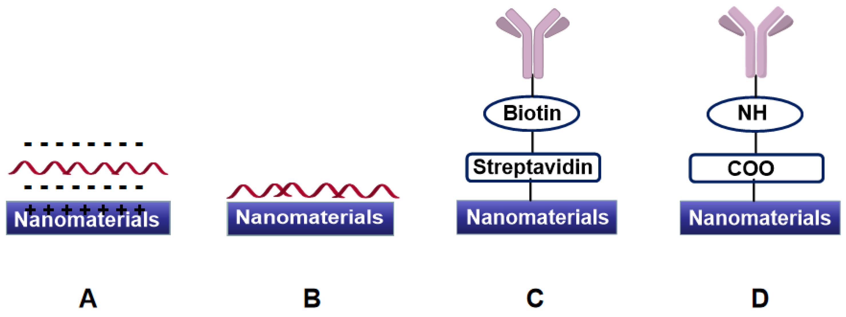
Figure 4.
Several schemes of colorimetric sensors based on Au NPs’ aggregation.
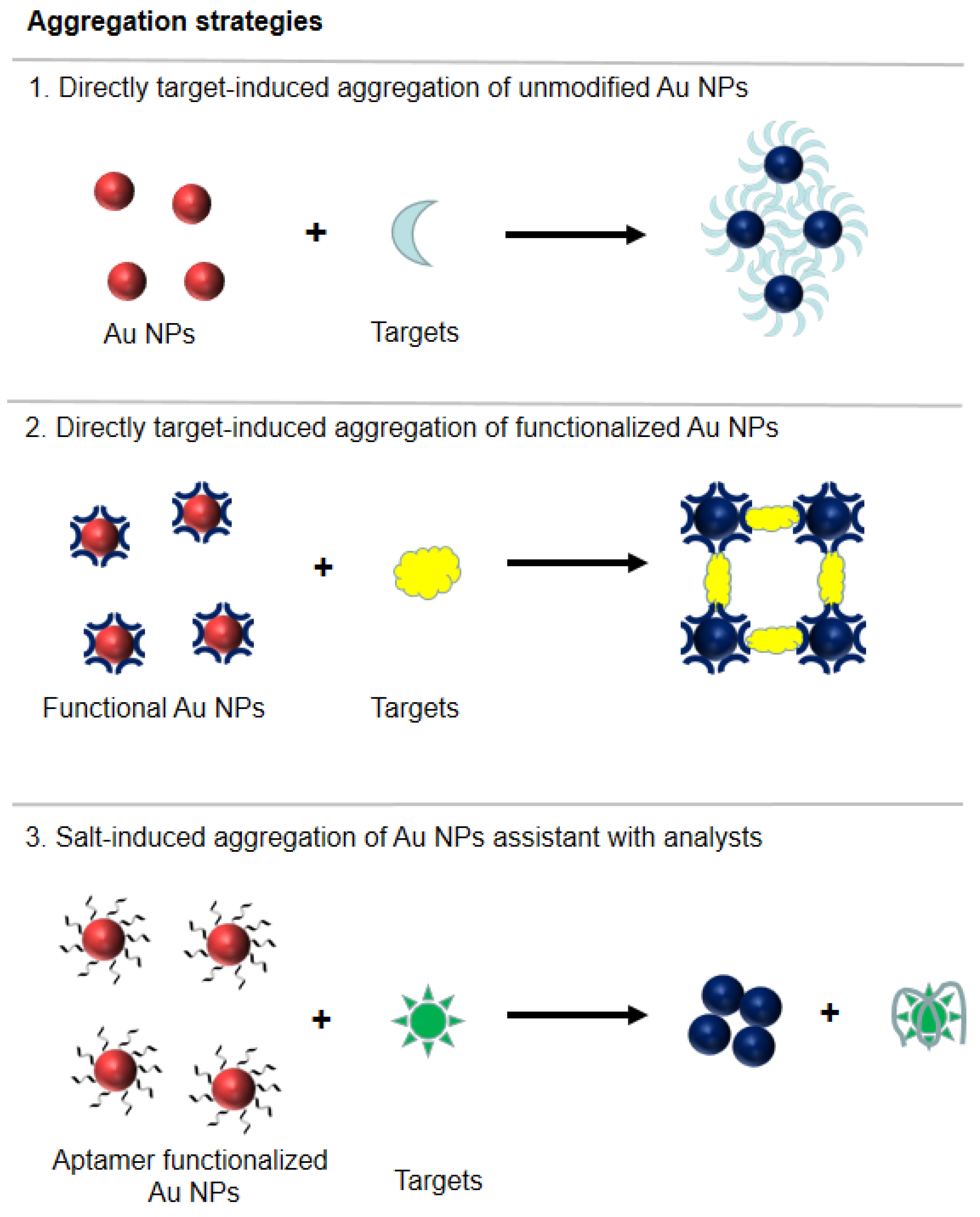
Figure 5.
Several schemes of colorimetric sensors based on Au NPs’ redispersion.
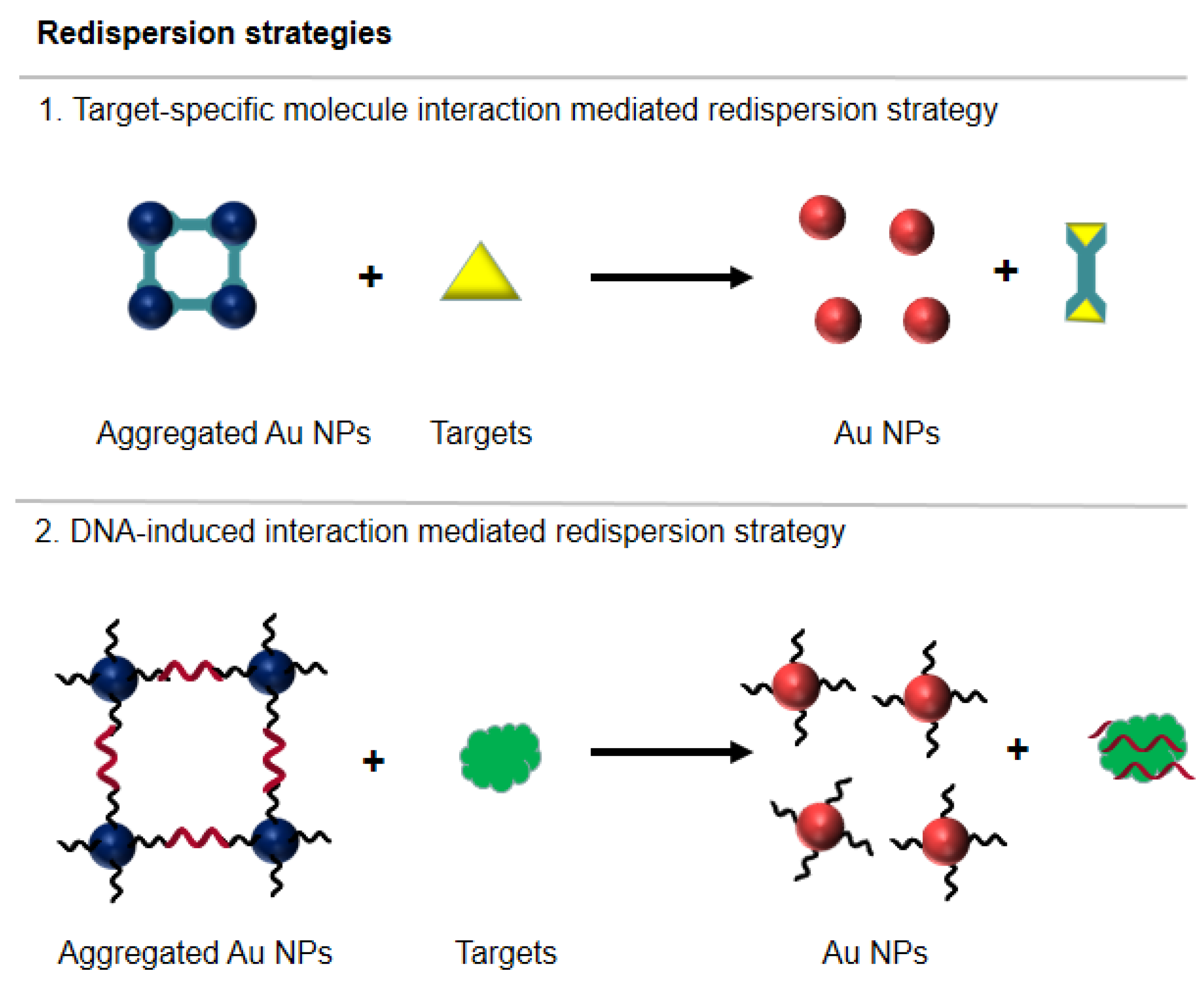
Figure 6.
Schematic diagram of the principle of food freshness detection based on AuNPs’ colorimetry [61].
Figure 6.
Schematic diagram of the principle of food freshness detection based on AuNPs’ colorimetry [61].

Figure 7.
(A) The scheme of the label-free fluorescence aptasensor for AFB1 detection based on TPE-Z/AFB1 aptamer/GO [104]; (B) the assembling mode of fluorophore with Ag+ [106]; (C) the mechanism of Cu NCs in histamine detection [107].

Figure 8.
Schematic illustrations of SPR biosensor-based platforms with advantages for food analysis.
Figure 8.
Schematic illustrations of SPR biosensor-based platforms with advantages for food analysis.

Figure 9.
(A) Electrochemical impedance sensor scheme for detecting DDT by EIS, including synthesis of PDA@Fe3O4 MIP MNPs (a) and sensor construction (b); (B) four microelectrode array modes (A) and platform assembly mode (B) of sensing modules in a label-free miniaturized platform protocol for the detection of L. monocytogenes by EIS [132].
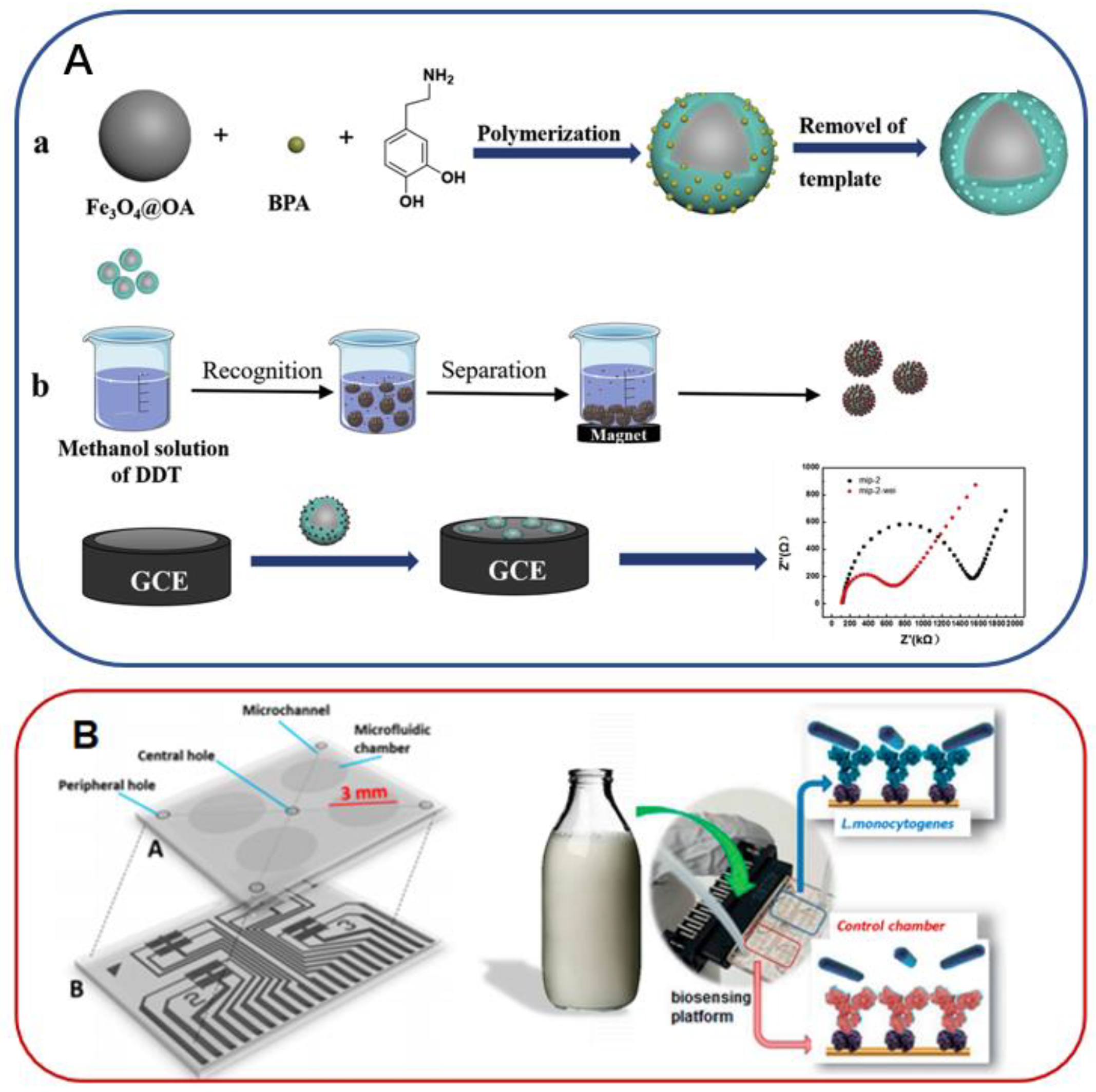
Figure 10.
The scheme of electrochemical aptasensors for monitoring OTA. The sensing interface consists of methylene blue-modified DNA aptamers (A), and sensor current changes are observed at higher (B) and lower (C) square wave frequencies [133].
Figure 10.
The scheme of electrochemical aptasensors for monitoring OTA. The sensing interface consists of methylene blue-modified DNA aptamers (A), and sensor current changes are observed at higher (B) and lower (C) square wave frequencies [133].

Figure 11.
Schematic diagram of exonuclease III-driven dual-amplification electrochemical aptamer sensor for chloramphenicol detection [135].
Figure 11.
Schematic diagram of exonuclease III-driven dual-amplification electrochemical aptamer sensor for chloramphenicol detection [135].
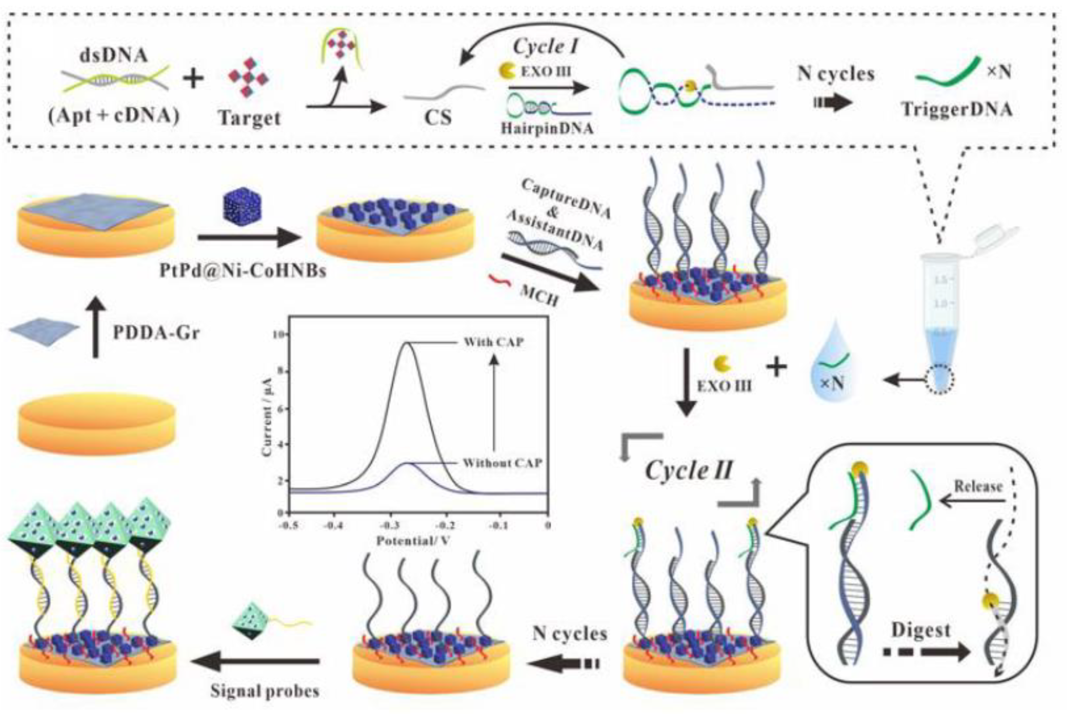
Figure 12.
The scheme of dual-signal aptasensor for detection of malathion [136].
Figure 12.
The scheme of dual-signal aptasensor for detection of malathion [136].

Figure 13.
Various nanomaterials and the enzymes nanomaterials mimicked.

Figure 14.
Schematic diagram of 2D MOF nanoenzymes for the detection of Staphylococcus aureus [147].
Figure 14.
Schematic diagram of 2D MOF nanoenzymes for the detection of Staphylococcus aureus [147].

Figure 15.
(A) Schematic diagram of a microfluidic colorimetric immunoassay device based on Au NPs [181]; (B) schematic diagram of bidirectional lateral flow immunoassay equipment based on NSs amplification [182].
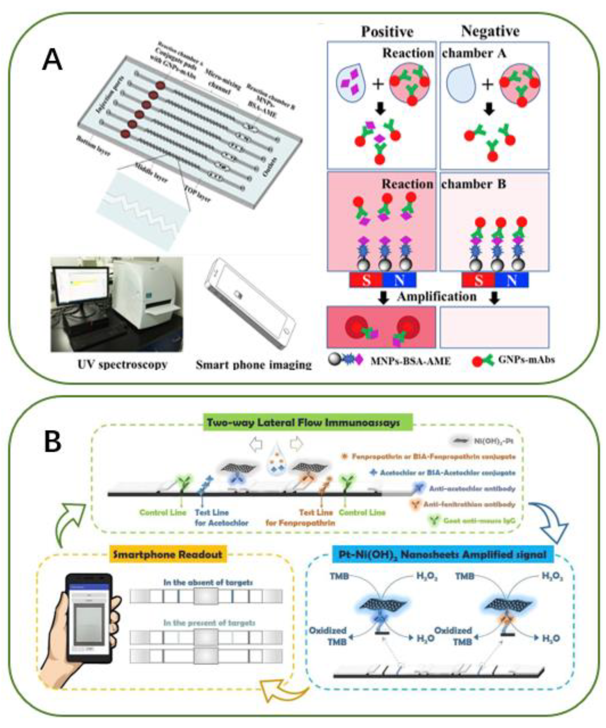
Figure 16.
The novel smartphone-based colorimetric biosensor for detection of S. Enteritidis [190].
Figure 16.
The novel smartphone-based colorimetric biosensor for detection of S. Enteritidis [190].

Figure 17.
(A) Schematic diagram (a) and profile (b) of a smartphone-based fluorescence biosensors for E. coli O157:H7 detection [191]; (B) smart biosensor for simultaneous detection of multiplex foodborne bacterial ssDNA [196].
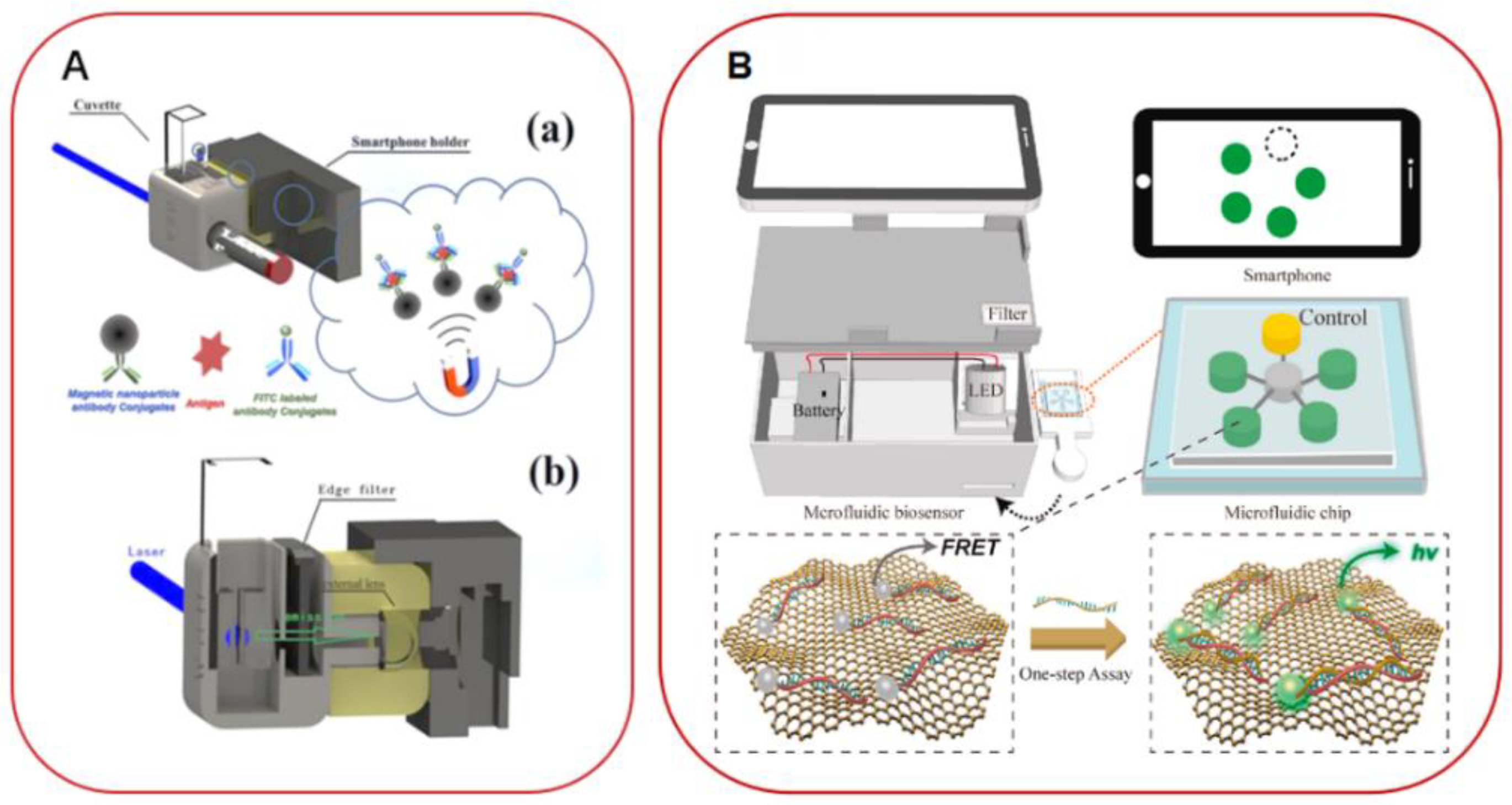

Table 1.
Several sensors for food analysis based on CDs produced from agriculture products.
| Carbon Precursor | Methods | Target | Limit of Detection | |
|---|---|---|---|---|
| Wang et al. [74] | Papaya flesh | Thermal/200 °C/5 h | Escherichia coil | 9.5 × 10⁴ cfu mL−1 |
| Das et al. [75] | Gram shells | Pyrolysis/315 °C/3 h | Escherichia coil | 107 cfu mL−1 |
| Hu et al. [76] | Orange peel | Microwave/900 W/1 min | Escherichia coil | 487 cfu mL−1 |
| Liu et al. [77] | Tomato puree | Microwave/10 min | Vanillin | 24.9 mg kg−1 |
| Bao et al. [78] | Eleocharis dulcis | Hydrothermal/120 °C/5 h | Fe3+ | 560 nM |
| Bandi et al. [79] | Onion waste | Hydrothermal/120 °C/2 h | Fe3+ | 310 nM |
| Shen et al. [80] | Sweet potatoes | Hydrothermal/180 °C/18 h | Fe3+ | 320 nM |
| Zhao et al. [81] | Corn bract | Reflux/100 °C/24 h | Hg2+ | 9.0 nM |
| Bano et al. [82] | Tamarindus indica leaves | Hydrothermal/210 °C/5 h | Hg2+ | 6 nM |
| Tyagi et al. [83] | Lemon peels | Hydrothermal/200 °C/12 h | Cr6+ | 73 nM |
| Kumar et al. [84] | Tulsi leaves | Hydrothermal/180 °C/4 h | Pb2+ | 0.59 nM |
| Bhatt et al. [85] | Tulsi leaves | Hydrothermal/200 °C/4 h | Cr6+ | 4.5 ppb |
| Wen et al. [86] | Pigskin | Hydrothermal/250 °C/2 h | Co2+ | 680 nM |
| Liu et al. [87] | Chocolate | Hydrothermal/200 °C/8 h | Pb2+ | 12.7 nM |
Table 2.
Examples for food analysis by (photo)electrochemical techniques.
| Analyte | Modified Electrode | Food Samples | Detection Techniques | LOD | |
|---|---|---|---|---|---|
| Rapini et al. [150] | Acetamiprid | PANI/AuNPs/GPSEs | Fruit juice | DPV | 86 nM |
| Jiao et al. [151] | Chlorpyrifos | Fc@MWCNTs/OMC/GCE | Fresh pakchoi, lettuce and leek | CV | 0.33 ng mL−1 |
| Liu et al. [152] | Acetamiprid | CdTe-MWCNTs/ rGONRs/ITO | Apples and tomatoes | PEC | 0.2 pM |
| Qiao et al. [153] | BPA | Au/ZnO/ITO | Milk and drinking water | PEC | 0.5 nM |
| Chen et al. [154] | β-agonists: RAC, CLB, PHL, SAL and PRO | AP-Ago/AuE | Pork sample | DPV | 40 (RAC), 0.35 (CLB), 1.0 (PHL), 0.53 (SAL) and 1.73 (PRO) pg mL−1 |
| Li et al. [155] | Acetamiprid | Co-doped ZnO/ITO | Cucumber | PEC | 0.18 nM |
| Prabhakar et al. [156] | Malathion | CHIT-IO/FTO | Lettuce leaves | DPV | 0.001 ng/mL |
| Song et al. [157] | KMY | HRP-AuNPCdna/Aptamer/AuE | Milk | DPV | 0.005 µg L−1 |
| Chen et al. [158] | KMY and OTC | Dynabead | Milk | SWV | 0.15 and 0.18 pM |
| Chen et al. [159] | CAP | cDNA/GCE | Fish samples | SWV | 0.33 pg mL−1 |
| Wang et al. [160] | KMY | AuE | Milk | DPV | 1.3 f M |
| Yan et al. [161] | CAP and OTC | AuNPs/GCE | Milk | SWV | 0.15 and 0.1 ng mL−1 |
| Ge et al. [162] | TTC | BiPO4/3DNGH/ITO | Milk | PEC | 0.033 nM |
| Danesh et al. [163] | SMY | Apt-CHIT/SPGE | Milk | DPV | 11.4 nM |
| Yin et al. [164] | SMY | NP-PtTi/Au@MWCNTsFe3O4/GCE | Milk | DPV | 7.8 pg mL−1 |
| Wang et al. [165] | AMP | ITO electrode | Milk | DPV | 4.0 pM |
| Zhao et al. [166] | Penicillin | PEDOT-AuNPs/GRFe2O3NPs/GCE | Milk | DPV | 0.057 ng mL−1 |
| Li et al. [167] | OTA | CDs-BP | Wheat and grape juice | EIS | 0.03 fg mL−1 |
| Hao et al. [168] | OTA and FB1 | BFE | Maize | SWV | 5 (OTA); 20 (FB1) pg mL−1 |
| Zheng et al. [169] | AFB1 | TS-AuNPs-cDNA/AuE | Animal feed and food samples | SWV | 0.6 × 10⁻⁴ ppt |
| Goud et al. [170] | AFB1 | SPCE | Beer and wine sample | EIS | 0.12 (seqA) and 0.25 (seqB) ng mL−1 |
| Istamboulié et al. [171] | AFM1 | SPCE | Milk | EIS | 1.15 ng L−1 |
| Mishra et al. [172] | OTA | Apt/SPCE | Cocoa beans | DPV | 0.07 ng mL−1 |
| Bagheryan et al. [173] | S. typhimurium | Diazonium-grafting layer modified SPCE | Apple juice sample | EIS | 6 cfu mL−1 |
| Jia et al. [174] | Salmonella | rGO-MWCNT/GCE | Chicken sample | EIS | 25 cfu mL−1 |
| Sheikhzadeh er al. [175] | S. typhimurium | Poly[pyrrole-co-3-carboxylpyrrole]copolymer/AuE | Apple juice sample | EIS | 3 cfu mL−1 |
Publisher’s Note: MDPI stays neutral with regard to jurisdictional claims in published maps and institutional affiliations. |
© 2022 by the authors. Licensee MDPI, Basel, Switzerland. This article is an open access article distributed under the terms and conditions of the Creative Commons Attribution (CC BY) license (https://creativecommons.org/licenses/by/4.0/).
Share and Cite
MDPI and ACS Style
Qiao, X.; He, J.; Yang, R.; Li, Y.; Chen, G.; Xiao, S.; Huang, B.; Yuan, Y.; Sheng, Q.; Yue, T. Recent Advances in Nanomaterial-Based Sensing for Food Safety Analysis. Processes 2022, 10, 2576. https://doi.org/10.3390/pr10122576
AMA Style
Qiao X, He J, Yang R, Li Y, Chen G, Xiao S, Huang B, Yuan Y, Sheng Q, Yue T. Recent Advances in Nanomaterial-Based Sensing for Food Safety Analysis. Processes. 2022; 10(12):2576. https://doi.org/10.3390/pr10122576
Chicago/Turabian StyleQiao, Xiujuan, Jingyi He, Ruixi Yang, Yanhui Li, Gengjia Chen, Sanxiong Xiao, Bo Huang, Yahong Yuan, Qinglin Sheng, and Tianli Yue. 2022. "Recent Advances in Nanomaterial-Based Sensing for Food Safety Analysis" Processes 10, no. 12: 2576. https://doi.org/10.3390/pr10122576
Note that from the first issue of 2016, this journal uses article numbers instead of page numbers. See further details here.