Self-Assembled Matrix by Umbilical Cord Stem Cells


Abstract
:1. Introduction
2. Materials and Methods
2.1. Establishment of Umbilical Cord Mesenchymal Stem Cells
2.2. Fibroblast Assembled Extracellular Matrix
2.3. Thickness
2.4. Transmission Electron Microscopy
2.5. Cell Numbers and Matrix Production
2.6. Real Time RT-PCR
2.7. Cuprolinic Blue Staining and GAG Side Chain Measurements
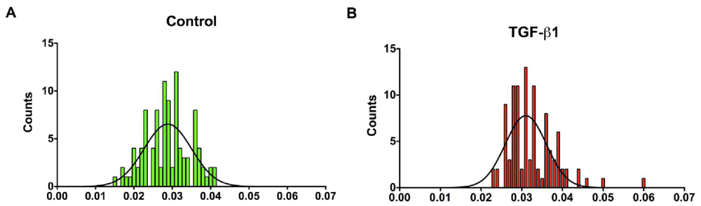
2.8. Collagen Fibril Diameter Measurements
2.9. Statistical Analysis
3. Results
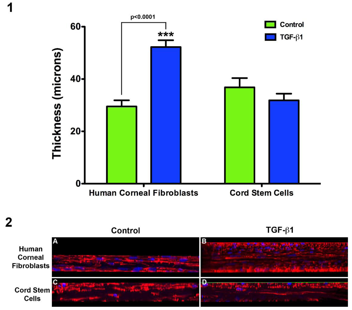
3.1. Construct Characterization

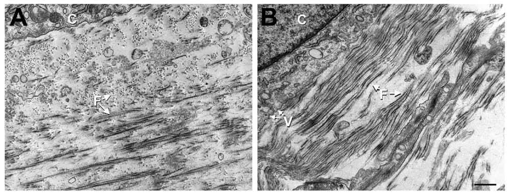
3.2. Ultrastructural Studies

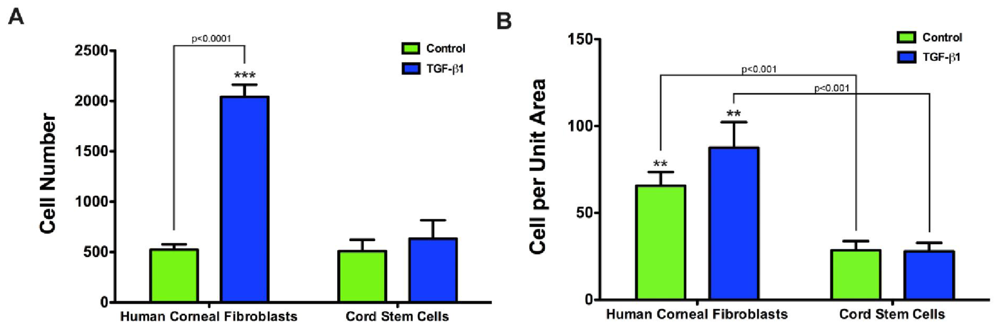
3.3. Cell Numbers and Extracellular Matrix Production
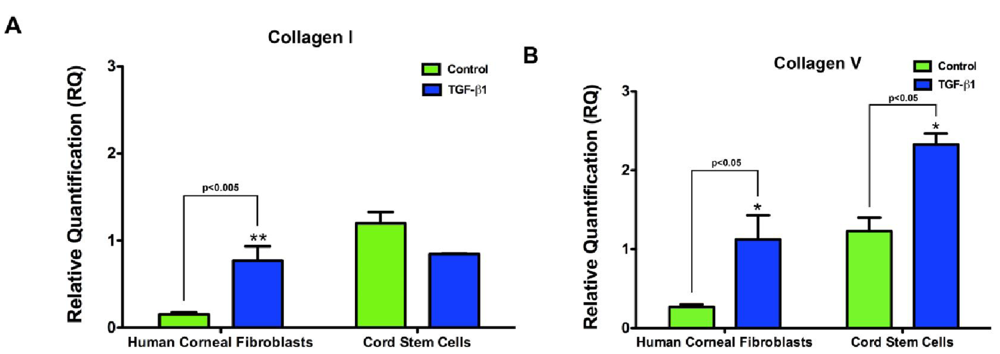
3.4. Collagen
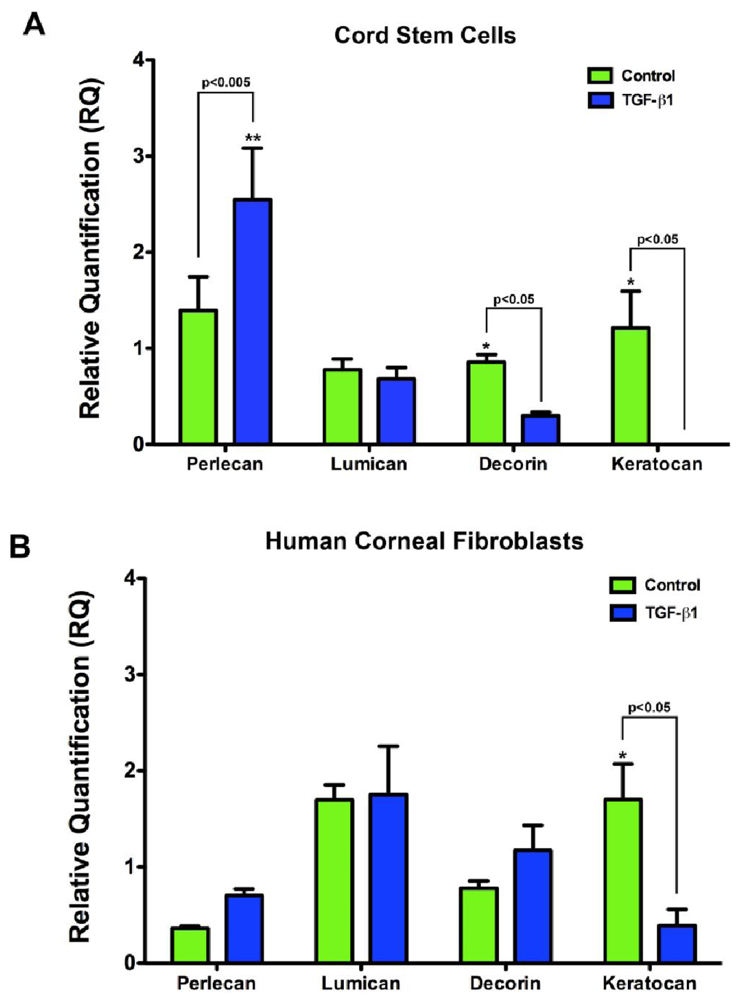
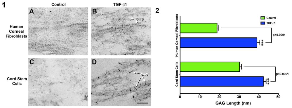
3.5. GAG Expression and Deposition


4. Discussion
5. Conclusions
References
- Daniels, J.T.; Dart, J.K.; Tuft, S.J.; Khaw, P.T. Corneal stem cells in review. Wound Repair Regen. 2001, 9, 483–494. [Google Scholar]
- Friedenstein, A.J.; Chailakhyan, R.K.; Gerasimov, U.V. Bone marrow osteogenic stem cells: In vitro cultivation and transplantation in diffusion chambers. Cell Tissue Kinet. 1987, 20, 263–272. [Google Scholar]
- Kuznetsov, S.A.; Krebsbach, P.H.; Satomura, K.; Kerr, J.; Riminucci, M.; Benayahu, D.; Robey, P.G. Single-colony derived strains of human marrow stromal fibroblasts form bone after transplantation in vivo. J. Bone Miner Res. 1997, 12, 1335–1347. [Google Scholar]
- Mueller, S.M.; Glowacki, J. Age-related decline in the osteogenic potential of human bone marrow cells cultured in three-dimensional collagen sponges. J. Cell Biochem. 2001, 82, 583–590. [Google Scholar]
- Harris, D.T.; Badowski, M.; Ahmad, N.; Gaballa, M.A. The potential of cord blood stem cells for use in regenerative medicine. Expert Opin. Biol. Ther. 2007, 7, 1311–1322. [Google Scholar]
- Koizumi, N.; Cooper, L.J.; Fullwood, N.J.; Nakamura, T.; Inoki, K.; Tsuzuki, M.; Kinoshita, S. An evaluation of cultivated corneal limbal epithelial cells, using cell-suspension culture. Invest. Ophthalmol. Vis. Sci. 2002, 43, 2114–2121. [Google Scholar]
- Shimazaki, J.; Yang, H.Y.; Tsubota, K. Amniotic membrane transplantation for ocular surface reconstruction in patients with chemical and thermal burns. Ophthalmology. Ophthalmology 1997, 104, 2068–2076. [Google Scholar]
- Tsai, R.J.F.; Li, L.M.; Chen, J.K. Reconstruction of damaged corneas by transplantation of autologous limbal epithelial cells. N. Engl. J. Med. 2000, 343, 86–93. [Google Scholar]
- Tseng, S.C.; Prabhasawat, P.; Barton, K.; Gray, T.; Meller, D. Amniotic membrane transplantation with or without limbal allografts for corneal surface reconstruction in patients with limbal stem cell deficiency. Arch. Ophthalmol. 1998, 116, 431–441. [Google Scholar]
- Mimura, T.; Amano, S.; Usui, T.; Araie, M.; Ono, K.; Akihiro, H.; Yokoo, S.; Yamagami, S. Transplantation of corneas reconstructed with cultured adult human corneal endothelial cells in nude rats. Exp. Eye Res. 2004, 79, 231–237. [Google Scholar]
- Mimura, T.; Yamagami, S.; Usui, T.; Honda, N.; Amano, S. Necessary prone position time for human corneal endothelial precursor transplantation in a rabbit endothelial deficiency model. Curr. Eye Res. 2007, 32, 617–623. [Google Scholar]
- Mimura, T.; Yamagami, S.; Yokoo, S.; Usui, T.; Tanaka, K.; Hattori, S.; Irie, S.; Miyata, K.; Araie, M.; Amano, S. Cultured human corneal endothelial cell transplantation with a collagen sheet in a rabbit model. Invest. Ophthal. Vis. Sci. 2004, 45, 2992–2997. [Google Scholar]
- Mimura, T.; Yokoo, S.; Araie, M.; Amano, S.; Yamagami, S. Treatment of rabbit bullous keratopathy with precursors derived from cultured human corneal endothelium. Invest. Ophthal. Vis. Sci. 2005, 46, 3637–3644. [Google Scholar]
- Amano, S.; Shimomura, N.; Kaji, Y.; Ishii, K.; Yamagami, S.; Araie, M. Antigenicity of porcine cornea as xenograft. Curr. Eye Res. 2003, 26, 313–318. [Google Scholar]
- Iozzo, R.V. Matrix proteoglycans: From molecular design to cellular function. Annu. Rev. Biochem. 1998, 67, 609–652. [Google Scholar]
- Knupp, C.; Pinali, C.; Lewis, P.N.; Parfitt, G.J.; Young, R.D.; Meek, K.M.; Quantock, A.J. The architecture of the cornea and structural basis of its transparency. Adv. Protein Chem. Struct. Biol. 2009, 78, 25–49. [Google Scholar]
- Cintron, C.; Covington, H.I.; Kublin, C.L. Morphologic analyses of proteoglycans in rabbit corneal scars. Invest. Ophthalmol. Vis. Sci. 1990, 31, 1789–1798. [Google Scholar]
- Funderburgh, J.L.; Chandler, J.W. Proteoglycans of rabbit corneas with nonperforating wounds. Invest. Ophthalmol. Vis. Sci. 1989, 30, 435–442. [Google Scholar]
- Brown, C.T.; Applebaum, E.; Banwatt, R.; Trinkaus-Randall, V. Synthesis of stromal glycosaminoglycans in response to injury. J. Cell Biochem. 1995, 59, 57–68. [Google Scholar]
- Markov, V.; Kusumi, K.; Tadesse, M.G.; William, D.A.; Hall, D.M.; Lounev, V.; Carlton, A.; Leonard, J.; Cohen, R.I.; Rappaport, E.F.; et al. Identification of cord blood-derived mesenchymal stem/stromal cell populations with distinct growth kinetics, differentiation potentials, and gene expression profiles. Stem Cells Dev. 2007, 16, 53–73. [Google Scholar]
- Karamichos, D.; Guo, X.Q.; Hutcheon, A.E.K.; Zieske, J.D. Human corneal fibrosis: An in vitro model. Invest. Ophthalmol. Vis. Sci. 2010, 51, 1382–1388. [Google Scholar]
- Karamichos, D.; Hutcheon, A.E.K.; Zieske, J.D. Transforming growth factor-beta3 regulates assembly of a non-fibrotic matrix in a 3d corneal model. J. Tissue Eng. Regen. Med. 2011, in press. [Google Scholar]
- Gipson, I.K.; Grill, S.M.; Spurr, S.J.; Brennan, S.J. Hemidesmosome formation in vitro. J. Cell Biol. 1983, 97, 849–857. [Google Scholar]
- Scott, J.E.; Haigh, M. Identification of specific binding sites for keratan sulphate proteoglycans and chondroitin-dermatan sulphate proteoglycans on collagen fibrils in cornea by the use of cupromeronic blue in critical-electrolyte-concentration techniques. Biochem. J. 1988, 253, 607–610. [Google Scholar]
- Mayo, C.; Ren, R.; Rich, C.; Stepp, M.A.; Trinkaus-Randall, V. Regulation by p2×7: Epithelial migration and stromal organization in the cornea. Invest. Ophthalmol. Vis. Sci. 2008, 49, 4384–4391. [Google Scholar]
- Ren, R.; Hong, Z.; Gong, H.; Laporte, K.; Skinner, M.; Seldin, D.C.; Costello, C.E.; Connors, L.H.; Trinkaus-Randall, V. Role of glycosaminoglycan sulfation in the formation of immunoglobulin light chain amyloid oligomers and fibrils. J. Biol. Chem. 2010, 285, 37672–37682. [Google Scholar]
- Ren, R.; Hutcheon, A.E.K.; Guo, X.Q.; Saeidi, N.; Melotti, S.A.; Ruberti, J.W.; Zieske, J.D.; Trinkaus-Randall, V. Human primary corneal fibroblasts synthesize and deposit proteoglycans in long-term 3-d cultures. Dev. Dyn. 2008, 237, 2705–2715. [Google Scholar]
- Rasband, W. Imagej, 1.5; National Institute of Mental Health: Bethesda, MD, USA, 2011. [Google Scholar]
- Guo, X.; Hutcheon, A.E.K.; Melotti, S.A.; Zieske, J.D.; Trinkaus-Randall, V.; Ruberti, J.W. Morphologic characterization of organized extracellular matrix deposition by ascorbic acid-stimulated human corneal fibroblasts. Invest. Ophthalmol. Vis. Sci. 2007, 48, 4050–4060. [Google Scholar]
- McCally, R.L.; Freund, D.E.; Zorn, A.; Bonney-Ray, J.; Grebe, R.; de la Cruz, Z.; Green, W.R. Light-scattering and ultrastructure of healed penetrating corneal wounds. Invest. Ophthalmol. Vis. Sci. 2007, 48, 157–165. [Google Scholar]
- Grigoropoulos, N.F.; Mathur, A. Stem cells in cardiac repair. Curr. Opin. Pharmacol. 2006, 6, 169–175. [Google Scholar]
- Kelly, D.J.; Prendergast, P.J. Mechano-regulation of stem cell differentiation and tissue regeneration in osteochondral defects. J. Biomech. 2005, 38, 1413–1422. [Google Scholar]
- Korbing, M.; Estrov, Z. Adult stem cells for tissue repair—A new therapeutic concept? N. Engl. J. Med. 2003, 349, 570–582. [Google Scholar]
- Krause, D.S.; Theise, N.D.; Collector, M.I.; Henegariu, O.; Hwang, S.; Gardner, R.; Neutzel, S.; Sharkis, S.J. Multi-organ, multi-lineage engraftment by a single bone marrow-derived stem cell. Cell 2001, 105, 369–377. [Google Scholar]
- Kawase, Y.; Yanagi, Y.; Takato, T.; Fujimoto, M.; Okochi, H. Characterization of multipotent adult stem cells from the skin: Transforming growth factor-beta (tgf-beta) facilitates cell growth. Exp. Cell Res. 2004, 295, 194–203. [Google Scholar]
- Toma, J.G.; Akhavan, M.; Fernandes, K.J.; Barnabé-Heider, F.; Sadikot, A.; Kaplan, D.R.; Miller, F.D. Isolation of multipotent adult stem cells from the dermis of mammalian skin. Nat. Cell Biol. 2001, 3, 778–784. [Google Scholar]
- Coles, B.L.; Angénieux, B.I.T.; Del Rio-Tsonis, K.; Spence, J.R.M.R.R.; Arsenijevic, Y.; van der Kooy, D. Facile isolation and the characterization of human retinal stem cells. Proc. Natl. Acad. Sci. USA 2004, 101, 15772–15777. [Google Scholar]
- Yamagami, S.; Mimura, T.; Yokoo, S.; Takato, T.; Amano, S. Isolation of human corneal endothelial cell precursors and construction of cell sheets by precursors. Cornea 2006, 10, S90–S92. [Google Scholar]
- Yokoo, S.; Yamagami, S.; Yanagi, Y.; Uchida, S.; Mimura, T.; Usui, T.; Amano, S. Human corneal endothelial cell precursors isolated by sphere-forming assay. Invest. Ophthal. Vis. Sci. 2005, 46, 1626–1631. [Google Scholar]
- Barry, F.P.; Murphy, J.M. Mesenchymal stem cells: Clinical applications and biological characterization. Int. J. Biochem. Cell Biol. 2004, 36, 568–584. [Google Scholar]
- Jorgensen, C.; Djouad, F.; Apparailly, F.; Noel, D. Engineering mesenchymal stem cells for immunotherapy. Gene Ther. 2003, 10, 928–931. [Google Scholar]
- Pittenger, M.F.; Mackay, A.M.; Beck, S.C.; Jaiswal, R.K.; Douglas, R.; Mosca, J.D.; Moorman, M.A.; Simonetti, D.W.; Craig, S.; Marshak, D.R. Multilineage potential of adult human mesenchymal stem cells. Science 1999, 284, 143–147. [Google Scholar]
- Daxer, A.; Misof, K.; Grabner, B.; Ettl, A.; Fratzl, P. Collagen fibrils in the human corneal stroma: Structure and aging. Invest. Ophthalmol. Vis. Sci. 1998, 39, 644–648. [Google Scholar]
- Hamada, R.; Giraud, J.P.; Graf, B.; Pouliquen, Y. Analytical and statistical study of the lamellae, keratocytes and collagen fibrils of the central region of the normal human cornea. (light and electron microscopy). Arch. Ophtalmol. Rev. Gen. Ophtalmol. 1972, 32, 563–570. [Google Scholar]
- Liu, H.; Zhang, J.; Liu, C.Y.; Wang, I.J.; Sieber, M.; Chang, J.; Jester, J.V.; Kao, W.W. Cell therapy of congenital corneal diseases with umbilical mesenchymal stem cells: Lumican null mice. PLoS One 2010, 5, e10707. [Google Scholar]
- Ruberti, J.W.; Zieske, J.D. Prelude to corneal tissue engineering—gaining control of collagen organization. Prog. Retin. Eye Res. 2008, 27, 549–577. [Google Scholar]
- Cao, F.J.; Feng, S.Q. Human umbilical cord mesenchymal stem cells and the treatment of spinal cord injury. Chin. Med. J. (Engl.) 2009, 122, 225–231. [Google Scholar]
- Lee, M.W.; Choi, J.; Yang, M.S.; Moon, Y.J.; Park, J.S.; Kim, H.C.; Kim, Y.J. Mesenchymal stem cells from cryopreserved human umbilical cord blood. Biochem. Biophys. Res. Commun. 2004, 320, 273–278. [Google Scholar]
- Baksh, D.; Yao, R.; Tuan, R.S. Comparison of proliferative and multilineage differentiation potential of human mesenchymal stem cells derived from umbilical cord and bone marrow. Stem Cells 2007, 25, 1384–1392. [Google Scholar]
- Wang, L.; Tran, I.; Seshareddy, K.; Weiss, M.L.; Detamore, M.S. A comparison of human bone marrow-derived mesenchymal stem cells and human umbilical cord-derived mesenchymal stromal cells for cartilage tissue engineering. Tissue Eng. Part A 2009, 15, 2259–2266. [Google Scholar]
- Cintron, C.; Gregory, J.D.; Damle, S.P.; Kublin, C.L. Biochemical analyses of proteoglycans in rabbit corneal scars. Invest. Ophthalmol. Vis. Sci. 1990, 31, 1975–1981. [Google Scholar]
- Midura, R.J.; Hascall, V.C. Analysis of the proteoglycans synthesized by corneal explants from embryonic chicken. II. Structural characterization of the keratan sulfate and dermatan sulfate proteoglycans from corneal stroma. J. Biol. Chem. 1989, 264, 1423–1430. [Google Scholar]
- Young, R.D.; Tudor, D.; Hayes, A.J.; Kerr, B.; Hayashida, Y.; Nishida, K.; Meek, K.M.; Caterson, B.; Quantock, A.J. Atypical composition and ultrastructure of proteoglycans in the mouse corneal stroma. Invest. Ophthalmol. Vis. Sci. 2005, 46, 1973–1978. [Google Scholar]
- Cintron, C.; Hassinger, L.C.; Kublin, C.L.; Cannon, D.J. Biochemical and ultrastructural changes in collagen during corneal wound healing. J. Ultrastruct. Res. 1978, 65, 13–22. [Google Scholar]
- Du, Y.; Roh, D.S.; Funderburgh, M.L.; Mann, M.M.; Marra, K.G.; Rubin, J.P.; Li, X.; Funderburgh, J.L. Adipose-derived stem cells differentiate to keratocytes in vitro. Mol. Vis. 2010, 16, 2680–2689. [Google Scholar]
- Brown, C.T.; Nugent, M.A.; Lau, F.W.; Trinkaus-Randall, V. Characterization of proteoglycans synthesized by cultured corneal fibroblasts in response to transforming growth factor beta and fetal calf serum. J. Biol. Chem. 1999, 274, 7111–7119. [Google Scholar]
- Muller, L.J.; Pels, E.; Schurmans, L.R.; Vrensen, G.F. A new three-dimensional model of the organization of proteoglycans and collagen fibrils in the human corneal stroma. Exp. Eye Res. 2004, 78, 493–501. [Google Scholar]
© 2011 by the authors; licensee MDPI, Basel, Switzerland. This article is an open access article distributed under the terms and conditions of the Creative Commons Attribution license (http://creativecommons.org/licenses/by/3.0/).
Share and Cite
Karamichos, D.; Rich, C.B.; Hutcheon, A.E.K.; Ren, R.; Saitta, B.; Trinkaus-Randall, V.; Zieske, J.D. Self-Assembled Matrix by Umbilical Cord Stem Cells. J. Funct. Biomater. 2011, 2, 213-229. https://doi.org/10.3390/jfb2030213
Karamichos D, Rich CB, Hutcheon AEK, Ren R, Saitta B, Trinkaus-Randall V, Zieske JD. Self-Assembled Matrix by Umbilical Cord Stem Cells. Journal of Functional Biomaterials. 2011; 2(3):213-229. https://doi.org/10.3390/jfb2030213
Chicago/Turabian StyleKaramichos, Dimitrios, Celeste B. Rich, Audrey E.K. Hutcheon, Ruiyi Ren, Biagio Saitta, Vickery Trinkaus-Randall, and James D. Zieske. 2011. "Self-Assembled Matrix by Umbilical Cord Stem Cells" Journal of Functional Biomaterials 2, no. 3: 213-229. https://doi.org/10.3390/jfb2030213