1. Introduction
It is important for oral and maxillofacial surgeons to repair craniofacial defects on oral cancer patients or patients with congenital problems [
1], but the treatment course for bone repair is time-consuming and expensive. Thus, surgeons want to solve this difficult situation by using bone grafting (transplantation) and implantation [
2]. Artificial bone replacements are not like natural bones and cause obvious side effects. The biomaterials have the ability to promote the regeneration of bones and would be the potential materials for bone repair [
3].
Silicate bioceramics are considered to possess great prospects in clinical application for orthopedic tissue regeneration due to their excellent osteogenesis and angiogenesis properties [
4]. Moreover, tissue engineering using single-phased silicate-containing calcium phosphate bioceramic scaffolds is a promising and highly effective approach for periodontal repair. This material provides appropriate mechanical properties, proper degradation, and good manufacturability [
5].
In the last years, sol–gel processes for bioceramics have been incorporated for chemical composition tuning and size control at the nanometric scale [
6]. Various elements were used to add to the bioactivity of bioceramics, such as silver (Ag) with antibacterial features [
7], magnesium (Mg) that induces the formation of an apatite layer, strontium (Sr) that stimulates the cellular response of HUVECs [
8], zinc (Zn) for the inhibition microbial adhesions [
9], titanium (Ti) for the killing or growth inhibition of bacteria, etc. In particular, the nanostructured TiO
2-incorporated materials have gained much attention in the field of tissue reconstruction because of their osteoconductivity, excellent biocompatibility, and favorable mechanical strength [
10].
In this study, the proposed bioceramics composed of silica and titanium oxide with various concentrations of titanium oxide were fabricated using the sol–gel method for tuning the formula, examined for their feasibility for biomimetic mineral deposition through immersion in simulated body fluid (SBF), and evaluated for their bioactivity using the cell toxicity test for the human osteosarcoma cells (MG63) in vitro.
2. Materials and Methods
Fabrication of SiO
2 powders was done by mixing tetraethoxysilane (TEOS), ethanol, water, and ammonium. Bioactive glass (BG) was synthesized by mixing TEOS, ethanol, and citric acid with various Ca and P ions added using the sol–gel method. In order to enhance bioactivity, calcium nitrate tetrahydrate (Ca(NO
3)
2) and diammonium hydrogen phosphate ((NH₄)₂HPO₄) were added. The formula of SiO
2 and BG was the same as that described in our previous study [
11]. The titanium oxide added was in the form of tetrabutyl titanate (TBOT), and it was directly added into the solution to form TiO
2 composition from the hydrolysis of TBOT. The samples of BG_Ti100 were prepared with an Si/Ti molar ratio of 10:1 for raw materials. The titanium oxide amount in the samples of BG_Ti75, BG_Ti50, and BG_Ti25 were sequentially reduced to 75%, 50%, and 25% of BG_Ti100, respectively.
After the sol–gel process, all the samples were calcinated at 500 °C for 6 h to get the bioceramics composed of silica and titanium oxide powders. Field-emission scanning electron microscopy (FE-SEM, AURIGA, Zeiss, Germany) was used to observe the microstructure and morphology of samples. The chemical composition was measured by using an energy dispersive X-ray spectrometer (EDS) [
12]. X-ray diffraction (XRD) spectra were used to identify the crystal structure through an XRD diffractometer (Bruker AXS Gmbh, Karlsruhe, Germany) operating at 40 kV and 40 mA (Cu Kα = 1.54184 Å as the radiation source). The diffraction patterns were collected over a 2θ range from 10° to 50°, with an incremental step size of 0.02°.
In the in vitro bioactivity test, 250 mg samples were soaked in 50 mL of simulated body fluid (SBF) [
13] to evaluate the ability for biomimetic mineral deposition. The SBF was prepared by dissolving NaCl, KCl, CaCl
2, MgCl
2, HCl, NaHCO
3, K
2HPO
4, and Na
2SO
4 in deionized water with a molar concentration of 142, 5.0, 2.5, 1.5, 148, 4.2, 1.0, and 0.5 mM for Na
+, K
+, Ca
2+, Mg
2+, Cl
−, HCO
3−, HPO
42−, and SO
42+, respectively. The SBF was buffered to a pH of 7.40 with a 37 wt.% HCl solution [
14]. After soaking in SBF for 7 days, the samples were taken out and dried at 60 °C for morphology observation and chemical composition measurement. The SBF solution immersed with various samples for 0, 1, 4, and 7 days were taken to measure the pH value and ion release concentrations. Inductively coupled plasma mass spectrometry (ICP-MS, ELEMENT XR, Thermo Fisher Scientific Inc, Waltham, MA, USA) was used for the measurement of precise elemental ratios at low concentrations, so the solution was required to be diluted 100 times at the µg/g (ppm) level.
The proposed silica-based bioceramics have a high reactivity with water solutions, and ions released from materials rapidly exchanged with the surrounding environment to lead to the incremental increase of the pH value. It is a positive effect for hydroxyl-carbonate-apatite formation, but it makes the pH-dependent cytotoxicity become a significant issue to evaluate the bioactivity of materials [
15]. Therefore, the human osteosarcoma cells (MG63, ATCC
® CRL-1427TM) were used in this study to evaluate cell viability [
16]. Dulbecco’s modified Eagle medium (DMEM) was used for MG63 cells. The medium contained 10% fetal bovine serum (FBS, Gibco
®, Thermo Fisher Scientific Inc, Waltham, MA, USA), and cells were incubated in an incubator at 37 °C with 5 % CO
2. The culture medium was changed every other day. Cell subculture was prepared using trypsin-EDTA solution. The testing samples were immersed in medium at 37 °C for 1 day to extract the samples. The ratio of samples and medium was 0.2 g samples per 1 mL medium according to the ISO 10993-5 [
17]. The cells were incubated with 100 μL MG63 solution with 1 × 100,000 cells/ mL in a 96-well plate for 24 h at 37 °C. Subsequently, the medium was replaced as 100 μL of the samples were extracted into a 96-well cell culture plate. Then, DMEM was used as the blank control group. Dimethyl sulfoxide (DMSO) was used as the positive control reagent. The ratio of DMSO and medium was 0.2 mL of DMSO per 1 mL of medium. Phosphate buffered saline (PBS) was used as the negative control reagent. The ratio of PBS and medium was 0.2 mL of PBS per 1 mL of medium. After incubation for 24 h, the cells were observed using optical microscopy.
An MTT assay was used to evaluate the cell viability while in the indirect method and the protocol was modified by the previous study [
18]. The human osteosarcoma cells, MG63, were quantitatively assessed using a tetrazolium compound (MTT; Sigma-Aldrich, Inc., Saint Louis, MO, USA) for a 24-h culture period. This tetrazolium compound, 3-(4,5-cimethylthiazol-2-yl)-2,5-diphenyl tetrazolium bromide (MTT) produces a colored product that has an absorbance (i.e., optical density (OD) value). The amount of colored product can be proportional to the cell number. The OD value of the color produced in the solution was measured by an ELISA reader (Sunrise-basic, Tecan, Männedorf, Switzerland). Data were collected and averaged from six different wells per condition. The data were statistically analyzed, and the results are expressed as the mean ± standard deviation. In order to evaluate the cell viability for various samples, the statistical differences between the samples and DMEM (the blank control group) were evaluated using a one-way analysis of variance (ANOVA) technique. In evaluating the test results, a *
p value of <0.05 was considered to be statistically significant, a **
p value of <0.01 was considered to be very statistically significant, and a ***
p value of <0.001 was considered to be highly statistically significant [
19].
3. Results
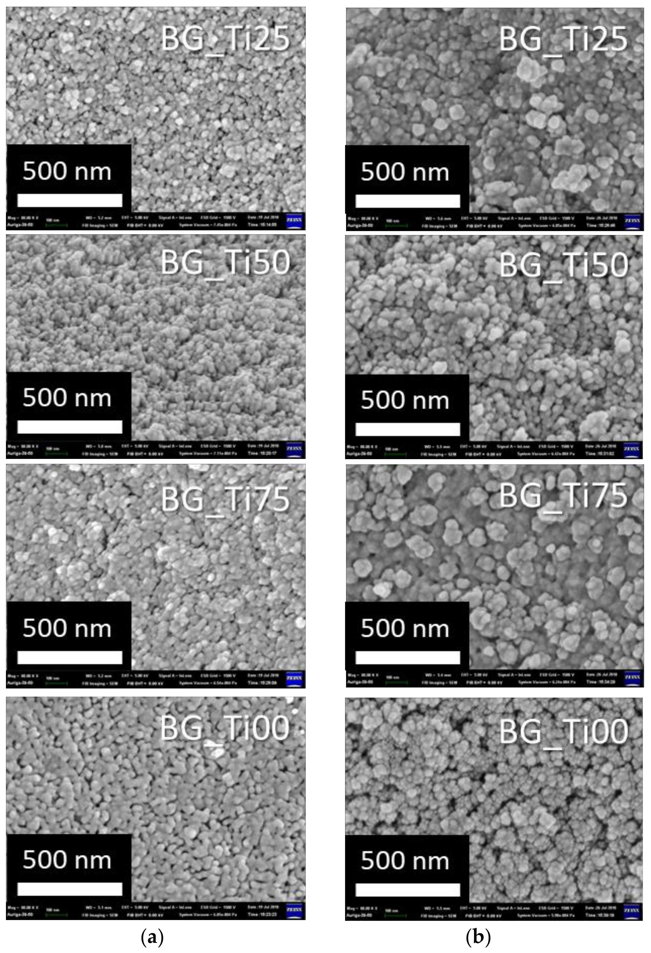
The microstructure of bioceramics composed of silica and titanium oxide with various concentrations of titanium oxide soaking in SBF for 0 and 7 days are shown in
Figure 1. At 0 days of soaking in SBF, the size of the bioceramic particles, though not very evident, become bigger from BG_Ti25 to BG_Ti00. At 7 days of soaking in SBF, it is obvious that mineral deposits on the sample surface and the particles also become bigger from BG_Ti25 to BG_Ti00. The particle also formed a cluster-like deposition on the BG_Ti00 surface. It showed the various microstructures on the sample surface after soaking in SBF for 7 days.
Table 1 shows that the atomic ratio of bioceramics composed of silica and titanium oxide with various concentrations of titanium oxide soaking in SBF for 7 days. The atomic ratio of various elements was described as follows. The Si atomic ratio of all the samples decreased after soaking for 7 days. Moreover, the Si atomic ratio decreased more drastically after 7 days if samples originally contained a higher Si atomic ratio. This showed the solubility of silica in the samples. For BG_Ti25, BG_Ti50, and BG_Ti75, the Ca atomic ratio increased after soaking for 7 days. However, Ca atomic ratio of BG_Ti100 decreased after soaking for 7 days. This showed that BG_Ti25, BG_Ti50, and BG_Ti75 can form calcium salt on their surface but BG_Ti100 can be the Ca ion provider. All of the samples’ P atomic ratios increased after soaking for 7 days. This showed that all samples can form the phosphate compound on their surface. In all samples, the Ti atomic ratio increased drastically after soaking for 7 days. The main reason was that the titanium oxide was not soluble in water. It is worth noting that the Si/Ti ratios were very high, and the Si/Ti ratio at 0 days decreased going from BG_Ti25 to BG_Ti00. However, the Si/Ti ratio decreased drastically after soaking in SBF for 7 days.
Figure 2 shows the phase identification results using XRD. An obvious peak appearing around 30° for BG_Ti100 at 0 days can be due to the calcium and phosphate compound formed after calcination at 500 °C. After soaking in SBF for 7 days, all samples appeared to peak in terms of apatite levels (HA). This was most obvious for the BG sample. The samples in decreasing order of apatite levels are as follows: BG_Ti75, BG_Ti50, BG_Ti25, BG_Ti100, and SiO
2. This demonstrated that all samples had the ability to deposit HA.
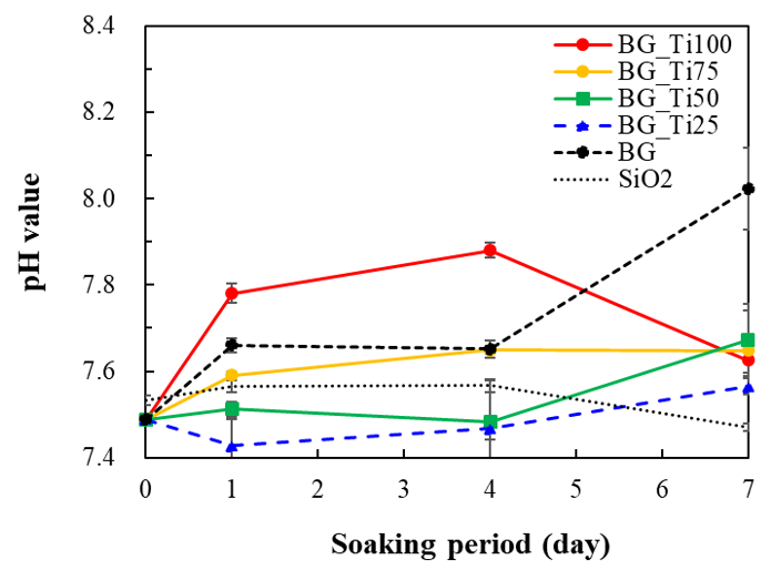
Figure 3 shows the pH value of SBF after the immersion of bioceramics composed of silica and titanium oxide with various concentrations of titanium oxide for 7 days. The pH value of BG dramatically changed after soaking in SBF for 7 days, and the pH value increased to 8.0. This can be due to the basic ions being released in great amounts. The pH value of SiO
2 was stable and decreased slightly after soaking in SBF for 7 days, according to the HA deposition on the samples. The pH value of BG_Ti100 obviously increased to 7.8 at the first stage due to the basic ions being released. For BG_Ti25, BG_Ti50, and BG_Ti75, pH values were very close to this, ranging from 7.4 to 7.6, after 7 days soaking in SBF.
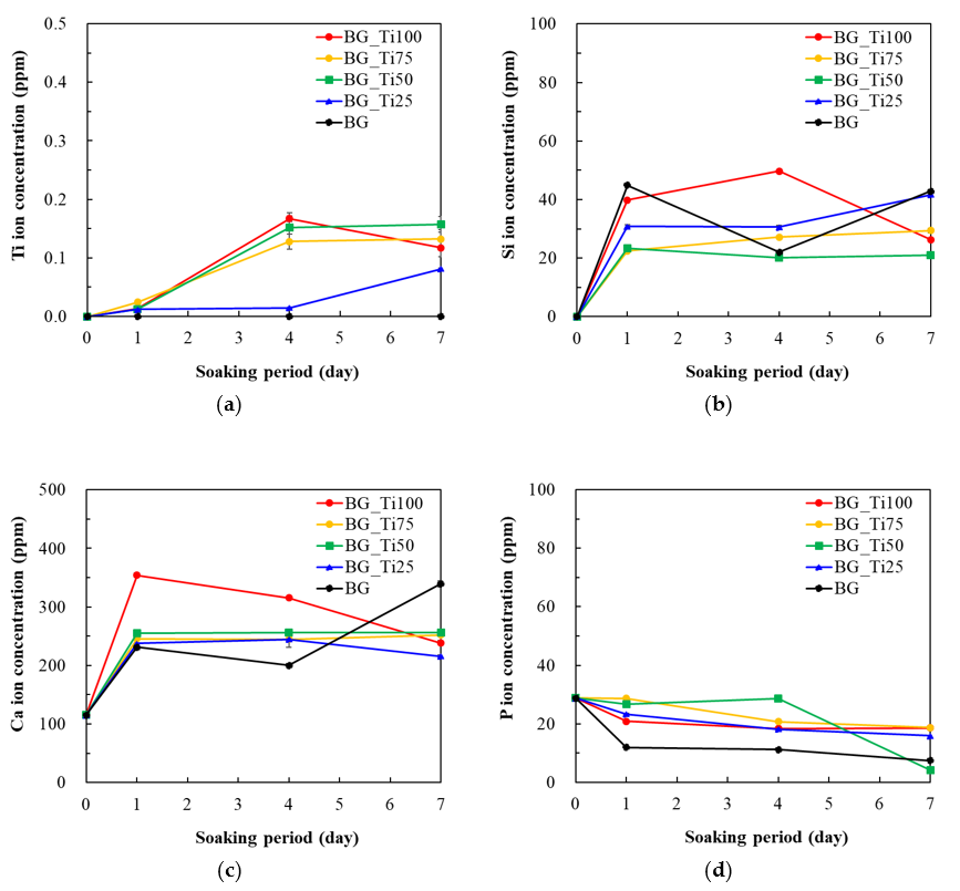
Figure 4 shows several types of ion released in SBF after the immersion of bioceramics composed of silica and titanium oxide with various concentrations of titanium oxide for 7 days, measured using ICP-MS. As shown in
Figure 4a, BG released no titanium ions. The BG_Ti25 sample released very little titanium ions (approximately 0.07 ppm) after 7 days of soaking; BG_Ti50, BG_Ti75, and BG_Ti100 released approximately similar concentrations of titanium ions after 7 days soaking. As shown in
Figure 4b, BG and BG_Ti25 released the highest concentration of silica ion after 7 days, and BG_Ti50, BG_Ti75, and BG_Ti100 released a similar amount of silica ions at 7 days, and it was worth noting that the concentration of silica of BG_Ti100 varied drastically from time to time. As shown in
Figure 4c, BG released the most calcium ion at 7 days. The other bioceramics composed of titanium oxide and silica released similar amounts of calcium ion at 7 days, except BG_Ti100, which released calcium concentrations that varied drastically. The others had a released calcium concentration that remained stable after 1 day. As shown in
Figure 4d, BG_Ti25, BG_Ti75, and BG_Ti100 released approximately similar concentrations of phosphate ion. In this regard, the sample that released the lowest concentration was BG_Ti50, followed by BG, at 7 days.
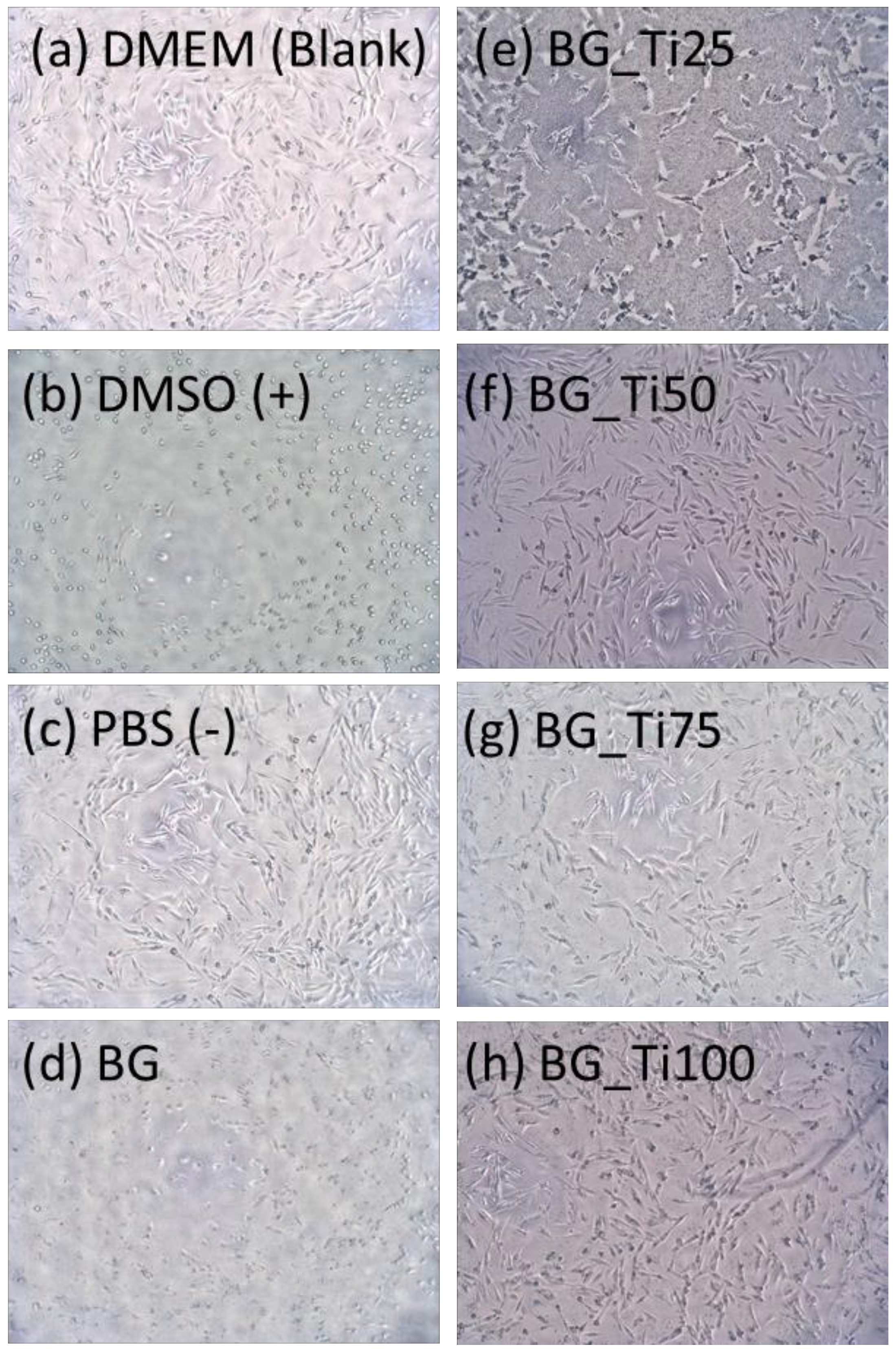
Figure 5 shows the morphology of MG63 cells incubating for 1 day in each type of extraction. Cells incubating in DMEM (
Figure 5a) and PBS (
Figure 5c) grew normally. In DMSO (
Figure 5b), for BG (
Figure 5d) and BG_Ti25 (
Figure 5e), almost all the cells died and changed their shape. Cells incubating in BG_Ti50 (
Figure 5f), BG_Ti75 (
Figure 5g), and BG_Ti100 (
Figure 5h) were alive and showed similar cell morphology.
Figure 6 shows the cell toxicity test results of MG63 cells incubating for 1 day in each type extraction. The OD value of PBS did not show an evident difference with DMEM, and this means that PBS was a successful negative control group. Whereas the OD value of BG_Ti75 was higher than DMEM in one deviation, but the OD value of the other extractions were far lower than DMEM.
4. Discussion
The atomic ratio of silica in all the samples decreased after 7 days of soaking in SBF, but the atomic ratio of titanium in all samples increased greatly after 7 days due to a large amount of silica dissolution [
20] and hardly any dissolution of titanium oxide. The proposed materials were silica-based bioceramics, and the O–Si–O bonds were breaking off due to OH
− groups, which were formed by the ionic exchange between Ca
2+ ions in the material and H
+ and H
3O
+ ions coming from the surrounding environment. This caused the silica network to break and the soluble silica to be released from the material and directly into the SBF solution [
21]. However, titanium oxide is not a water-soluble material, but its hydrophilicity can help the titanium oxide nanoparticle dispersion and suspension in the water solution [
22]. The proposed materials were bioceramics composed of silica and titanium oxide with various concentrations of titanium oxide. After soaking in the SBF solution, the soluble silica was released from the material and made the composites decompose to release titanium oxide nanoparticles into the SBF solution. This might mean that these bioceramics can be controlled by tuning the Si/Ti ratio to modify the dissolution rate of samples. However, the titanium precursor (TBOT) was rapidly formed titanium oxide due to the hydrolysis of TBOT [
23], so the reaction environment of the solution used in the sol–gel process was not stable. In this study, the solution used in the sol–gel process was controlled in an acid environment to fabricate nanoscale particles [
24], but titanium oxide formed by hydrolysis of TBOT was slightly dissolved in the solution. As evident in
Figure 1a, the added amount of titanium oxide caused the formation of coarse micro-structures due to the slightly basic solution. A large amount of titanium oxide due to the hydrolysis of TBOT not only changed the reaction environment but also formed a lot of TiO
2 precipitate in the sol–gel process. As in
Table 1, the much lower Si/Ti ratio of samples at 0 days showed that only a little titanium oxide could be formed inside silica networks.
After 7 days of soaking in SBF, the atomic ratio of calcium was an important factor for bioceramics as a bioactive bone graft material. The calcium atomic ratio of all samples was increased, besides BG_Ti100, based on
Table 1. This means that BG_Ti100 became the main calcium supplier to the environment but was not superior for calcium salt being deposited onto the surface. It also can be seen in
Figure 4c that the calcium concentration of BG_Ti100 measured by ICP-MS suddenly rose high after 1 day. A lot of calcium ions were released into the environment in vitro, and this sample displayed a weak ability to deposit HA, as shown in
Figure 2b. However, the samples of BG_Ti25, BG_Ti50, and BG_Ti75 have several advantages in the in vitro tests, such as the increase of both the atomic ratios of calcium and phosphate after 7 days soaking in SBF, increased deposition on the sample surfaces to make bigger particle sizes (shown in
Figure 1b), increased formation of HA crystal structures (shown in
Figure 2b), keeping a stable pH value of the soaking environment (shown in
Figure 3), and released calcium ions under a stable state (shown in
Figure 4c).
The cell morphology of MG63, which incubated in the extraction of BG_Ti25 for 1 day, showed obvious cell deformation (shown in
Figure 5e) and some precipitation on the bottom of culture plate. This might be the acid residue of the sol–gel process for fabricating nanoscale bioceramics, and the pH value of SBF after the immersion of BG_Ti25 for 1 day also displayed the phenomenon of a decreasing pH value (shown in
Figure 3). In contrast, BG_Ti50 and BG_Ti75 did not show evident differences in MG63 cell morphology after 1 day of incubating. Additionally, only BG_Ti75 showed an OD value higher than that of DMEM groups in the cell toxicity test of MG63 cells. The OD value of BG_Ti75 and DMEM groups were 0.790 ± 0.115 and 0.665 ± 0.053, respectively. These OD values can be compared to the cell concentrations to evaluate the bioactivity of samples. The MG63 cell number of the DMEM group was set as 100%, and the cell number of the BG_Ti75 group can be increased to 120%. This shows that BG_Ti75 is a bioceramic material with a biomimetic deposition effect and can respectably promote MG63 cell growth.

 and
and 


