Review

Iron, Aging, and Neurodegeneration

Department of Biology and Biochemistry, University of Bath, Bath BA2 7AY, UK
*
Author to whom correspondence should be addressed.
Metals 2015, 5(4), 2070-2092; https://doi.org/10.3390/met5042070
Submission received: 5 October 2015 / Revised: 30 October 2015 / Accepted: 2 November 2015 / Published: 6 November 2015
(This article belongs to the Special Issue Metallomics)

Abstract

:
Iron is a trace element of considerable interest to both chemistry and biology. In a biological context its chemistry is vital to the roles it performs. However, that same chemistry can contribute to a more deleterious role in a variety of diseases. The brain is a very sensitive organ due to the irreplaceable nature of neurons. In this regard regulation of brain iron chemistry is essential to maintaining neuronal viability. During the course of normal aging, the brain changes the way it deals with iron and this can contribute to its susceptibility to disease. Additionally, many of the known neurodegenerative diseases have been shown to be influenced by changes in brain iron. This review examines the role of iron in the brain and neurodegenerative diseases and the potential role of changes in brain iron caused by aging.

1. Introduction

While the atomic nature of matter might be an absolute given in the 21st century, the role of metal atoms in biological systems remains a developing field. In particular, the difficulty of translating between chemical and biological systems remains central to advancing concepts that could actually lead to a better understanding of how our minds work. At the fundamental level, we also still need to understand how the movement of single electrons can have a significant impact on cellular mechanisms that influence the way we age. In biological systems the movement of electrons is often dependent on metal ions and their role in enzyme activities or more fundamentally as co-factors in catalysis of various reactions. The regulation of such reactions can be both positive and negative as one results in maintaining cellular activity essential for life while the opposite can result in the generation of harmful reactive chemical species of oxygen or nitrogen. The production of reactive oxygen species (ROS) or nitrogen species (RNS) has been linked to changes in the brain associated with normal aging and also to diseases that can occur during aging such as the neurodegenerative diseases [1,2]. For many neurodegenerative diseases, especially the most common ones like Alzheimer’s disease and Parkinson’s disease, aging is a prerequisite for developing them [3,4]. While we can measure changes in various chemicals or reactions in the brain, the actual mechanism by which growing older makes us more susceptible to neurodegenerative diseases is a mystery. Yet, it has quite clearly emerged that changes in certain metals, especially iron, might be very important to both how the brain ages and to neurodegeneration.
Iron is the second most abundant metal on earth and has multiple oxidation states [5]. This makes it a leading candidate as a co-factor for enzyme-catalyzed reactions that require electron transfer. Iron can exist as Fe2+ (highly water soluble) or Fe3+ (much less soluble) in biological systems. Higher oxidation states are generated by some reactions but are much less stable than Fe2+ and Fe3+. The redox potential of Fe2+/Fe3+ is highly variable in biological systems and depends largely on the way it is coordinated within the structure of the ligands it binds [6]. This means that its utility is very high. Iron binding proteins can have different iron centers with specific classes of co-ordination [7]. The main classes are heme proteins, iron-sulfur proteins, and a class that is neither. This latter group includes iron storage and iron transport proteins, proteins coordinating a single Fe atom to histidine, glutamate or aspartate, or two Fe atoms in an oxygen-bridged center. The latter family includes many oxygen-binding proteins as well as Fe-oxidizing proteins such as ferritins.
Iron enters the body through the diet and is taken up via two main mechanisms [8]. The first (major) source of iron is through the metabolism of heme, which is absorbed at the intestinal apical membrane where the iron is released by heme oxygenase. Iron released in this way can directly enter the cellular free iron pool, where it can either enter storage in ferritin, or be transported outside the intestinal cell. The second uptake mechanism is at the brush border of the duodenum where it is first reduced to Fe2+ by the reductase Dcytb and then transported into the enterocytes by divalent metal transporter 1 (DMT-1) [9]. Iron taken up by this route enters the same pathway as heme iron. Once iron reaches the blood it is largely transported by transferrin. The main uptake mechanism involves the interaction of transferrin with its receptor at the cell surface. Internalization is through clathrin-coated pits, which directs transferrin to endosomes. There, the transferrin undergoes a conformational change as a result of the low pH of the endosomes causing iron release. The iron is reduced and transported to the cytosol. Iron uptake can also occur through DMT-1, which also transports other divalent metals [10].
Within cells, iron is associated with a variety of proteins as a co-factor, but is also stored. The main storage protein is ferritin, but other storage proteins exist such as hemosiderin [11,12]. Additionally, cells possess a pool of free iron known as the labile iron pool [13]. This pool is considered transient but necessary as the source of iron for newly synthesized proteins that require iron as a co-factor. The need to tightly regulate this pool of Fe2+ is considerable given the potential of free iron to catalyze radical generating reactions such as the Haber-Weiss reaction [14]. The production of radicals in the form of ROS and RSN has the potential to result in unwanted protein modifications and lipid peroxidation. These modifications of proteins can also lead to misfolding of the proteins. Aggregation of proteins in the brain can potentially give rise to neurodegenerative diseases. The necessity of iron for many biological activities and the potential of iron to cause damage to cells must obviously be balanced. These processes deteriorate as we age. The consequences for the brain are particularly severe in this regard. However, the exact role of iron in either aging or neurodegeneration is still under investigation and remains uncertain. The evidence for its role is explored in this review.

2. Iron in the Brain

The brain contains significant amounts of iron. However, the distribution is not uniform. Regions such as the substantia nigra and the globus pallidus have the highest levels, exceeding that of the liver, the main site of iron storage in the body [15,16,17]. According to cell type, oligodendrocytes have the highest iron content and astrocytes have very low cellular iron [18]. The main storage protein for iron is ferritin, which is composed of a mixture of H- or L-ferritin monomers (heavy or light chain), and varies between cell types as to which is expressed predominantly. Neurons express predominantly H-ferritin while microglia express L-ferritin [19,20]. Some neurons, such as those of the substantia nigra, express neuromelanin. Neuromelanin is synthesized from L-Dopa in dopaminergic cells and forms stable complexes with Fe(III). Thus cells expressing neuromelanin are likely to have increased iron storage [21,22].
Iron storage increases with age in the brain [23]. Again, this is not uniform and some regions show greater increases than others. Studies of ferritin levels have indicated high increases in the cortex, globus pallidus, and substantia nigra. In other regions such as the locus coeruleus the iron concentrations remain low throughout life [24]. The main cell types accumulating iron with age are microglia and astrocytes [23]. These changes are found in many brain regions including the cortex, cerebellum, hippocampus, and basal ganglia. In contrast, there is little change in iron in oligodendrocytes despite their higher concentrations of cellular iron. The origin of increased brain iron remains unclear. However, increased vascularization of the brain may increase the chance of iron exchange between the blood and tissues of the brain, thus increasing the concentration of iron [25,26]. Whatever the reason, an increase in a potentially oxidative metal in the brain increases the chances of deleterious reactions. It is probably no coincidence that the regions associated with changes in iron are also those associated with several neurodegenerative diseases.

3. Neurodegenerative Diseases Linked to Iron

This review is aimed at understanding the potential role of iron in aging and neurodegeneration. However, despite a potential involvement in more common diseases, there are instances of quite specific diseases directly linked to iron and its metabolism. Some of these diseases have successful treatments that involve the use of iron chelators.
Friedreich’s Ataxia is the most common of the ataxias and affects adolescents [27]. The disease is caused by mutations in the gene for frataxin, a protein found in the mitochondria that is associated with the assembly of Fe-S clusters and may act as an iron chaperone [28]. Mitochondria are also considered to be the main site of iron accumulation in Friedreich’s Ataxia [29]. Recent research has suggested that frataxin acts as an allosteric mediator of Fe-S cluster assembly [30]. It is found in tissue with high metabolism such as the heart and dorsal root ganglia parallel to the spinal cord [31]. The mutation in the gene involves trinucleotide repeat insertions (GAA) in the first intron. Severity of the disease appears to be linked to the number of insertions, which decrease the expression level of frataxin [28]. Patients have severe neurological problems that seem to be linked to problems with excess iron. Treatment of patients with the iron chelator deferiprone caused marked reduction in iron levels within the dentate nuclei and alleviated symptoms including manipulative dexterity, speech fluency, reduction in neuropathy, and ataxia gait [32].
Neuroferritinopathy is a rare, dominantly inherited disease associated with mutations in the gene for ferritin [33]. The disease has early and late onset forms and is associated with motor symptoms, spasticity and cognitive deficits [34]. The cause is usually a frameshift mutation in exon 4 for the L-ferritin gene, which causes a conformational change in the C-terminus of the molecule and alters its ability to store iron [35,36]. The mutation results in iron/ferritin-rich aggregates forming in cells [37]. Mouse transgenic models show a strong relation between functional changes and abnormal iron metabolism [38]. However, attempts to treat the disease with chelation therapy have so far been unsuccessful [39].
Many of the iron-storage diseases are inherited and pantothenate kinase-associated neurodegeneration (PKAN, formerly known as Hallervorden-Spatz syndrome) is another such disease [40]. It is characterized by Parkinson’s-like symptoms as well as significant mental abnormalities. It develops in childhood and is usually fatal. It is associated with mutations in the gene encoding pantothenate kinase 2 (PANK2) [41]. For this reason the name was changed from Hallervorden-Spatz syndrome. PKAN, now the more common term for the disease, is considered one of a family of diseases called NBIA (neurodegeneration with brain iron accumulation) [42,43]. PKAN brains show specific areas with high levels of iron accumulation, including the globus pallidus and the substantia nigra [44]. The exact relation of iron accumulation to disease progress is unclear. It has been suggested that the disease results in coenzyme A deficiency and subsequently increased cysteine, which is then able to chelate the iron [45]. However, treatment of patients with iron chelation therapy caused reduced iron accumulation but no change in symptoms. Therefore, the role of iron in the disease is unclear and may just be a symptom rather than a cause [46].
NBIA diseases also include neuroferritinopathy, aceruloplasminemia, beta-propeller protein-associated neurodegeneration, and a number of inherited diseases [43,47,48]. The incidence of NBIA disease continues to increase and clearly shows the importance of understanding iron metabolism and its potential link to neuronal loss. Many NBIA diseases are childhood diseases and are clearly not linked to aging. However, they are illustrative of the potential impact that accumulation of iron in the brain due to natural aging processes has on the vulnerability of neurons to iron-associated cell death.

4. Iron in Alzheimer’s Disease

Alzheimer’s disease (AD) is both the most common dementia and also the most common neurodegenerative disease. The greatest risk factor for the disease remains aging. The risk of developing the disease accelerates as we grow older, with those over 85 having almost a 1 in 2 change of developing it. AD is progressive and irreversible and results in memory loss, cognitive decline, a variety of psychological changes including anxiety, depression, and aggression, and eventual loss of physiological functions due to dementia [49]. Several brain areas are affected including the hippocampus, temporal lobe, and frontal cortex. Particularly, the cholinergic innervation is severely disrupted [50]. The exact cause of the disease remains contentious. There are inherited and sporadic forms of the disease. The inherited forms, although much rarer, point towards the proteins most likely to be at the heart of the disease [51]. The lead hypothesis of the cause of AD is the amyloid cascade hypothesis [52]. The formation of aggregated forms of β-amyloid (Aβ) is a common hallmark of the disease. Aβ is formed from a large precursor called APP (amyloid precursor protein). Inherited mutations linked to AD are found in the APP gene or in the genes of proteins associated with processing APP to form Aβ. The formation of Aβ aggregates in the brain is likely to play a significant role in the disease and may be associated with the neuronal loss observed [52]. The other significant protein in AD is tau, a microtubule-associated protein [53]. In AD tau becomes hyper-phosphorylated and forms paired-helical filaments (PHF), also known as tangles. There are suggestions that PHF are the true cause of AD. However, it could also be that some interplay between Aβ and tau may be important [54].
APP is a transmembrane protein and as the parent protein for Aβ there is significant debate over its role in the cell and whether altering its metabolism could be the significant causal effect that initiates AD [55]. A significant consideration in this debate is that APP binds metals [56]. APP contains a classical type II copper-binding domain in its N-terminus. The residues His147, His151, and Tyr168 were identified as the copper coordination sphere of the binding region within the E1 region of the protein [57]. The His-X-His motif is similar to that seen in Cu/Zn superoxide dismutase. APP has the potential to reduce Cu2+ to Cu+. The E2 domain of APP has also been suggested to have two metal binding sites [58]. Of these, Zn2+ is thought to be coordinated between His382, His432, and His436. Copper can also bind this site with the addition of His313 to the three His of the Zn site. A second, low-affinity site is coordinated by Glu387, Asp429, and His458. There has also been a report that the E2 site interacts with iron. A suggested site for iron binding around Glu337 and Glu340 is still debated [59]. The idea that APP could function as a ferroxidase [60] has been dismissed in favor of the suggestion that APP can regulate iron export from the cell (Figure 1) [59,61].
As well as physical interactions between metals and APP, there has been considerable research on the cellular implications of those possible interactions. Of considerable interest are those that might alter or initiate the disease (Figure 2).
Interaction of APP with copper is thought to alter both its dimerization and the rate of cleavage to form Aβ [62,63]. However, the aggregation of Aβ is also thought to be influenced by its interaction with copper and zinc [64,65,66]. Cu, in particular, is considered to accelerate aggregation. This led the research group of Ashley Bush to investigate whether copper chelators such as clioquinol could inhibit Aβ aggregate accumulation and possibly be a treatment for AD itself. While clioquinol proved effective in depletion of Aβ in transgenic mice [67], it unfortunately did not progress in clinical trials [68].
Figure 1. Several of the key proteins associated with neurodegeneration have been suggested to have roles associated with iron. This figure summaries these suggestions. α-synuclein is associated with PD and has been shown to bind iron and reduce it, thus converting Fe3+ to Fe2+ which is utilized for many cellular activities. α-synuclein, like most ferrireductases, is associated with the inner face of the cell membrane. There have also been reports that the prion protein, associated with diseases like CJD, can also act to reduce iron as a ferrireductase. Lastly, the amyloid precursor protein (APP) the precursor to beta-amyloid known from AD, has been shown to enhance iron export out of cells. This process requires the presence of ferroportin, which mediates the iron export process.
Metals 05 02070 g001
While the relation of copper and zinc to AD and the APP protein has proven quite solid, evidence supporting a link between iron and AD has taken much longer to emerge [69]. Elevated brain iron has been reported in patients with AD [70,71]. However, as is clear from this review, this is not unique to AD. Iron accumulation in AD occurs without a parallel increase in ferritin [72]. IRP proteins normally respond to increased levels of iron by interacting with genes containing the IRE element and causing regulation of a number of proteins in a coordinated fashion. This usually ensures that the transferrin receptor and ferritin are regulated together. In AD it appears there is a dysfunction of this system that leads to increased free iron that would otherwise be stored in proteins such as ferritin [73]. In a recent clinical study of cerebral spinal fluid (CSF) from AD patients, it was found that ferritin levels were predictive of cognitive status [74]. While not being distinctly elevated in all cases of AD, declining cognitive status from mild cognitive impairment (MCI) to AD was associated with altered ferritin levels and was also greater with younger onset of AD. This would suggest a relationship between high iron burden in the brain and an early age for AD onset. Additionally, it was found that ferritin levels were higher in individuals with the APOE protein allele ε4 [74]. This allele is an epigenetic marker for greater risk of AD. However, the link between APOE and iron metabolism cannot currently be explained.
Figure 2. While numerous proteins associated with neurodegeneration may have a role in iron metabolism in the healthy cell, under certain conditions these processes might be disturbed. This diagram represents a summary of the potential changes that can occur during aging which might disturb the way iron is either utilized or its interactions with other proteins. Changes in the protein might result in changes in levels of iron, particularly Fe2+. The protein may become overexpressed, resulting in the formation of excess Fe2+, or some kind of damage to the protein might result in lost activity and reduced Fe2+. Additionally, aging effects might result in the misfolding of the protein, either as a result of an increase or a loss of interaction with iron or simply through another process. Misfolded protein could then aggregate and cause toxic damage to cells. In the case of excess generation of Fe2+, there could be abnormal interaction with other proteins such as neuromelanin or the Fe2+ could result in oxidative stress. In both cases the end result could be the generation of toxic species that lead to cell death. Lastly, in some cases the lack of Fe2+ might result in a breakdown of essential cellular process necessary for cell survival.
Metals 05 02070 g002
As mentioned above, Aβ binds metals. While there has been a focus on copper, there is also evidence for an interaction with iron [72]. While this could mediate toxicity or oxidation events, there has also been a suggestion that the interaction could be protective. In this case the Aβ would sequester the iron and prevent it mediating toxic damage. While this may seem paradoxical given the “bad” reputation of Aβ, it is actually true that free iron is highly toxic and Aβ can accumulate without any apparent cell loss [75,76]. This observation, although suggesting a positive role for Aβ in AD, does not then explain how iron might be toxic.
Mitochondria are the organelles most likely to show damage in AD [77]. As the site of heme synthesis in the cell, mitochondria are a major site for iron handling. Mitochondria are the major source of oxygen radical generation in cells. Damage to mitochondrial-specific proteins, which can increase free iron, has been shown [78]. Studies of mitochondrial DNA suggest that there is a greater turnover of the organelle in AD [79]. This supports the fact that there is mitochondrial abnormality in AD [80]. There is no direct evidence that this dysfunction is causal rather than a result of the damage that leads to cell death in AD or that there is a direct link between mitochondria and iron. Further research may bring into focus the causal links between these different factors. However, it is clear that iron disturbance is one important part of trying to decipher the mystery behind AD.

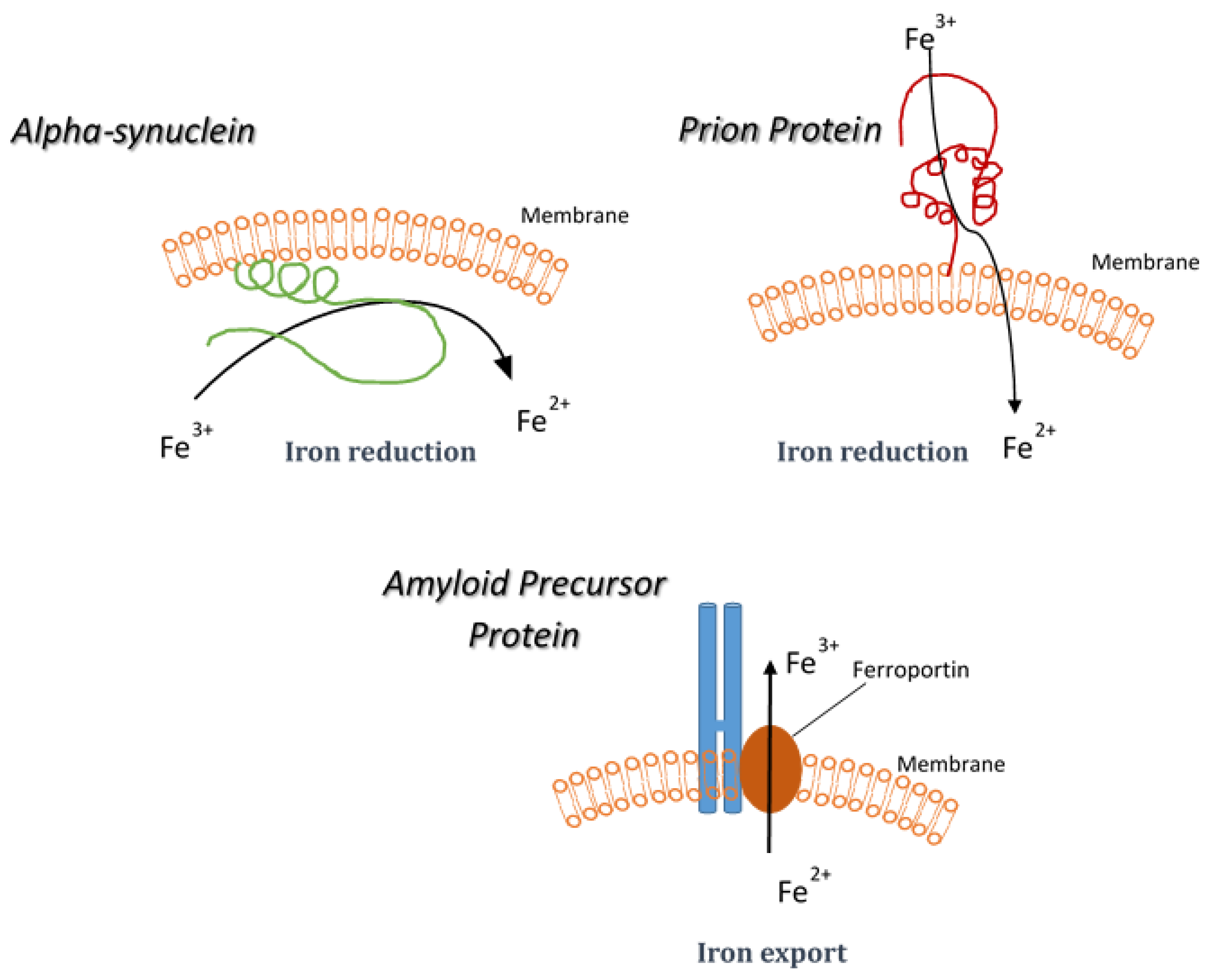
5. Synucleinopathies and Iron Reduction

Synucleinopathies are a family of diseases associated with the deposition of the protein α-synuclein in an aggregated form in CNS tissue [81]. These diseases include Parkinson’s disease (PD), Multiple System Atrophy, and Dementia with Lewy Bodies (DLB) [82,83]. Parkinson’s is very well known, being the most common of the non-dementing neurodegenerative diseases. It is associated with loss of dopaminergic neurons from the substantia nigra, which results in a movement disorder [84]. PD has a long association with disturbances to iron metabolism [85]. However, any mechanistic relation between the disease and iron metabolism has yet to be firmly established. PD, like AD has sporadic and inherited forms. The inherited forms are related to mutations in the genes of a long list of proteins which include α-synuclein, parkin, leucine-rich repeat kinase (LRRK), PINK-1, and DJ-1 [86]. The inherited forms can be either early or late onset (depending on the mutation). The clinical symptoms of PD include resting tremors, muscle rigidity, and bradykinesia [87]. Treatments are generally focused on restoring some amount of dopamine to the patient as the loss of dopaminergic neurons is the principal change that results in the majority of symptoms. These treatments range from supplying the dopamine precursor (L-Dopa) to altering dopamine transport and breakdown [88].
Changes in PD patient brains include increased levels of Fe(III) and reduced levels of the Fe(III)-binding protein ferritin [89]. Increased iron deposits in the substantia nigra are associated with α-synuclein-positive Lewy bodies in PD and neurodegeneration with brain iron accumulation [90,91]. However, there are also changes in other metals in PD such as zinc in the substantia nigra and high levels of copper in the cerebrospinal fluid [92]. There is also evidence from epidemiological studies that increased incidence of PD is associated with environmental metal exposure. Individuals with chronic industrial exposure to copper, manganese, or iron have an increased rate of PD [93]. In experimental models, alterations in metal homeostasis were observed with toxin-induced animal models of PD. An accumulation of iron was observed in the substantia nigra of MPTP-treated mice, which is likely a result of the observed upregulation of transferrin receptor expression and iron uptake [94,95]. Although these observations may be consequences of the disease progression, experimental studies using FeCl3 injected directly into the substantia nigra of rats resulted in a 95% reduction in striatal dopamine and altered behavior, supporting the idea that iron initiates dopaminergic degeneration in PD [96].
While observation of changes in metal levels in a disease might be interesting, without a mechanistic link the findings remain at best a marker of change. The increased cellular iron is likely to come about through changes either as a result of intake or release. However, with regards to the death of dopaminergic neurons, there is also the possibility that it is not the absolute amounts that are important but the ratio of Fe(II) to Fe(III). Similarly, alteration in the interactions of iron with various proteins may also be critical. For example, the substantia nigra is characterized by high levels of neuromelanin, the dark pigmented protein that gives the substantia nigra its name. Studies of PD patients have shown high levels of iron associated with neuromelanin granules [97]. This could suggest that neuromelanin traps redox-active Fe2+, which is then able to initiate the oxidative process [98,99]. However, another study suggests that the iron leaves the neuromelanin and migrates to the cytosol in PD [100].
Cellular responses to fluctuating iron levels are regulated by iron regulatory proteins (IRPs) that bind to iron response elements (IRE) in RNA [101]. However, they also respond to increased levels of oxidative stress and can then alter cellular protein expression to cause an increase in the free iron pool [102]. The implication is that oxidative stress can cause a dysregulation of iron metabolism. The read-out for such a change would be increased iron levels without a corresponding change in iron-binding proteins such as ferritin. This has been observed in PD [103]. As well as potential interactions of iron with other proteins, iron could also alter dopamine specifically, causing the generation of toxic dopamine byproducts that then kill dopaminergic cells specifically. One of the metabolic products of dopamine 3,4-dihydroxyphenylacetaldehyde (DOPAL) can induce aggregation of α-synuclein in the presence of iron [104]. DOPAL can also generate reactive oxygen species in the presence of iron [105].
Synucleins have been linked to metals. Firstly, all three synucleins bind copper [106,107]. α-synuclein also binds other metals including iron [108,109]. Exposure of α-synuclein to metals during extensive shaking can also accelerate its aggregation [110]. This creates a dichotomy with regards to α-synuclein in terms of both its potential normal cellular activity and its aggregation, which is associated with pathological states such as in PD. α-synuclein toxicity is mostly associated with copper [111,112]. During the aggregation process, copper induces the formation of a unique stellate oligomer that is highly toxic to neuronal cells in culture [111]. Exposure to iron does not have this effect, although some studies have shown that iron can accelerate aggregation of α-synuclein [113]. The aggregation of α-synuclein in cells is associated with the increased expression levels of the protein. However, increased expression of α-synuclein has also been shown to increase cellular iron concentrations [114].
Recent studies have shown that α-synuclein can bind both copper and iron simultaneously [109]. When α-synuclein binds copper it is able to undergo redox cycling, as shown by cyclic voltammetry [106]. The implication is that α-synuclein can use copper to move electrons. Further studies have shown that α-synuclein can cause the reduction of iron (Figure 1) [109]. Kinetic analysis with purified recombinant protein has shown that this activity is enzymatic, potentially making α-synuclein a ferrireductase. The implication is that the potential of α-synuclein to generate Fe2+ in cells may be its normal cellular role. What this means for the pathology of the synucleinopathies currently remains unclear. However, there are two distinct possibilities. First, increased expression of α-synuclein could result in excess production of Fe2+, which could then initiate catastrophic oxidative processes in the cell, interact with other proteins such as neuromelanin, or simply be neurotoxic (Figure 2). The alternative is that when α-synuclein is highly expressed in cells and begins to aggregate the protein becomes functionally inactive but may still bind Fe3+, thus sequestering iron that is needed for normal cellular activities such as the synthesis of dopamine. Give the potential role of Fe2+ in numerous aberrant processes, the former possibility is more likely. However, as with the general role of iron in PD, the role of iron reduction in PD is still unclear and further research is needed to understand what causative role there is, if any.

6. Prion Diseases

Also known as transmissible spongriform encephalopathies, prion diseases are rare [115]. They are more widely known because of the concurrent outbreaks of both bovine spongiform encephalopathy (BSE) in cattle and variant Creutzfeldt-Jakob disease (vCJD) in humans [116,117]. The potential transmissibility of prion diseases remains an ever-present concern. However, the major form of human prion disease (sporadic form of Creutzfeldt-Jakob disease) is still very rare and is not naturally transmissible [118]. Prion diseases can either be inherited (through mutations in the Prnp gene), sporadic (with no known cause) or transmissible. The transmissibility has largely been demonstrated experimentally, but may be a result of misadventure such as with vCJD or Kuru (a disease spread by ritual cannibalism). The disease transmissibility is associated with an abnormal isoform of the prion protein [119]. The prion protein (PrPc) is a cellular copper-binding glycoprotein expressed at the cell surface [120,121]. The function of PrPc remains controversial, but the strongest evidence supports its role as an antioxidant protein associated with increased cell viability [122,123,124]. The protease-resistant isoform of PrPc often accumulates at high levels in the central nervous tissue of patients and animals with prion diseases. Therefore, this abnormal isoform (PrPSc) is a hallmark of the disease. It is tightly associated with the infectious agent and may be a direct cause of the neurodegeneration seen in the disease [125].
Following the discovery of the copper binding capability of PrPc, extensive studies were carried out to assess the relation of PrPc and prion diseases to metal homeostasis. The strongest associations were found between prion diseases and copper metabolism but also manganese metabolism [126]. The latter was somewhat unexpected and remains controversial. While copper has been associated with normal PrPc activities, the data suggest that manganese binds to PrPc in disease and can cause its conformational change [127,128]. Manganese binding to PrP also increases its survival in the environment and increases its ability to cause prion infection in cells [129].
Studies looking at the levels of trace elements in the brains of animals with BSE, sheep scrapie, patients with CJD or vCJD, and rodents experimentally infected with scrapie all show a similar trend [130,131,132]. They indicate reduced levels of copper and increased levels of manganese. Similar changes have also been observed in the blood. A recent study has shown increased manganese in prion plaques in experimental hamsters [133]. Other trace elements were also studied and very little difference was observed for any other metal. These studies included Fe, which showed no changes in any of the tissues or diseases analyzed.
Despite these studies showing no changes in iron, there are others that suggest a strong role for PrP in iron metabolism [134]. An initial study suggested increases in both ferritin and iron response proteins in astrocytes in rodent scrapie models [135]. The research group of Nina Singh has extensively studied the links between Fe and prion disease. Their findings initially suggested that PrP could influence iron uptake [136]. Cells overexpressing PrP showed increased levels of intracellular iron. Further studies suggested that CJD patients have increased levels of iron, while PrP knockout mice have decreased iron levels [137,138]. A number of studies have shown that the divalent metal transporter-1 (DMT-1) is altered in both PrP-knockout and prion disease [139]. This protein is linked to the transport of multiple divalent metals and not just iron. However, changes in this protein could explain changes in intracellular iron. Despite the changes in uptake and storage of iron suggested by these studies, it has also been suggested that PrPc itself is a ferrireductase (Figure 1) [140]. The implication is that PrPc causes conversion of Fe3+ to Fe2+, which is then more likely to be transported into the cell, thus resulting in increased cellular iron. However, unlike alpha-synuclein, for which kinetic studies on purified protein suggested iron reduction is a result of true enzymatic catalysis109], studies on the ferrireductase activity of PrPc have only been performed on cell extracts [140]. As PrPc has already been suggested to be associated with redox balance in cells [123], this observation may only be reporting a secondary effect rather than a direct function of PrPc in iron reduction.
Unlike other diseases where the association with iron metabolism has been robustly established, the link between iron and prion diseases remains unclear. The majority of the data do not support this link. Iron binding to PrP is weak and highly pH dependent, implying there is not likely to be a physiological interaction of PrPc and iron. However, it will be interesting to see if further data confirm a role of PrPc in iron metabolism.

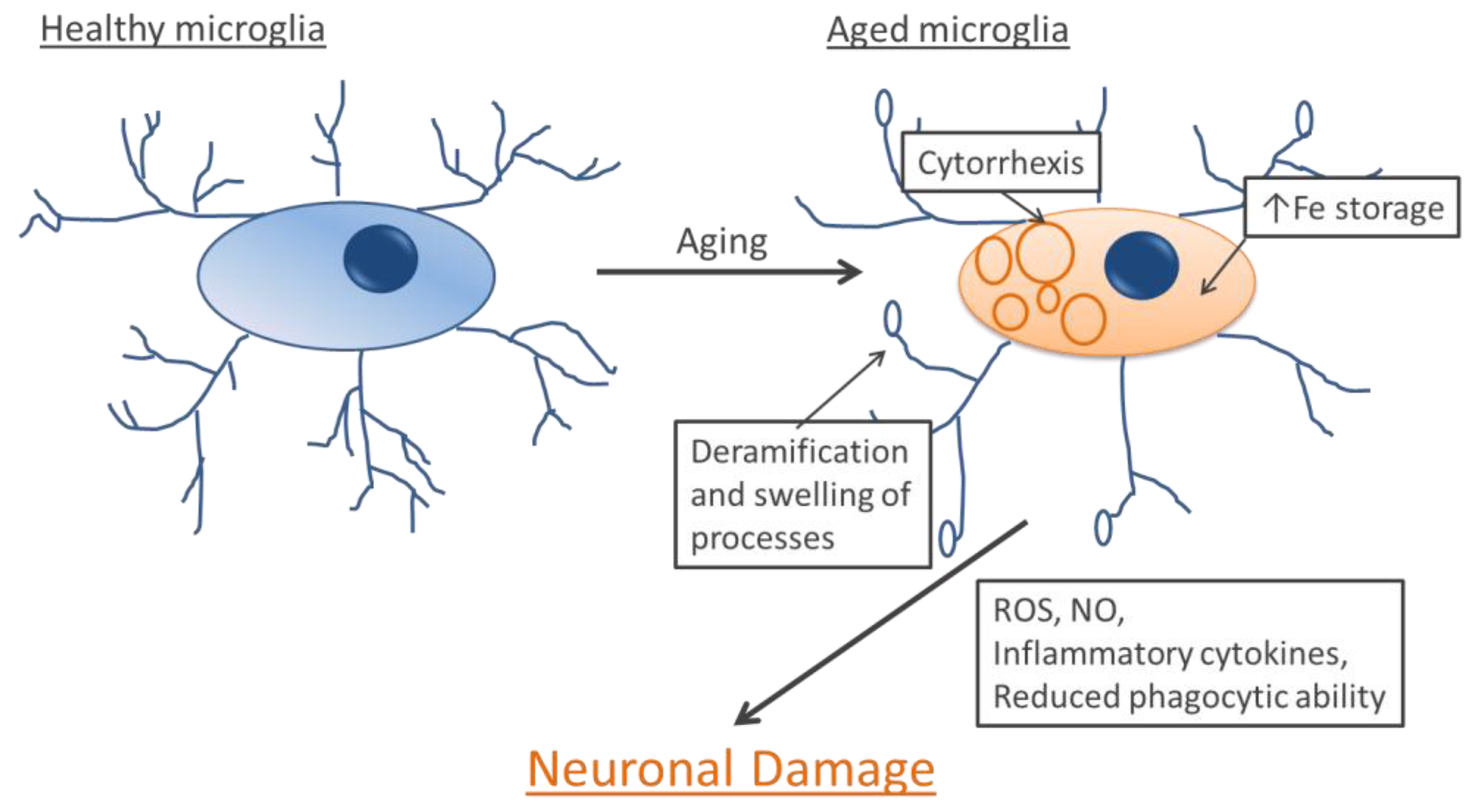
7. Microglia and Aging

While iron metabolism is of interest to specific diseases, general aging can cause other changes upstream that also impact on the same diseases. Here again, changes in the way iron is handled by the brain impact on the cellular environment. An example of the changing environment in the aging brain is a change in the supporting cells in the brain, including microglia [141]. Microglia are the resident macrophages of the brain and are its first and main form of active immune defense. Healthy microglia are very sensitive to their environment, constantly surveying for and phagocytosing any foreign material or cellular debris that they encounter. Additionally, they are capable of releasing cytotoxic substances that can kill neurons that are damaged or infected. Such substances include H2O2, nitric oxide, inflammatory cytokines, proteases, and neurotransmitters. Activated microglia can act as antigen-presenting cells and activate T-cells. After an infection has been dealt with, microglia can recruit cells involved in neuronal repair and secrete anti-inflammatory cytokines [142]. The idea of aging microglia stems from histological observations of healthy aged brains where the cells often develop dystrophic phenotypic characteristics [143]. Resting microglia have a ramified morphology with many fine processes extending from the cell body. Dystrophic microglia found in aging brains lose this fine process of ramification (Figure 3). Dystrophic microglia often develop abnormally shaped processes with spheroidal swellings and cytoplasmic fragmentation (cytorrhexis) [143]. Dystrophic microglia have also been associated with increased release of toxic ROS and inflammatory cytokines and impaired phagocytic ability [144,145]. Cytokines are small proteins involved in intercellular signaling. They are released by a multitude of cell types including immune cells like microglia and bind to cell surface receptors on other cells where, through signaling cascades, they alter the transcriptional profile of the target cells. The proinflammatory cytokines found to be released by dystrophic microglia include IL-6, TNF-α, and IL-1β [145].
Figure 3. Age-related changes in microglia. When microglia age they lose some of their processes and develop abnormalities in others. Additionally, they often exhibit cytoplasmic fragmentation. They also store more iron. Their increased release of neurotoxic substances and reduced ability to phagocytose debris and toxic protein aggregates leaves neurons vulnerable.
Metals 05 02070 g003
Ferritin, the main iron storage protein in the brain, is highly expressed in microglia [19]. Microglia are the main cells in the brain that store iron. One way to identify dystrophic microglia, apart from their morphology, is their higher levels of iron storage, demonstrated by the expression of the iron storage protein ferritin. Additionally, an outward rectifier K+ channel called Kv1.3 has been found to increase expression in dystrophic microglia in aged mouse brains [146]. Dystrophic microglia become more prevalent with human aging and have been found to increase in a variety of diseases including AD [147] and Huntington’s disease [148]. There is also evidence that the chronic inflammation that accompanies neurodegeneration leads to local increases in microglia with high iron and ferritin content, possibly due to iron scavenging [149]. The association between increased iron storage and an altered microglia phenotype, particularly a dystrophic one, suggests a possible causative role for iron. As iron can damage cells, dystrophic microglia possibly develop as a direct result of increased iron storage.

8. Microglia and Neurodegenerative Diseases

The presence of healthy glial cells is critically important to neuronal wellbeing. Microglia maintain homeostasis in the healthy brain and fight infection when it is present through a complicated system of signaling molecules. The importance of microglia to neurons is supported by a higher incidence of dystrophic microglia and microglial apoptosis in AD [150]. The inflammation of the nervous system in neurodegenerative disease was thought to be due to activated microglia. However, dystrophic microglia also have impaired neuroprotective ability and generate the low but sustained release of inflammatory factors seen during neurodegeneration.
The effect of dystrophic microglia on the pathogenic changes occurring in Alzheimer’s disease is not well understood. Over the progression of the disease, microglia seem to change from exerting a neuroprotective function to being closer to a classically activated state. This change in phenotype may result in microglial neurotoxicity or alternatively in dysfunction that prevents the cells from fulfilling their protective role [150]. Dystrophic microglia have been co-localized with neurofibrillary tangles in AD brains [151]. Microglia with impaired phagocytic and motility functions have been co-localized with Aβ deposits in mouse models of Alzheimer’s disease [152]. Healthy microglia have been shown to take up Aβ and also to release enzymes that degrade it [153]. However, dystrophic microglia have impaired Aβ phagocytic ability [144]. If healthy microglia are activated with LPS, they help reduce the Aβ burden in the brains of mice [154]. Pathologically activated microglia release pro-inflammatory cytokines and reactive oxygen species that can make neurons more sensitive to Aβ toxicity [155]. Dystrophic microglia have been found to be hyper-responsive to stimulation [145]. Inflammatory cytokines have been shown to increase the expression of APP in neurons, which can result in increased production of Aβ through also favoring the amyloidogenic APP processing pathway [156,157,158]. The microglial p40 subunit of IL-12 and IL-23 has been found to be elevated in AD brains and to correlate with a worse Aβ pathology [159].

9. Conclusions

Iron is a two-edged sword for biological systems—essential for many cellular activities, but also able to cause damage to macromolecules or disrupt sensitive processes. In the brain this balance is even more delicate given the irreplaceable nature of neurons. Research into the role of iron in both disease and normal activities in the brain will continue. However, the changes and impact of iron during aging and within neurodegenerative diseases is now well established.

Author Contributions

DMA and DRB wrote the manuscript.

Conflicts of Interest

The authors declare no conflict of interest.

References

  1. Oliveira, B.F.; Nogueira-Machado, J.A.; Chaves, M.M. The role of oxidative stress in the aging process. Sci. World J. 2010, 10, 1121–1128. [Google Scholar] [CrossRef] [PubMed]
  2. Cobb, C.A.; Cole, M.P. Oxidative and nitrative stress in neurodegeneration. Neurobiol. Dis. 2015. [Google Scholar] [CrossRef] [PubMed]
  3. Phillipson, O.T. Management of the aging risk factor for Parkinson’s disease. Neurobiol. Aging 2014, 35, 847–857. [Google Scholar] [CrossRef] [PubMed]
  4. Mao, P.; Reddy, P.H. Aging and amyloid beta-induced oxidative DNA damage and mitochondrial dysfunction in Alzheimer’s disease: Implications for early intervention and therapeutics. Biochim. Biophys. Acta 2011, 1812, 1359–1370. [Google Scholar] [CrossRef] [PubMed]
  5. Ward, R.J.; Zucca, F.A.; Duyn, J.H.; Crichton, R.R.; Zecca, L. The role of iron in brain ageing and neurodegenerative disorders. Lancet Neurol. 2014, 13, 1045–1060. [Google Scholar] [CrossRef]
  6. Crichton, R.R.; Ward, R.J. Iron homeostasis. Met. Ions Biol. Syst. 1998, 35, 633–665. [Google Scholar] [PubMed]
  7. Drennan, C.L.; Peters, J.W. Surprising cofactors in metalloenzymes. Curr. Opin. Struct. Biol. 2003, 13, 220–226. [Google Scholar] [CrossRef]
  8. Dunn, L.L.; Rahmanto, Y.S.; Richardson, D.R. Iron uptake and metabolism in the new millennium. Trends Cell Biol. 2007, 17, 93–100. [Google Scholar] [CrossRef] [PubMed]
  9. Sargent, P.J.; Farnaud, S.; Evans, R.W. Structure/function overview of proteins involved in iron storage and transport. Curr. Med. Chem. 2005, 12, 2683–2693. [Google Scholar] [CrossRef] [PubMed]
  10. Skjorringe, T.; Burkhart, A.; Johnsen, K.B.; Moos, T. Divalent metal transporter 1 (DMT1) in the brain: Implications for a role in iron transport at the blood-brain barrier, and neuronal and glial pathology. Front. Mol. Neurosci. 2015. [Google Scholar] [CrossRef] [PubMed]
  11. Koorts, A.M.; Viljoen, M. Ferritin and ferritin isoforms II: Protection against uncontrolled cellular proliferation, oxidative damage and inflammatory processes. Arch. Physiol. Biochem. 2007, 113, 55–64. [Google Scholar] [CrossRef] [PubMed]
  12. Winter, W.E.; Bazydlo, L.A.; Harris, N.S. The molecular biology of human iron metabolism. Lab. Med. 2014, 45, 92–102. [Google Scholar] [CrossRef] [PubMed]
  13. Hider, R.C.; Kong, X. Iron speciation in the cytosol: An overview. Dalton Trans. 2013, 42, 3220–3229. [Google Scholar] [CrossRef] [PubMed]
  14. Kruszewski, M. Labile iron pool: The main determinant of cellular response to oxidative stress. Mutat. Res. 2003, 531, 81–92. [Google Scholar] [CrossRef] [PubMed]
  15. Koeppen, A.H. A brief history of brain iron research. J. Neurol. Sci. 2003, 207, 95–97. [Google Scholar] [CrossRef]
  16. Koeppen, A.H. The history of iron in the brain. J. Neurol. Sci. 1995, 134, 1–9. [Google Scholar] [CrossRef]
  17. Gotz, M.E.; Double, K.; Gerlach, M.; Youdim, M.B.; Riederer, P. The relevance of iron in the pathogenesis of Parkinson’s disease. Ann. N. Y. Acad. Sci. 2004, 1012, 193–208. [Google Scholar] [CrossRef] [PubMed]
  18. Burdo, J.R.; Martin, J.; Menzies, S.L.; Dolan, K.G.; Romano, M.A.; Fletcher, R.J.; Garrick, M.D.; Garrick, L.M.; Connor, J.R. Cellular distribution of iron in the brain of the belgrade rat. Neuroscience 1999, 93, 1189–1196. [Google Scholar] [CrossRef]
  19. Connor, J.R.; Boeshore, K.L.; Benkovic, S.A.; Menzies, S.L. Isoforms of ferritin have a specific cellular distribution in the brain. J. Neurosci. Res. 1994, 37, 461–465. [Google Scholar] [CrossRef] [PubMed]
  20. Moos, T.; Morgan, E.H. The significance of the mutated divalent metal transporter (DMT1) on iron transport into the belgrade rat brain. J. Neurochem. 2004, 88, 233–245. [Google Scholar] [CrossRef] [PubMed]
  21. Zecca, L.; Casella, L.; Albertini, A.; Bellei, C.; Zucca, F.A.; Engelen, M.; Zadlo, A.; Szewczyk, G.; Zareba, M.; Sarna, T. Neuromelanin can protect against iron-mediated oxidative damage in system modeling iron overload of brain aging and Parkinson’s disease. J. Neurochem. 2008, 106, 1866–1875. [Google Scholar] [PubMed]
  22. Zucca, F.A.; Giaveri, G.; Gallorini, M.; Albertini, A.; Toscani, M.; Pezzoli, G.; Lucius, R.; Wilms, H.; Sulzer, D.; Ito, S.; et al. The neuromelanin of human substantia nigra: Physiological and pathogenic aspects. Pigment Cell Res. 2004, 17, 610–617. [Google Scholar] [CrossRef] [PubMed]
  23. Zecca, L.; Gallorini, M.; Schunemann, V.; Trautwein, A.X.; Gerlach, M.; Riederer, P.; Vezzoni, P.; Tampellini, D. Iron, neuromelanin and ferritin content in the substantia nigra of normal subjects at different ages: Consequences for iron storage and neurodegenerative processes. J. Neurochem. 2001, 76, 1766–1773. [Google Scholar] [CrossRef] [PubMed]
  24. Hallgren, B.; Sourander, P. The effect of age on the non-haemin iron in the human brain. J. Neurochem. 1958, 3, 41–51. [Google Scholar] [CrossRef] [PubMed]
  25. Faucheux, B.A.; Bonnet, A.M.; Agid, Y.; Hirsch, E.C. Blood vessels change in the mesencephalon of patients with Parkinson’s disease. Lancet 1999, 353, 981–982. [Google Scholar] [CrossRef]
  26. Brun, A.; Englund, E. Brain changes in dementia of Alzheimer’s type relevant to new imaging diagnostic methods. Prog. Neuro-Psychopharmacol. Biol. Psychiatry 1986, 10, 297–308. [Google Scholar] [CrossRef]
  27. Pandolfo, M. Friedreich’s ataxia: Clinical aspects and pathogenesis. Semin. Neurol. 1999, 19, 311–321. [Google Scholar] [CrossRef] [PubMed]
  28. Campuzano, V.; Montermini, L.; Molto, M.D.; Pianese, L.; Cossee, M.; Cavalcanti, F.; Monros, E.; Rodius, F.; Duclos, F.; Monticelli, A.; et al. Friedreich’s ataxia: Autosomal recessive disease caused by an intronic GAA triplet repeat expansion. Science 1996, 271, 1423–1427. [Google Scholar] [CrossRef] [PubMed]
  29. Napier, I.; Ponka, P.; Richardson, D.R. Iron trafficking in the mitochondrion: Novel pathways revealed by disease. Blood 2005, 105, 1867–1874. [Google Scholar] [CrossRef] [PubMed]
  30. Tsai, C.L.; Barondeau, D.P. Human frataxin is an allosteric switch that activates the Fe-S cluster biosynthetic complex. Biochemistry 2010, 49, 9132–9139. [Google Scholar] [CrossRef] [PubMed]
  31. Jiralerspong, S.; Liu, Y.; Montermini, L.; Stifani, S.; Pandolfo, M. Frataxin shows developmentally regulated tissue-specific expression in the mouse embryo. Neurobiol. Dis. 1997, 4, 103–113. [Google Scholar] [CrossRef] [PubMed]
  32. Boddaert, N.; Sang, K.H.L.Q.; Rotig, A.; Leroy-Willig, A.; Gallet, S.; Brunelle, F.; Sidi, D.; Thalabard, J.C.; Munnich, A.; Cabantchik, Z.I. Selective iron chelation in friedreich ataxia: Biologic and clinical implications. Blood 2007, 110, 401–408. [Google Scholar] [CrossRef] [PubMed]
  33. Levi, S.; Rovida, E. Neuroferritinopathy: From ferritin structure modification to pathogenetic mechanism. Neurobiol. Dis. 2015. [Google Scholar] [CrossRef] [PubMed]
  34. Levi, S.; Finazzi, D. Neurodegeneration with brain iron accumulation: Update on pathogenic mechanisms. Front. Pharmacol. 2014. [Google Scholar] [CrossRef] [PubMed]
  35. Curtis, A.R.; Fey, C.; Morris, C.M.; Bindoff, L.A.; Ince, P.G.; Chinnery, P.F.; Coulthard, A.; Jackson, M.J.; Jackson, A.P.; McHale, D.P.; et al. Mutation in the gene encoding ferritin light polypeptide causes dominant adult-onset basal ganglia disease. Nat. Genet. 2001, 28, 350–354. [Google Scholar] [CrossRef] [PubMed]
  36. Baraibar, M.A.; Muhoberac, B.B.; Garringer, H.J.; Hurley, T.D.; Vidal, R. Unraveling of the e-helices and disruption of 4-fold pores are associated with iron mishandling in a mutant ferritin causing neurodegeneration. J. Biol. Chem. 2010, 285, 1950–1956. [Google Scholar] [CrossRef] [PubMed]
  37. Mancuso, M.; Davidzon, G.; Kurlan, R.M.; Tawil, R.; Bonilla, E.; Mauro, S.D.; Powers, J.M. Hereditary ferritinopathy: A novel mutation, its cellular pathology, and pathogenetic insights. J. Neuropathol. Exp. Neurol. 2005, 64, 280–294. [Google Scholar] [PubMed]
  38. Barbeito, A.G.; Garringer, H.J.; Baraibar, M.A.; Gao, X.; Arredondo, M.; Nunez, M.T.; Smith, M.A.; Ghetti, B.; Vidal, R. Abnormal iron metabolism and oxidative stress in mice expressing a mutant form of the ferritin light polypeptide gene. J. Neurochem. 2009, 109, 1067–1078. [Google Scholar] [CrossRef] [PubMed]
  39. Burn, J.; Chinnery, P.F. Neuroferritinopathy. Semin. Pediatr. Neurol. 2006, 13, 176–181. [Google Scholar] [CrossRef] [PubMed]
  40. Hayflick, S.J. Unraveling the hallervorden-spatz syndrome: Pantothenate kinase-associated neurodegeneration is the name. Curr. Opin. Pediatr. 2003, 15, 572–577. [Google Scholar] [CrossRef] [PubMed]
  41. Zhou, B.; Westaway, S.K.; Levinson, B.; Johnson, M.A.; Gitschier, J.; Hayflick, S.J. A novel pantothenate kinase gene (pank2) is defective in hallervorden-spatz syndrome. Nat. Genet. 2001, 28, 345–349. [Google Scholar] [CrossRef] [PubMed]
  42. Zeidman, L.A.; Pandey, D.K. Declining use of the hallervorden-spatz disease eponym in the last two decades. J. Child Neurol. 2012, 27, 1310–1315. [Google Scholar] [CrossRef] [PubMed]
  43. Hogarth, P. Neurodegeneration with brain iron accumulation: Diagnosis and management. J. Mov. Disord. 2015, 8, 1–13. [Google Scholar] [CrossRef] [PubMed]
  44. Szumowski, J.; Bas, E.; Gaarder, K.; Schwarz, E.; Erdogmus, D.; Hayflick, S. Measurement of brain iron distribution in hallevorden-spatz syndrome. J. Magn. Reson. Imaging 2010, 31, 482–489. [Google Scholar] [CrossRef] [PubMed]
  45. Gordon, N. Pantothenate kinase-associated neurodegeneration (hallervorden-spatz syndrome). Eur. J. Paediatr. Neurol. 2002, 6, 243–247. [Google Scholar] [CrossRef] [PubMed]
  46. Hayflick, S.J.; Hogarth, P. As iron goes, so goes disease? Haematologica 2011, 96, 1571–1572. [Google Scholar] [CrossRef] [PubMed]
  47. Ghosh, M.C.; Zhang, L.; Rouault, T.A. Iron misregulation and neurodegenerative disease in mouse models that lack iron regulatory proteins. Neurobiol. Dis. 2015. [Google Scholar] [CrossRef] [PubMed]
  48. Rouault, T.A. Mammalian iron-sulphur proteins: Novel insights into biogenesis and function. Nat. Rev. Mol. Cell Biol. 2015, 16, 45–55. [Google Scholar] [CrossRef] [PubMed]
  49. Hardy, J.; Selkoe, D.J. The amyloid hypothesis of Alzheimer’s disease: Progress and problems on the road to therapeutics. Science 2002, 297, 353–356. [Google Scholar] [CrossRef] [PubMed]
  50. Lombardo, S.; Maskos, U. Role of the nicotinic acetylcholine receptor in Alzheimer’s disease pathology and treatment. Neuropharmacology 2015, 96, 255–262. [Google Scholar] [CrossRef] [PubMed]
  51. Guerreiro, R.; Hardy, J. Genetics of Alzheimer’s disease. Neurotherapeutics 2014, 11, 732–737. [Google Scholar] [CrossRef] [PubMed]
  52. Hardy, J.; Allsop, D. Amyloid deposition as the central event in the aetiology of Alzheimer’s disease. Trends Pharmacol. Sci. 1991, 12, 383–388. [Google Scholar] [CrossRef]
  53. Iqbal, K.; Liu, F.; Gong, C.X.; Grundke-Iqbal, I. Tau in alzheimer disease and related tauopathies. Curr. Alzheimer Res. 2010, 7, 656–664. [Google Scholar] [CrossRef] [PubMed]
  54. Barbato, C.; Canu, N.; Zambrano, N.; Serafino, A.; Minopoli, G.; Ciotti, M.T.; Amadoro, G.; Russo, T.; Calissano, P. Interaction of Tau with Fe65 links tau to APP. Neurobiol. Dis. 2005, 18, 399–408. [Google Scholar] [CrossRef] [PubMed]
  55. Nalivaeva, N.N.; Turner, A.J. The amyloid precursor protein: A biochemical enigma in brain development, function and disease. FEBS Lett. 2013, 587, 2046–2054. [Google Scholar] [CrossRef] [PubMed]
  56. Hesse, L.; Beher, D.; Masters, C.L.; Multhaup, G. The beta A4 amyloid precursor protein binding to copper. FEBS Lett. 1994, 349, 109–116. [Google Scholar] [CrossRef]
  57. Spoerri, L.; Vella, L.J.; Pham, C.L.; Barnham, K.J.; Cappai, R. The amyloid precursor protein copper binding domain histidine residues 149 and 151 mediate App stability and metabolism. J. Biol. Chem. 2012, 287, 26840–26853. [Google Scholar] [CrossRef] [PubMed]
  58. Dahms, S.O.; Konnig, I.; Roeser, D.; Guhrs, K.H.; Mayer, M.C.; Kaden, D.; Multhaup, G.; Than, M.E. Metal binding dictates conformation and function of the amyloid precursor protein (APP) E2 domain. J. Mol. Biol. 2012, 416, 438–452. [Google Scholar] [CrossRef] [PubMed]
  59. Ebrahimi, K.H.; Dienemann, C.; Hoefgen, S.; Than, M.E.; Hagedoorn, P.L.; Hagen, W.R. The amyloid precursor protein (APP) does not have a ferroxidase site in its E2 domain. PLoS ONE 2013. [Google Scholar] [CrossRef]
  60. Duce, J.A.; Tsatsanis, A.; Cater, M.A.; James, S.A.; Robb, E.; Wikhe, K.; Leong, S.L.; Perez, K.; Johanssen, T.; Greenough, M.A.; et al. Iron-export ferroxidase activity of beta-amyloid precursor protein is inhibited by zinc in Alzheimer’s disease. Cell 2010, 142, 857–867. [Google Scholar] [CrossRef] [PubMed]
  61. Wong, B.X.; Tsatsanis, A.; Lim, L.Q.; Adlard, P.A.; Bush, A.I.; Duce, J.A. β-amyloid precursor protein does not possess ferroxidase activity but does stabilize the cell surface ferrous iron exporter ferroportin. PLoS ONE 2014. [Google Scholar] [CrossRef] [PubMed]
  62. Borchardt, T.; Camakaris, J.; Cappai, R.; Masters, C.L.; Beyreuther, K.; Multhaup, G. Copper inhibits β-amyloid production and stimulates the non-amyloidogenic pathway of amyloid-precursor-protein secretion. Biochem. J. 1999, 344, 461–467. [Google Scholar] [CrossRef] [PubMed]
  63. Baumkotter, F.; Schmidt, N.; Vargas, C.; Schilling, S.; Weber, R.; Wagner, K.; Fiedler, S.; Klug, W.; Radzimanowski, J.; Nickolaus, S.; et al. Amyloid precursor protein dimerization and synaptogenic function depend on copper binding to the growth factor-like domain. J. Neurosci. 2014, 34, 11159–11172. [Google Scholar] [CrossRef] [PubMed]
  64. Garzon-Rodriguez, W.; Yatsimirsky, A.K.; Glabe, C.G. Binding of Zn(II), Cu(II), and Fe(II) ions to Alzheimer’s A beta peptide studied by fluorescence. Bioorg. Med. Chem. Lett. 1999, 9, 2243–2248. [Google Scholar] [CrossRef]
  65. Bush, A.I.; Pettingell, W.H., Jr.; Paradis, M.D.; Tanzi, R.E. Modulation of a beta adhesiveness and secretase site cleavage by zinc. J. Biol. Chem. 1994, 269, 12152–12158. [Google Scholar] [PubMed]
  66. Streltsov, V.A.; Titmuss, S.J.; Epa, V.C.; Barnham, K.J.; Masters, C.L.; Varghese, J.N. The structure of the amyloid-beta peptide high-affinity copper II binding site in alzheimer disease. Biophys. J. 2008, 95, 3447–3456. [Google Scholar] [CrossRef] [PubMed]
  67. Cherny, R.A.; Atwood, C.S.; Xilinas, M.E.; Gray, D.N.; Jones, W.D.; McLean, C.A.; Barnham, K.J.; Volitakis, I.; Fraser, F.W.; Kim, Y.; et al. Treatment with a copper-zinc chelator markedly and rapidly inhibits β-amyloid accumulation in Alzheimer’s disease transgenic mice. Neuron 2001, 30, 665–676. [Google Scholar] [CrossRef]
  68. Faux, N.G.; Ritchie, C.W.; Gunn, A.; Rembach, A.; Tsatsanis, A.; Bedo, J.; Harrison, J.; Lannfelt, L.; Blennow, K.; Zetterberg, H.; et al. PBT2 rapidly improves cognition in Alzheimer’s disease: Additional phase II analyses. J. Alzheimers Dis. 2010, 20, 509–516. [Google Scholar] [PubMed]
  69. Ayton, S.; Lei, P.; Bush, A.I. Biometals and their therapeutic implications in Alzheimer’s disease. Neurotherapeutics 2015, 12, 109–120. [Google Scholar] [CrossRef] [PubMed]
  70. Dedman, D.J.; Treffry, A.; Candy, J.M.; Taylor, G.A.; Morris, C.M.; Bloxham, C.A.; Perry, R.H.; Edwardson, J.A.; Harrison, P.M. Iron and aluminium in relation to brain ferritin in normal individuals and Alzheimer’s-disease and chronic renal-dialysis patients. Biochem. J. 1992, 287, 509–514. [Google Scholar] [CrossRef] [PubMed]
  71. Bartzokis, G.; Sultzer, D.; Mintz, J.; Holt, L.E.; Marx, P.; Phelan, C.K.; Marder, S.R. In vivo evaluation of brain iron in Alzheimer’s disease and normal subjects using mri. Biol. Psychiatry 1994, 35, 480–487. [Google Scholar] [CrossRef]
  72. Castellani, R.J.; Honda, K.; Zhu, X.; Cash, A.D.; Nunomura, A.; Perry, G.; Smith, M.A. Contribution of redox-active iron and copper to oxidative damage in alzheimer disease. Ageing Res. Rev. 2004, 3, 319–326. [Google Scholar] [CrossRef] [PubMed]
  73. Bonda, D.J.; Lee, H.G.; Blair, J.A.; Zhu, X.; Perry, G.; Smith, M.A. Role of metal dyshomeostasis in Alzheimer’s disease. Metallomics 2011, 3, 267–270. [Google Scholar] [CrossRef] [PubMed]
  74. Ayton, S.; Faux, N.G.; Bush, A.I. Alzheimer’s Disease Neuroimaging Initiative. Ferritin levels in the cerebrospinal fluid predict Alzheimer’s disease outcomes and are regulated by APOE. Nat. Commun. 2015. [Google Scholar] [CrossRef] [PubMed]
  75. Rottkamp, C.A.; Raina, A.K.; Zhu, X.; Gaier, E.; Bush, A.I.; Atwood, C.S.; Chevion, M.; Perry, G.; Smith, M.A. Redox-active iron mediates amyloid-beta toxicity. Free Radic. Biol. Med. 2001, 30, 447–450. [Google Scholar] [CrossRef]
  76. Stephenson, D.T.; Clemens, J.A. In vivo effects of beta-amyloid implants in rodents: Lack of potentiation of damage associated with transient global forebrain ischemia. Brain Res. 1992, 586, 235–246. [Google Scholar] [CrossRef]
  77. Friedland-Leuner, K.; Stockburger, C.; Denzer, I.; Eckert, G.P.; Muller, W.E. Mitochondrial dysfunction: Cause and consequence of Alzheimer’s disease. Prog. Mol. Biol. Transl. Sci. 2014, 127, 183–210. [Google Scholar] [PubMed]
  78. Atamna, H. Heme, iron, and the mitochondrial decay of ageing. Ageing Res. Rev. 2004, 3, 303–318. [Google Scholar] [CrossRef] [PubMed]
  79. Hirai, K.; Aliev, G.; Nunomura, A.; Fujioka, H.; Russell, R.L.; Atwood, C.S.; Johnson, A.B.; Kress, Y.; Vinters, H.V.; Tabaton, M.; et al. Mitochondrial abnormalities in Alzheimer’s disease. J. Neurosci. 2001, 21, 3017–3023. [Google Scholar] [PubMed]
  80. Perry, G.; Taddeo, M.A.; Petersen, R.B.; Castellani, R.J.; Harris, P.L.; Siedlak, S.L.; Cash, A.D.; Liu, Q.; Nunomura, A.; Atwood, C.S.; et al. Adventiously-bound redox active iron and copper are at the center of oxidative damage in alzheimer disease. Biometals 2003, 16, 77–81. [Google Scholar] [CrossRef] [PubMed]
  81. Spillantini, M.G.; Schmidt, M.L.; Lee, V.M.; Trojanowski, J.Q.; Jakes, R.; Goedert, M. α-synuclein in lewy bodies. Nature 1997, 388, 839–840. [Google Scholar] [CrossRef] [PubMed]
  82. Saito, Y.; Ruberu, N.N.; Sawabe, M.; Arai, T.; Kazama, H.; Hosoi, T.; Yamanouchi, H.; Murayama, S. Lewy body-related α-synucleinopathy in aging. J. Neuropathol. Exp. Neurol. 2004, 63, 742–749. [Google Scholar] [CrossRef] [PubMed]
  83. Goedert, M. α-synuclein and neurodegenerative diseases. Nat. Rev. Neurosci. 2001, 2, 492–501. [Google Scholar] [CrossRef] [PubMed]
  84. Dickson, D.W. Parkinson’s disease and parkinsonism: Neuropathology. Cold Spring Harb. Perspect. Med. 2012. [Google Scholar] [CrossRef] [PubMed]
  85. Dexter, D.T.; Wells, F.R.; Agid, F.; Agid, Y.; Lees, A.J.; Jenner, P.; Marsden, C.D. Increased nigral iron content in postmortem parkinsonian brain. Lancet 1987, 2, 1219–1220. [Google Scholar] [CrossRef]
  86. Singleton, A.B.; Farrer, M.J.; Bonifati, V. The genetics of Parkinson’s disease: Progress and therapeutic implications. Mov. Disord. 2013, 28, 14–23. [Google Scholar] [CrossRef] [PubMed]
  87. Leenders, K.L.; Oertel, W.H. Parkinson’s disease: Clinical signs and symptoms, neural mechanisms, positron emission tomography, and therapeutic interventions. Neural Plast. 2001, 8, 99–110. [Google Scholar] [CrossRef] [PubMed]
  88. Cenci, M.A. Presynaptic mechanisms of L-DOPA-induced dyskinesia: The findings, the debate, and the therapeutic implications. Front Neurol 2014. [Google Scholar] [CrossRef] [PubMed]
  89. Dexter, D.T.; Carayon, A.; Javoy-Agid, F.; Agid, Y.; Wells, F.R.; Daniel, S.E.; Lees, A.J.; Jenner, P.; Marsden, C.D. Alterations in the levels of iron, ferritin and other trace metals in Parkinson’s disease and other neurodegenerative diseases affecting the basal ganglia. Brain 1991, 114, 1953–1975. [Google Scholar] [CrossRef] [PubMed]
  90. Hirsch, E.C.; Brandel, J.P.; Galle, P.; Javoy-Agid, F.; Agid, Y. Iron and aluminum increase in the substantia nigra of patients with Parkinson’s disease: An X-ray microanalysis. J. Neurochem. 1991, 56, 446–451. [Google Scholar] [CrossRef] [PubMed]
  91. Tofaris, G.K.; Revesz, T.; Jacques, T.S.; Papacostas, S.; Chataway, J. Adult-onset neurodegeneration with brain iron accumulation and cortical α-synuclein and tau pathology: A distinct clinicopathological entity. Arch. Neurol. 2007, 64, 280–282. [Google Scholar] [CrossRef] [PubMed]
  92. Pall, H.S.; Williams, A.C.; Blake, D.R.; Lunec, J.; Gutteridge, J.M.; Hall, M.; Taylor, A. Raised cerebrospinal-fluid copper concentration in Parkinson’s disease. Lancet 1987, 2, 238–241. [Google Scholar] [CrossRef]
  93. Gorrell, J.M.; DiMonte, D.; Graham, D. The role of the environment in Parkinson’s disease. Environ. Health Perspect. 1996, 104, 652–654. [Google Scholar] [CrossRef] [PubMed]
  94. Kalivendi, S.V.; Cunningham, S.; Kotamraju, S.; Joseph, J.; Hillard, C.J.; Kalyanaraman, B. α-synuclein up-regulation and aggregation during mpp+-induced apoptosis in neuroblastoma cells: Intermediacy of transferrin receptor iron and hydrogen peroxide. J. Biol. Chem. 2004, 279, 15240–15247. [Google Scholar] [CrossRef] [PubMed]
  95. Mandel, S.; Maor, G.; Youdim, M.B. Iron and α-synuclein in the substantia nigra of mptp-treated mice: Effect of neuroprotective drugs R-apomorphine and green tea polyphenol (−)-epigallocatechin-3-gallate. J. Mol. Neurosci. 2004, 24, 401–416. [Google Scholar] [CrossRef]
  96. Sengstock, G.J.; Olanow, C.W.; Menzies, R.A.; Dunn, A.J.; Arendash, G.W. Infusion of iron into the rat substantia nigra: Nigral pathology and dose-dependent loss of striatal dopaminergic markers. J. Neurosci. Res. 1993, 35, 67–82. [Google Scholar] [CrossRef] [PubMed]
  97. Double, K.L.; Halliday, G.M. New face of neuromelanin. J. Neural Transm. Suppl. 2006, 119–123. [Google Scholar]
  98. Li, J.; Yang, J.; Zhao, P.; Li, S.; Zhang, R.; Zhang, X.; Liu, D.; Zhang, B. Neuromelanin enhances the toxicity of alpha-synuclein in SK-N-SH cells. J. Neural Transm. 2012, 119, 685–691. [Google Scholar] [CrossRef] [PubMed]
  99. Gerlach, M.; Riederer, P.; Double, K.L. Neuromelanin-bound ferric iron as an experimental model of dopaminergic neurodegeneration in Parkinson’s disease. Parkinsonism Relat. Disord. 2008, 14, S185–S188. [Google Scholar] [CrossRef] [PubMed]
  100. Fasano, M.; Bergamasco, B.; Lopiano, L. Is neuromelanin changed in Parkinson’s disease? Investigations by magnetic spectroscopies. J. Neural Transm. 2006, 113, 769–774. [Google Scholar] [CrossRef] [PubMed]
  101. Silva, B.; Faustino, P. An overview of molecular basis of iron metabolism regulation and the associated pathologies. Biochim. Biophys. Acta 2015, 1852, 1347–1359. [Google Scholar] [CrossRef] [PubMed]
  102. Hanson, E.S.; Leibold, E.A. Regulation of the iron regulatory proteins by reactive nitrogen and oxygen species. Gene Expr. 1999, 7, 367–376. [Google Scholar] [PubMed]
  103. Connor, J.R.; Snyder, B.S.; Arosio, P.; Loeffler, D.A.; LeWitt, P. A quantitative analysis of isoferritins in select regions of aged, parkinsonian, and Alzheimer’s diseased brains. J. Neurochem. 1995, 65, 717–724. [Google Scholar] [CrossRef] [PubMed]
  104. Jinsmaa, Y.; Sullivan, P.; Gross, D.; Cooney, A.; Sharabi, Y.; Goldstein, D.S. Divalent metal ions enhance dopal-induced oligomerization of alpha-synuclein. Neurosci. Lett. 2014, 569, 27–32. [Google Scholar] [CrossRef] [PubMed]
  105. Li, S.W.; Lin, T.S.; Minteer, S.; Burke, W.J. 3,4-dihydroxyphenylacetaldehyde and hydrogen peroxide generate a hydroxyl radical: Possible role in Parkinson’s disease pathogenesis. Brain Res. Mol. Brain Res. 2001, 93, 1–7. [Google Scholar] [CrossRef]
  106. Davies, P.; Wang, X.; Sarell, C.J.; Drewett, A.; Marken, F.; Viles, J.H.; Brown, D.R. The synucleins are a family of redox-active copper binding proteins. Biochemistry 2010, 50, 37–47. [Google Scholar] [CrossRef] [PubMed]
  107. Rasia, R.M.; Bertoncini, C.W.; Marsh, D.; Hoyer, W.; Cherny, D.; Zweckstetter, M.; Griesinger, C.; Jovin, T.M.; Fernandez, C.O. Structural characterization of copper(II) binding to α-synuclein: Insights into the bioinorganic chemistry of Parkinson’s disease. Proc. Natl. Acad. Sci. USA 2005, 102, 4294–4299. [Google Scholar] [CrossRef] [PubMed]
  108. Binolfi, A.; Rasia, R.M.; Bertoncini, C.W.; Ceolin, M.; Zweckstetter, M.; Griesinger, C.; Jovin, T.M.; Fernandez, C.O. Interaction of α-synuclein with divalent metal ions reveals key differences: A link between structure, binding specificity and fibrillation enhancement. J. Am. Chem. Soc. 2006, 128, 9893–9901. [Google Scholar] [CrossRef] [PubMed]
  109. Davies, P.; Moualla, D.; Brown, D.R. α-synuclein is a cellular ferrireductase. PLoS ONE 2011. [Google Scholar] [CrossRef]
  110. Uversky, V.N.; Li, J.; Fink, A.L. Metal-triggered structural transformations, aggregation, and fibrillation of human α-synuclein. A possible molecular nk between Parkinson’s disease and heavy metal exposure. J. Biol. Chem. 2001, 276, 44284–44296. [Google Scholar] [CrossRef] [PubMed]
  111. Wright, J.A.; Wang, X.; Brown, D.R. Unique copper-induced oligomers mediate α-synuclein toxicity. FASEB J. 2009, 23, 2384–2393. [Google Scholar] [CrossRef] [PubMed]
  112. Wang, X.; Moualla, D.; Wright, J.A.; Brown, D.R. Copper binding regulates intracellular α-synuclein localisation, aggregation and toxicity. J. Neurochem. 2010, 113, 704–714. [Google Scholar] [CrossRef] [PubMed]
  113. Levin, J.; Hogen, T.; Hillmer, A.S.; Bader, B.; Schmidt, F.; Kamp, F.; Kretzschmar, H.A.; Botzel, K.; Giese, A. Generation of ferric iron links oxidative stress to α-synuclein oligomer formation. J. Parkinson’s Dis. 2011, 1, 205–216. [Google Scholar] [CrossRef]
  114. Ortega, R.; Carmona, A.; Roudeau, S.; Perrin, L.; Ducic, T.; Carboni, E.; Bohic, S.; Cloetens, P.; Lingor, P. α-synuclein over-expression induces increased iron accumulation and redistribution in iron-exposed neurons. Mol. Neurobiol. 2015. [Google Scholar] [CrossRef] [PubMed]
  115. Prusiner, S.B. Prions. Proc. Natl. Acad. Sci. USA 1998, 95, 13363–13383. [Google Scholar] [CrossRef] [PubMed]
  116. Ironside, J.W.; Sutherland, K.; Bell, J.E.; McCardle, L.; Barrie, C.; Estebeiro, K.; Zeidler, M.; Will, R.G. A new variant of Creutzfeldt-Jakob disease: Neuropathological and clinical features. Cold Spring Harb. Symp. Quant. Biol. 1996, 61, 523–530. [Google Scholar] [CrossRef] [PubMed]
  117. Hope, J.; Reekie, L.J.; Hunter, N.; Multhaup, G.; Beyreuther, K.; White, H.; Scott, A.C.; Stack, M.J.; Dawson, M.; Wells, G.A. Fibrils from brains of cows with new cattle disease contain scrapie-associated protein. Nature 1988, 336, 390–392. [Google Scholar] [CrossRef] [PubMed]
  118. Knight, R. Creutzfeldt-Jakob disease: A rare cause of dementia in elderly persons. Clin. Infect. Dis. 2006, 43, 340–346. [Google Scholar] [CrossRef] [PubMed]
  119. Bolton, D.C.; McKinley, M.P.; Prusiner, S.B. Identification of a protein that purifies with the scrapie prion. Science 1982, 218, 1309–1311. [Google Scholar] [CrossRef] [PubMed]
  120. Endo, T.; Groth, D.; Prusiner, S.B.; Kobata, A. Diversity of oligosaccharide structures linked to asparagines of the scrapie prion protein. Biochemistry 1989, 28, 8380–8388. [Google Scholar] [CrossRef] [PubMed]
  121. Brown, D.R.; Qin, K.; Herms, J.W.; Madlung, A.; Manson, J.; Strome, R.; Fraser, P.E.; Kruck, T.; von Bohlen, A.; Schulz-Schaeffer, W.; et al. The cellular prion protein binds copper in vivo. Nature 1997, 390, 684–687. [Google Scholar] [CrossRef] [PubMed]
  122. Brown, D.R.; Wong, B.S.; Hafiz, F.; Clive, C.; Haswell, S.J.; Jones, I.M. Normal prion protein has an activity like that of superoxide dismutase. Biochem. J. 1999, 344, 1–5. [Google Scholar] [CrossRef] [PubMed]
  123. Brown, D.R. Prion and prejudice: Normal protein and the synapse. Trends Neurosci. 2001, 24, 85–90. [Google Scholar] [CrossRef]
  124. Davies, P.; Brown, D.R. The chemistry of copper binding to PrP: Is there sufficient evidence to elucidate a role for copper in protein function? Biochem. J. 2008, 410, 237–244. [Google Scholar] [CrossRef] [PubMed]
  125. Brown, D.R. Metalloproteins and neuronal death. Metallomics 2010, 2, 186–194. [Google Scholar] [CrossRef] [PubMed]
  126. Brown, D.R. Prions and manganese: A maddening beast. Metallomics 2011, 3, 229–238. [Google Scholar] [CrossRef] [PubMed]
  127. Brown, D.R.; Hafiz, F.; Glasssmith, L.L.; Wong, B.S.; Jones, I.M.; Clive, C.; Haswell, S.J. Consequences of manganese replacement of copper for prion protein function and proteinase resistance. EMBO J. 2000, 19, 1180–1186. [Google Scholar] [CrossRef] [PubMed]
  128. Brazier, M.W.; Davies, P.; Player, E.; Marken, F.; Viles, J.H.; Brown, D.R. Manganese binding to the prion protein. J. Biol. Chem. 2008, 283, 12831–12839. [Google Scholar] [CrossRef] [PubMed]
  129. Davies, P.; Brown, D.R. Manganese enhances prion protein survival in model soils and increases prion infectivity to cells. PLoS ONE 2009. [Google Scholar] [CrossRef] [PubMed] [Green Version]
  130. Hesketh, S.; Sassoon, J.; Knight, R.; Hopkins, J.; Brown, D.R. Elevated manganese levels in blood and central nervous system occur before onset of clinical signs in scrapie and bovine spongiform encephalopathy. J. Anim. Sci. 2007, 85, 1596–1609. [Google Scholar] [CrossRef] [PubMed]
  131. Hesketh, S.; Sassoon, J.; Knight, R.; Brown, D.R. Elevated manganese levels in blood and CNS in human prion disease. Mol. Cell. Neurosci. 2008, 37, 590–598. [Google Scholar] [CrossRef] [PubMed]
  132. Thackray, A.M.; Knight, R.; Haswell, S.J.; Bujdoso, R.; Brown, D.R. Metal imbalance and compromised antioxidant function are early changes in prion disease. Biochem. J. 2002, 362, 253–258. [Google Scholar] [CrossRef] [PubMed]
  133. Johnson, C.J.; Gilbert, P.U.; Abrecht, M.; Baldwin, K.L.; Russell, R.E.; Pedersen, J.A.; Aiken, J.M.; McKenzie, D. Low copper and high manganese levels in prion protein plaques. Viruses 2013, 5, 654–662. [Google Scholar] [CrossRef] [PubMed]
  134. Singh, N.; Haldar, S.; Tripathi, A.K.; McElwee, M.K.; Horback, K.; Beserra, A. Iron in neurodegenerative disorders of protein misfolding: A case of prion disorders and Parkinson’s disease. Antioxid. Redox Signal. 2014, 21, 471–484. [Google Scholar] [CrossRef] [PubMed]
  135. Kim, B.H.; Jun, Y.C.; Jin, J.K.; Kim, J.I.; Kim, N.H.; Leibold, E.A.; Connor, J.R.; Choi, E.K.; Carp, R.I.; Kim, Y.S. Alteration of iron regulatory proteins (IRP1 and IRP2) and ferritin in the brains of scrapie-infected mice. Neurosci. Lett. 2007, 422, 158–163. [Google Scholar] [CrossRef] [PubMed]
  136. Singh, A.; Mohan, M.L.; Isaac, A.O.; Luo, X.; Petrak, J.; Vyoral, D.; Singh, N. Prion protein modulates cellular iron uptake: A novel function with implications for prion disease pathogenesis. PLoS ONE 2009. [Google Scholar] [CrossRef]
  137. Singh, A.; Isaac, A.O.; Luo, X.; Mohan, M.L.; Cohen, M.L.; Chen, F.; Kong, Q.; Bartz, J.; Singh, N. Abnormal brain iron homeostasis in human and animal prion disorders. PLoS Pathog. 2009. [Google Scholar] [CrossRef] [PubMed]
  138. Singh, A.; Kong, Q.; Luo, X.; Petersen, R.B.; Meyerson, H.; Singh, N. Prion protein (PrP) knock-out mice show altered iron metabolism: A functional role for PrP in iron uptake and transport. PLoS ONE 2009. [Google Scholar] [CrossRef] [PubMed]
  139. Kralovicova, S.; Fontaine, S.N.; Alderton, A.; Alderman, J.; Ragnarsdottir, K.V.; Collins, S.J.; Brown, D.R. The effects of prion protein expression on metal metabolism. Mol. Cell. Neurosci. 2009, 41, 135–147. [Google Scholar] [CrossRef] [PubMed]
  140. Singh, A.; Haldar, S.; Horback, K.; Tom, C.; Zhou, L.; Meyerson, H.; Singh, N. Prion protein regulates iron transport by functioning as a ferrireductase. J. Alzheimers Dis. 2013, 35, 541–552. [Google Scholar] [PubMed]
  141. Brown, D.R. Role of microglia in age-related changes to the nervous system. Sci. World J. 2009, 9, 1061–1071. [Google Scholar] [CrossRef] [PubMed]
  142. Aloisi, F. Immune function of microglia. Glia 2001, 36, 165–179. [Google Scholar] [CrossRef] [PubMed]
  143. Streit, W.J.; Sammons, N.W.; Kuhns, A.J.; Sparks, D.L. Dystrophic microglia in the aging human brain. Glia 2004, 45, 208–212. [Google Scholar] [CrossRef] [PubMed]
  144. Njie, E.G.; Boelen, E.; Stassen, F.R.; Steinbusch, H.W.; Borchelt, D.R.; Streit, W.J. Ex vivo cultures of microglia from young and aged rodent brain reveal age-related changes in microglial function. Neurobiol. Aging 2012, 33, 195.e1–195.e12. [Google Scholar] [CrossRef] [PubMed]
  145. Solito, E.; Sastre, M. Microglia function in Alzheimer’s disease. Front. Pharmacol. 2012. [Google Scholar] [CrossRef] [PubMed]
  146. Schilling, T.; Eder, C. Microglial K+ channel expression in young adult and aged mice. Glia 2015, 63, 664–672. [Google Scholar] [CrossRef] [PubMed]
  147. Lopes, K.O.; Sparks, D.L.; Streit, W.J. Microglial dystrophy in the aged and Alzheimer’s disease brain is associated with ferritin immunoreactivity. Glia 2008, 56, 1048–1060. [Google Scholar] [CrossRef] [PubMed]
  148. Simmons, D.A.; Casale, M.; Alcon, B.; Pham, N.; Narayan, N.; Lynch, G. Ferritin accumulation in dystrophic microglia is an early event in the development of Huntington’s disease. Glia 2007, 55, 1074–1084. [Google Scholar] [CrossRef] [PubMed]
  149. Thomsen, M.S.; Andersen, M.V.; Christoffersen, P.R.; Jensen, M.D.; Lichota, J.; Moos, T. Neurodegeneration with inflammation is accompanied by accumulation of iron and ferritin in microglia and neurons. Neurobiol. Dis. 2015. [Google Scholar] [CrossRef] [PubMed]
  150. Streit, W.J.; Xue, Q.S.; Tischer, J.; Bechmann, I. Microglial pathology. Acta Neuropathol. Commun. 2014. [Google Scholar] [CrossRef] [PubMed]
  151. Streit, W.J.; Braak, H.; Xue, Q.S.; Bechmann, I. Dystrophic (senescent) rather than activated microglial cells are associated with tau pathology and likely precede neurodegeneration in Alzheimer’s disease. Acta Neuropathol. 2009, 118, 475–485. [Google Scholar] [CrossRef] [PubMed]
  152. Krabbe, G.; Halle, A.; Matyash, V.; Rinnenthal, J.L.; Eom, G.D.; Bernhardt, U.; Miller, K.R.; Prokop, S.; Kettenmann, H.; Heppner, F.L. Functional impairment of microglia coincides with β-amyloid deposition in mice with alzheimer-like pathology. PLoS ONE 2013. [Google Scholar] [CrossRef] [PubMed]
  153. Qiu, W.Q.; Ye, Z.; Kholodenko, D.; Seubert, P.; Selkoe, D.J. Degradation of amyloid β-protein by a metalloprotease secreted by microglia and other neural and non-neural cells. J. Biol. Chem. 1997, 272, 6641–6646. [Google Scholar] [CrossRef] [PubMed]
  154. DiCarlo, G.; Wilcock, D.; Henderson, D.; Gordon, M.; Morgan, D. Intrahippocampal LPS injections reduce Aβ load in APP + PS1 transgenic mice. Neurobiol. Aging 2001, 22, 1007–1012. [Google Scholar] [CrossRef]
  155. Li, M.; Pisalyaput, K.; Galvan, M.; Tenner, A.J. Macrophage colony stimulatory factor and interferon-γ trigger distinct mechanisms for augmentation of β-amyloid-induced microglia-mediated neurotoxicity. J. Neurochem. 2004, 91, 623–633. [Google Scholar] [CrossRef] [PubMed]
  156. Mrak, R.E.; Griffin, W.S. The role of activated astrocytes and of the neurotrophic cytokine S100B in the pathogenesis of Alzheimer’s disease. Neurobiol. Aging 2001, 22, 915–922. [Google Scholar] [CrossRef]
  157. Erta, M.; Quintana, A.; Hidalgo, J. Interleukin-6, a major cytokine in the central nervous system. Int. J. Biol. Sci. 2012, 8, 1254–1266. [Google Scholar] [CrossRef] [PubMed]
  158. Cartier, N.; Lewis, C.A.; Zhang, R.; Rossi, F.M. The role of microglia in human disease: Therapeutic tool or target? Acta Neuropathol. 2014, 128, 363–380. [Google Scholar] [CrossRef] [PubMed]
  159. Prokop, S.; Miller, K.R.; Heppner, F.L. Microglia actions in Alzheimer’s disease. Acta Neuropathol. 2013, 126, 461–477. [Google Scholar] [CrossRef] [PubMed]

