<?xml version="1.0" encoding="UTF-8"?>
<!DOCTYPE article PUBLIC "-//NLM//DTD Journal Publishing DTD v2.3 20070202//EN" "journalpublishing.dtd">
<article xmlns:xlink="http://www.w3.org/1999/xlink" xml:lang="en" article-type="review-article">
<front>
<journal-meta>
<journal-id journal-id-type="publisher-id">Cancers</journal-id>
<journal-title>Cancers</journal-title>
<issn pub-type="epub">2072-6694</issn>
<publisher>
<publisher-name>Molecular Diversity Preservation International (MDPI)</publisher-name></publisher></journal-meta>
<article-meta>
<article-id pub-id-type="doi">10.3390/cancers3011158</article-id>
<article-id pub-id-type="publisher-id">cancers-03-01158</article-id>
<article-categories>
<subj-group>
<subject>Review</subject></subj-group></article-categories>
<title-group>
<article-title>Implication of Heat Shock Factors in Tumorigenesis: Therapeutical Potential</article-title></title-group>
<contrib-group>
<contrib contrib-type="author">
<name><surname>de Thonel</surname><given-names>Aurelie</given-names></name><xref ref-type="aff" rid="af1-cancers-03-01158"><sup>1</sup></xref><xref ref-type="aff" rid="af2-cancers-03-01158"><sup>2</sup></xref></contrib>
<contrib contrib-type="author">
<name><surname>Mezger</surname><given-names>Valerie</given-names></name><xref ref-type="aff" rid="af3-cancers-03-01158"><sup>3</sup></xref><xref ref-type="aff" rid="af4-cancers-03-01158"><sup>4</sup></xref><xref ref-type="author-notes" rid="fn1-cancers-03-01158"><sup>†</sup></xref><xref ref-type="corresp" rid="c1-cancers-03-01158"><sup>*</sup></xref></contrib>
<contrib contrib-type="author">
<name><surname>Garrido</surname><given-names>Carmen</given-names></name><xref ref-type="aff" rid="af1-cancers-03-01158"><sup>1</sup></xref><xref ref-type="aff" rid="af2-cancers-03-01158"><sup>2</sup></xref><xref ref-type="aff" rid="af5-cancers-03-01158"><sup>5</sup></xref><xref ref-type="author-notes" rid="fn1-cancers-03-01158"><sup>†</sup></xref><xref ref-type="corresp" rid="c1-cancers-03-01158"><sup>*</sup></xref></contrib></contrib-group>
<aff id="af1-cancers-03-01158">
<label>1</label> INSERM U866, Dijon, France; E-Mail: <email>aurelie.dethonel@u-bourgogne.fr</email> (A.D.T.)</aff>
<aff id="af2-cancers-03-01158">
<label>2</label> Faculty of Medicine and Pharmacy, University of Burgundy, 21033 Dijon, France</aff>
<aff id="af3-cancers-03-01158">
<label>3</label> CNRS, UMR7216 Epigenetics and Cell Fate, Paris, France</aff>
<aff id="af4-cancers-03-01158">
<label>4</label> University Paris Diderot, 75013 Paris, France</aff>
<aff id="af5-cancers-03-01158">
<label>5</label> CHU, Dijon BP1542, Dijon, France</aff>
<author-notes><fn id="fn1-cancers-03-01158" fn-type="equal">
<label>†</label>
<p>These two authors contributed equally to this work.</p></fn>
<corresp id="c1-cancers-03-01158">
<label>*</label> Author to whom correspondence should be addressed; E-Mails: <email>valerie.mezger@univ-paris-diderot.fr</email> (V.M.); <email>cgarrido@u-bourgogne.fr</email> (C.G.); Tel.: +33-1-57-27-8914 or 8925 (V.M.); +33-3-80-39-3284 (C.G.); Fax: +33-3-80-39-3434 (C.G.).</corresp></author-notes>
<pub-date pub-type="collection">
<year>2011</year></pub-date>
<pub-date pub-type="epub">
<day>07</day>
<month>03</month>
<year>2011</year></pub-date>
<volume>3</volume>
<issue>1</issue>
<fpage>1158</fpage>
<lpage>1181</lpage>
<history>
<date date-type="received">
<day>30</day>
<month>01</month>
<year>2011</year></date>
<date date-type="accepted">
<day>23</day>
<month>02</month>
<year>2011</year></date></history>
<permissions>
<copyright-statement>© 2011 by the authors; licensee MDPI, Basel, Switzerland.</copyright-statement>
<copyright-year>2011</copyright-year>
<license>
<p>This article is an open access article distributed under the terms and conditions of the Creative Commons Attribution license (http://creativecommons.org/licenses/by/3.0/).</p></license></permissions>
<abstract>
<p>Heat Shock Factors (HSF) form a family of transcription factors (four in mammals) which were named according to the discovery of their activation by a heat shock. HSFs trigger the expression of genes encoding Heat Shock Proteins (HSPs) that function as molecular chaperones, contributing to establish a cytoprotective state to various proteotoxic stresses and in pathological conditions. Increasing evidence indicates that this ancient transcriptional protective program acts genome-widely and performs unexpected functions in the absence of experimentally defined stress. Indeed, HSFs are able to re-shape cellular pathways controlling longevity, growth, metabolism and development. The most well studied HSF, HSF1, has been found at elevated levels in tumors with high metastatic potential and is associated with poor prognosis. This is partly explained by the above-mentioned cytoprotective (HSP-dependent) function that may enable cancer cells to adapt to the initial oncogenic stress and to support malignant transformation. Nevertheless, HSF1 operates as major multifaceted enhancers of tumorigenesis through, not only the induction of classical heat shock genes, but also of “non-classical” targets. Indeed, in cancer cells, HSF1 regulates genes involved in core cellular functions including proliferation, survival, migration, protein synthesis, signal transduction, and glucose metabolism, making HSF1 a very attractive target in cancer therapy. In this review, we describe the different physiological roles of HSFs as well as the recent discoveries in term of non-cogenic potential of these HSFs, more specifically associated to the activation of “non-classical” HSF target genes. We also present an update on the compounds with potent HSF1-modulating activity of potential interest as anti-cancer therapeutic agents.</p></abstract>
<kwd-group>
<kwd>Heat Shock Factors</kwd>
<kwd>cancer</kwd>
<kwd>therapeutical approaches</kwd></kwd-group></article-meta></front>
<body>
<sec sec-type="intro">
<label>1.</label>
<title>Introduction</title>
<p>The cellular response to proteotoxic stress, historically called “Heat Shock Response” (HSR), is strongly conserved through evolution and involves the activation of transcriptional regulators named Heat Shock Factors (HSFs). The mammalian HSF family consists of four members (HSF1, HSF2, HSF3, and HSF4) and was first characterized as master transcription factors by their abilities to regulate the expression of a set of highly conserved proteins, called Heat Shock Proteins (HSPs). HSPs function mainly as molecular chaperones and display strong cytoprotective effects against stress-induced proteotoxic damage by preventing protein aggregation, targeting misfolded proteins for degradation, or blocking the apoptotic pathway. Beyond the regulation of <italic>Hsp</italic> genes, HSFs have been involved in the regulation of numerous other genes activated by heat and other stresses [<xref ref-type="bibr" rid="b1-cancers-03-01158">1</xref>–<xref ref-type="bibr" rid="b4-cancers-03-01158">4</xref>]. In addition, under non stressful conditions, HSFs target a wide spectrum of genes involved in a variety of biological processes including cell maintenance, differentiation and development [<xref ref-type="bibr" rid="b2-cancers-03-01158">2</xref>–<xref ref-type="bibr" rid="b6-cancers-03-01158">6</xref>]. Last but not least, HSF1 is a conserved regulator of aging by promoting longevity, and has been involved in age-related neurodegeneration (Reviewed in [<xref ref-type="bibr" rid="b7-cancers-03-01158">7</xref>–<xref ref-type="bibr" rid="b9-cancers-03-01158">9</xref>]).</p>
<p>The universally conserved abilities of HSFs to promote cellular adaptation and survival in response to environmental stress are accompanied by a Janus-like behavior, which can favor oncogenic transformation [<xref ref-type="bibr" rid="b10-cancers-03-01158">10</xref>]. The first link between HSR and cancer was provided by the observation that aberrant expression of HSPs is frequently associated with cancer. But, the role of HSF1 in cancer far exceeds the sole regulation of <italic>Hsp</italic> genes since there is not always a correlation between the high HSF activation and/or expression levels frequently found in cancer cells, and those of HSPs [<xref ref-type="bibr" rid="b11-cancers-03-01158">11</xref>–<xref ref-type="bibr" rid="b13-cancers-03-01158">13</xref>]. In fact, there is growing evidence that the role of HSFs in tumorigenesis involves the induction of, at least some, classical heat shock genes, as well as many “non-classical” targets. In this review, we present recent discoveries in terms of oncogenic potential of HSFs, and notably those associated with the activation of “non classical” HSF target genes.</p></sec>
<sec>
<label>2.</label>
<title>HSF Structure, Expression and Function</title>
<sec>
<label>2.1.</label>
<title>Structure/Activity</title>
<p>
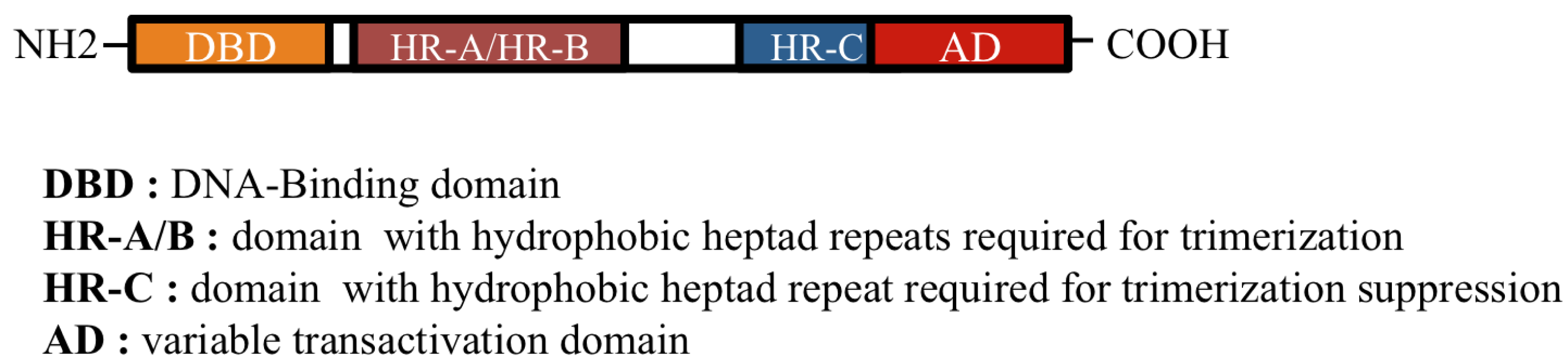
HSF family members (HSF1, HSF2, HSF3, and HSF4) display unique as well as overlapping functions, show tissue-specific patterns of expression, are submitted to numerous post-translational modifications, and interact with many protein partners [<xref ref-type="bibr" rid="b14-cancers-03-01158">14</xref>–<xref ref-type="bibr" rid="b18-cancers-03-01158">18</xref>]. HSF proteins are composed of functional domains, among which the most conserved is the amino terminal DNA-binding domain (DBD). Upon heat shock, thanks to its oligomerization domain (heptad repeats HR-A/B), adjacent to the DBD, the latent inactive monomeric HSF1 forms a trimer and is subjected to extensive post-translational modifications. In normal conditions, this assembling is prevented by another heptad repeat domain HR-C [<xref ref-type="bibr" rid="b19-cancers-03-01158">19</xref>,<xref ref-type="bibr" rid="b20-cancers-03-01158">20</xref>], which specifically interacts with HR-A/B domains and maintains HSF1 in a monomeric conformation (<xref ref-type="fig" rid="f1-cancers-03-01158">Figure 1</xref>). The deletion of the HR-C domain results in constitutive trimerization which may explain why HSF4, that lacks this domain, has a constitutive DNA-binding ability [<xref ref-type="bibr" rid="b19-cancers-03-01158">19</xref>]. HSF2 and HSF3 are present as a dimer [<xref ref-type="bibr" rid="b21-cancers-03-01158">21</xref>] and share the ability to bind DNA sequences, called Heat Shock Elements (HSE), originally defined as succession of several inverted repeats of the pentanucleotide motif NGAAN and recently refined by genome-wide analysis [<xref ref-type="bibr" rid="b3-cancers-03-01158">3</xref>]. HSF family members exhibit different binding site preferences in terms of architecture and composition of the HSE, thereby allowing a great diversity in the regulation of specific target genes [<xref ref-type="bibr" rid="b4-cancers-03-01158">4</xref>,<xref ref-type="bibr" rid="b22-cancers-03-01158">22</xref>,<xref ref-type="bibr" rid="b23-cancers-03-01158">23</xref>]. In this respect, HSFs are able to act as both activators and repressors depending on the environmental conditions and the targeting genes [<xref ref-type="bibr" rid="b1-cancers-03-01158">1</xref>–<xref ref-type="bibr" rid="b3-cancers-03-01158">3</xref>]. In addition, subtle crosstalks exist between HSFs, which, together with the combinatorial possibilities allowed by the different post-translational modifications, multiply the levels of fine-tuning the expression of HSF target genes. Furthermore, in specific conditions, HSF1 and HSF2 can form heterotrimers through which HSF2 transiently modulates the very potent and dominant HSF1 activity on its target genes [<xref ref-type="bibr" rid="b24-cancers-03-01158">24</xref>–<xref ref-type="bibr" rid="b28-cancers-03-01158">28</xref>].</p>
<p>Upon stress, HSF1 and HSF4 are able to recruit chromatin remodelers SWI/SNF [<xref ref-type="bibr" rid="b29-cancers-03-01158">29</xref>,<xref ref-type="bibr" rid="b30-cancers-03-01158">30</xref>] and to target stress-specific histone modifications [<xref ref-type="bibr" rid="b31-cancers-03-01158">31</xref>]. In human cells, HSF1 was shown to direct the transcription of satellite III DNA in pericentromeric heterochromatic regions of particular human chromosomes that are strikingly characterized by an epigenetical status euchromatin. Therefore, stress-activated HSF1 can trigger the conversion of a partially constitutive heterochromatin status to a transcription competent status [<xref ref-type="bibr" rid="b32-cancers-03-01158">32</xref>–<xref ref-type="bibr" rid="b34-cancers-03-01158">34</xref>]. Likewise, HSF2 has an epigenetic role, strictly speaking, in the heritability of a decondensed chromatin status on the <italic>Hsp70</italic> gene. During mitosis, HSF2 inhibits chromatin condensation through interaction with condensin, in a process called ≪bookmarking≫ [<xref ref-type="bibr" rid="b35-cancers-03-01158">35</xref>].</p></sec>
<sec>
<label>2.2.</label>
<title>Expression and Physiological Functions</title>
<p>Besides their role in stress response, HSFs assume specific and non-overlapping functions which enlighten their role in cancer. They regulate proliferation, asymmetric division, differentiation, migration, and survival by either activating or repressing their target genes (<xref ref-type="table" rid="t1-cancers-03-01158">Table 1</xref>).</p>
<list list-type="simple">
<list-item>
<label>-</label>
<p><bold>HSF1.</bold> The ubiquitously expressed HSF1, known as a master controller of the cellular-response to different stress conditions, is a maternal factor, stored in the oocyte, and essential for oogenesis and preimplantation development through the regulation of <italic>Hsp90</italic>α gene expression [<xref ref-type="bibr" rid="b36-cancers-03-01158">36</xref>–<xref ref-type="bibr" rid="b38-cancers-03-01158">38</xref>]; reviewed in [<xref ref-type="bibr" rid="b5-cancers-03-01158">5</xref>]. It is also required for the development and maintenance of tissues including the adult brain [<xref ref-type="bibr" rid="b39-cancers-03-01158">39</xref>–<xref ref-type="bibr" rid="b41-cancers-03-01158">41</xref>] germ cells [<xref ref-type="bibr" rid="b42-cancers-03-01158">42</xref>–<xref ref-type="bibr" rid="b44-cancers-03-01158">44</xref>], ciliated cells [<xref ref-type="bibr" rid="b45-cancers-03-01158">45</xref>], and immune cells [<xref ref-type="bibr" rid="b46-cancers-03-01158">46</xref>,<xref ref-type="bibr" rid="b47-cancers-03-01158">47</xref>], regulating both <italic>Hsp</italic> and non-Hs<italic>p</italic> target genes.</p></list-item>
<list-item>
<label>-</label>
<p><bold>HSF2.</bold> As mentioned above, HSF2 is able to finely tune the HSR mediated by HSF1. However, its role in stress is more striking upon proteasome inhibition, because it regulates the expression of proteasome subunits [<xref ref-type="bibr" rid="b6-cancers-03-01158">6</xref>]. In the context of differentiation, as in stress, a pool of nuclear HSF2 can associate with HSF1 and modulate the expression of heat-shock proteins [<xref ref-type="bibr" rid="b24-cancers-03-01158">24</xref>,<xref ref-type="bibr" rid="b26-cancers-03-01158">26</xref>] as well as noncoding satellite III RNA in nuclear stress bodies (unique subnuclear organelles which form in response to heat shock) [<xref ref-type="bibr" rid="b27-cancers-03-01158">27</xref>]. Interestingly, HSF2 is mainly associated with brain development and gametogenesis [<xref ref-type="bibr" rid="b48-cancers-03-01158">48</xref>–<xref ref-type="bibr" rid="b50-cancers-03-01158">50</xref>] where it displays a spatiotemporal expression pattern [<xref ref-type="bibr" rid="b5-cancers-03-01158">5</xref>,<xref ref-type="bibr" rid="b51-cancers-03-01158">51</xref>]. In the developing brain cortex, HSF2 regulates multiple aspects of neuronal migration through gene expression, like p35, a crucial activator of the serine/threonine kinase, Cdk5. In spermatogenesis, HSF2 cell-specific expression is negatively regulated by microRNA miR18, which belongs to the Oncomir-1 cluster associated with tumorigenesis [<xref ref-type="bibr" rid="b52-cancers-03-01158">52</xref>]. HSF2 and HSF1 can form heterotrimers in testis [<xref ref-type="bibr" rid="b27-cancers-03-01158">27</xref>] and share common target genes during spermatogenesis, in particular sex chromosomal multicopy genes [<xref ref-type="bibr" rid="b15-cancers-03-01158">15</xref>,<xref ref-type="bibr" rid="b53-cancers-03-01158">53</xref>]. HSF2 binding to its target genes during spermatogenesis is associated with histone H4 acetylation [<xref ref-type="bibr" rid="b53-cancers-03-01158">53</xref>]. The inactivation of both HSF1 and HSF2 leads to completely male sterility, reinforcing their intertwined functions, which also rely on global chromatin remodeling.</p></list-item>
<list-item>
<label>-</label>
<p><bold>HSF3.</bold> Initially only identified in the chicken (cHSF3), this ubiquitously expressed factor has been recently identified in the mouse (mHSF3). In human, HSF3 might be present as a pseudogene, since no transcripts have been detected [<xref ref-type="bibr" rid="b54-cancers-03-01158">54</xref>]. In contrast to others HSFs, HSF3 does not display the same regulatory role in different species [<xref ref-type="bibr" rid="b55-cancers-03-01158">55</xref>]. Whereas cHSF3 is the major HSF in response to heat shock and therefore the main inducer of HSPs, mHSF3 does not induce classical <italic>hsp</italic> genes, but instead induces the expression of other stress-responsive genes, raising the possibility that it could regulate distinct sets of genes in development or longevity [<xref ref-type="bibr" rid="b18-cancers-03-01158">18</xref>].</p></list-item>
<list-item>
<label>-</label>
<p><bold>HSF4.</bold> Mainly expressed in the human heart, brain, skeletal muscle, and pancreas [<xref ref-type="bibr" rid="b56-cancers-03-01158">56</xref>], HSF4 has been shown to be required for the development and maintenance of sensory organs [<xref ref-type="bibr" rid="b5-cancers-03-01158">5</xref>,<xref ref-type="bibr" rid="b57-cancers-03-01158">57</xref>,<xref ref-type="bibr" rid="b58-cancers-03-01158">58</xref>]. During this process, HSF4 either cooperates or competes with HSF1 for common target genes, including members of the Fibroblast Growth Factor (FGF) family, in a cell-specific manner in lens and olfactory epithelium [<xref ref-type="bibr" rid="b45-cancers-03-01158">45</xref>,<xref ref-type="bibr" rid="b59-cancers-03-01158">59</xref>]. Recently, it has been reported that HSF4 binds to more flexible consensus HSE, which are not found in the promoter proximal regions of <italic>Hsp</italic> genes, but are preferentially located in introns, exons and distal regions. HSF4 binding is associated with reduced histone H3K9 methylation. Among the different targets of HSF4 after a heat shock, 33% are non-classical heat-shock genes [<xref ref-type="bibr" rid="b4-cancers-03-01158">4</xref>]. Substantial proportions of these regions are also bound by HSF1, reinforcing the concept of their mutual interactions.</p></list-item></list>
<p>Overall, not only HSF1, but also other HSF family members, play significant roles in the induction of non-classical genes in response to heat shock or in developmental conditions. In this respect, HSFs regulate new target genes which include proteostasis genes (proteasome subunits), growth factor genes (FGF, Leukemia Inhibitory Factor (LIF)), and genes that are directly or indirectly involved in cytoskeleton dynamics (<italic>Hsp90</italic> and cortical actin in eggs [<xref ref-type="bibr" rid="b37-cancers-03-01158">37</xref>,<xref ref-type="bibr" rid="b38-cancers-03-01158">38</xref>]; <italic>p35/p39/Cdk5</italic> [<xref ref-type="bibr" rid="b50-cancers-03-01158">50</xref>]; <italic>b<sub>IV</sub></italic> <italic>tubulin</italic> in ciliary beating activity [<xref ref-type="bibr" rid="b60-cancers-03-01158">60</xref>]; <italic>Bfsp</italic>, lens specific intermediate filaments [<xref ref-type="bibr" rid="b61-cancers-03-01158">61</xref>]). HSFs also direct the establishment of epigenetic marks and might impact genome structure, <italic>i.e.</italic> chromatin condensation state in spermatogenesis; histone methylation or acetylation status and likely retrotransposons [<xref ref-type="bibr" rid="b4-cancers-03-01158">4</xref>,<xref ref-type="bibr" rid="b53-cancers-03-01158">53</xref>,<xref ref-type="bibr" rid="b62-cancers-03-01158">62</xref>].</p></sec></sec>
<sec>
<label>3.</label>
<title>HSFs in Cancers</title>
<sec>
<label>3.1.</label>
<title>Multiple Roles of HSF1 in Cancer</title>
<sec>
<label>3.1.1.</label>
<title>Role of HSF1 in the Initiation and Maintenance of Transformed Phenotypes</title>
<p>HSF1 has been involved in the etiology of cancer by its multiple effects in facilitating transformation and tumor invasiveness in response to diverse oncogenic stimuli [<xref ref-type="bibr" rid="b10-cancers-03-01158">10</xref>]. In this respect, <italic>Hsf1</italic><sup>−/−</sup> mice show lower incidence of tumors induced by mutations of the RAS oncogene or a hot spot mutation in the tumor suppressor p53, and they show improved survival [<xref ref-type="bibr" rid="b10-cancers-03-01158">10</xref>]. HSF1 is also crucial for cell transformation and tumorigenesis induced by the human epidermal growth factor receptor-2 (HER2), an oncogene responsible for breast tumors aggressiveness. Knockdown of HSF1 leads to growth arrest and senescence of HER-2-expressing cells [<xref ref-type="bibr" rid="b63-cancers-03-01158">63</xref>]. Moreover, HSF1 is required for the maintenance of the transformed phenotype in: (i) established oncogenic cell lines; (ii) breast cells lines with progressive oncogenic states (<italic>i.e.</italic>, primary human mammary epithelial (PHME) cells; immortalized human mammary epithelial (HME) cells; fully transformed and tumorigenic HME cells (HMLER); [<xref ref-type="bibr" rid="b64-cancers-03-01158">64</xref>]); (iii) a collection of breast cell lines derived from spontaneous human tumors, with various p53 status (MCF-7 or various mutant alleles BT-474, MDAMB-231, and T47D); (iv) malignant cells of diverse histological origins either derived from human tumors (HeLa (cervix), PC-3 (prostate), and S462 and 90-8 (peripheral nerve sheath) or derived by <italic>in vitro</italic> transformation (293T; kidney). Furthermore, in immortalized MEFs, HSF1 is essential for basal and EGF-induced migration, a process crucial for tumor invasion and metastasis [<xref ref-type="bibr" rid="b65-cancers-03-01158">65</xref>].</p>
<p>HSF1 is often elevated in human cancer cells compare to normal cells. However, somatic mutations in <italic>Hsf1</italic> have not yet been identified in human cancers, and overexpression of HSF1 does not lead to transformation, as mutant RAS does. Here, HSF1 does not display typical oncogenic features <italic>per se</italic> but represents a mechanistic and therapeutic challenging target. It is possible that the core function pathways regulated by HSF1 in normal cells might be massively mobilized in cancer cells, like signal transduction, ribosome biogenesis, translation and glucose metabolism [<xref ref-type="bibr" rid="b10-cancers-03-01158">10</xref>]. Indeed, cancer cells growth and development highly depend on normal cellular functions governed by genes, which are not typical cancer related genes, and through which HSF1 can promote tumorigenesis.</p>
<p>The unique HSF present in fission yeast cells is essential for normal growth and drives the transcription of target genes that encode proteins with a broad range of biological functions, including protein folding and degradation, energy generation, protein trafficking, maintenance of cell integrity, small molecules transport, cell signaling and transcription [<xref ref-type="bibr" rid="b66-cancers-03-01158">66</xref>,<xref ref-type="bibr" rid="b67-cancers-03-01158">67</xref>]. Conversely, in murine cells, HSF1 is more dispensable for cell growth and survival, although <italic>Hsf1</italic><sup>−/−</sup> mice show defects in postnatal growth and placenta formation, suggesting that in normal cells HSF1, while a major actor in the heat shock response, is also important for other cellular process [<xref ref-type="bibr" rid="b68-cancers-03-01158">68</xref>].</p>
<p>In normal mammalian cells, HSF1 is known to regulate <italic>Hsp</italic> genes, but also a wide range of non-<italic>Hsp</italic> genes upon stress or differentiation [<xref ref-type="bibr" rid="b2-cancers-03-01158">2</xref>,<xref ref-type="bibr" rid="b3-cancers-03-01158">3</xref>]. In aggressive cancer cells, HSF1 is expressed at high levels, which could amplify its activity and broaden the spectrum of its targets. By analogy to what happens in yeast, HSF1 could regulate central functions of the cell biology, in addition to the already known HSP expression. However, although the levels of HSPs (especially HSP27, HSP70 and HSP90) are elevated in different types of cancers, there is not always a correlation between HSF1 constitutive activation and HSP expression. In this way, genetic knockdown of HSF1 fails to induce a decrease in the levels of HSPs in some cancer cell lines [<xref ref-type="bibr" rid="b69-cancers-03-01158">69</xref>]. It is believed that the elevated expression of HSF1 in cancer cells does not lead to a kind of global “heat shock response”, but only specific members of the HSP family are induced in a tumor-dependent manner. That explains why HSP expression in cancer cells displays considerable variation, depending on the HSP member and the tumor characteristics. For instance, in HER-2 (human epidermal growth factor receptor-2 oncogene) expressing cells, in aggressive breast cancer models, HSF1 knockdown is accompanied by a specific down-regulation of HSP27 and HSP70 expression [<xref ref-type="bibr" rid="b63-cancers-03-01158">63</xref>].</p></sec>
<sec>
<label>3.1.2.</label>
<title>HSF1 and Prostate Cancers</title>
<p>HSF1 expression has been shown to be elevated in prostate carcinoma compared to its normal counterpart [<xref ref-type="bibr" rid="b70-cancers-03-01158">70</xref>]. An elevated protein expression of HSF1 and some HSPs was reported in the aggressively malignant cell lines DU145 and CA-HPV-10. However, no difference in the RNA level was detected suggesting that HSP induction was not mediated by HSF1-dependent transcription. In addition, growth of PC3 cells <italic>in vivo</italic>, as tumor xenografts, was accompanied by a marked decrease in HSPs expression (HSP27, HSP70, HSP60, HSP90) whereas HSF level was not modulated. In a PC3 metastatic variant (PC3M), a strong increase in HSF1 mRNA was observed, as well as an increase in HSF1 protein and nuclear localization [<xref ref-type="bibr" rid="b71-cancers-03-01158">71</xref>]. Overexpression of HSF1 in non-metastatic PC-3 cells was accompanied by an upregulation of HSP27 at the protein level, but HSP70 and HSP90 were not affected, once again pointing out that the elevated expression of HSPs in cancer cells does not always depend on HSF1. Interestingly, in prostate cancer tissue, HSP27 upregulation, but not HSP70 or HSP90, was significantly associated with clinicopathological factors [<xref ref-type="bibr" rid="b72-cancers-03-01158">72</xref>]. In prostate cancer cells, HSF1 also influences the development of an aneuploid state and mitotic progression [<xref ref-type="bibr" rid="b73-cancers-03-01158">73</xref>].</p></sec>
<sec>
<label>3.1.3.</label>
<title>HSF1 and Breast Cancers</title>
<p>HSF1 may partly participate in breast cancer progression by inducing specific HSPs, mainly HSP27 [<xref ref-type="bibr" rid="b74-cancers-03-01158">74</xref>]. In breast cancer, the highly malignant factor heregulin beta-1 (HRGβ1) is a secreted factor, which binds to c-ErbB-3 and -4 receptors and provokes the recruitment of c-erbB-2 and receptors heterodimerization. The binding of HRGβ1 to the cell surface induces an increase in HSF1 levels that results in anchorage-independent growth and protection from cisplatin-induced apoptosis mediated by HRGβ1 [<xref ref-type="bibr" rid="b74-cancers-03-01158">74</xref>]. One aspect of this process relies on the inhibition of GSK3, a kinase that antagonized HSF1 activation. Part of the anti-apoptotic effects of HSF1 seems to operate <italic>via</italic> activation of the <italic>Hsp</italic>70 promoter and therefore likely involves HSPs. Remarkably a crosstalk between the beta-catenin/Wnt pathway and the heat shock cascade has been identified in breast cancer tumors with high metastatic potential. Indeed, the formation of a complex between beta-catenin, HSP27 and HSF1 has been detected in breast cancer biopsies. The striking presence of beta-catenin in the cytoplasm (and not only at the membrane) co-expressed with HSP27 and with HSF1 being also nuclear could have some clinical relevance in terms of prognosis [<xref ref-type="bibr" rid="b75-cancers-03-01158">75</xref>]. Notably, HSF1 plays an additional role in the etiology of breast cancer. Unlike normal cells, cancer cells preferentially catabolize glucose by glycolysis, thereby producing high levels of lactic acid. For instance, Zhao <italic>et al.</italic> have shown that ErbB2 promotes glycolysis in a HSF1-dependent manner. Overexpression of ErbB2 increases the expression of HSF1, which binds to <italic>lactate dehydrogenase A</italic> (<italic>LDH-A</italic>) promoter and activates its transcription [<xref ref-type="bibr" rid="b76-cancers-03-01158">76</xref>], allowing the metabolization of pyruvate to lactate.</p></sec>
<sec>
<label>3.1.4.</label>
<title>HSF1 and Gastro-Intestinal Cancers</title>
<p>The search for activation of signal transduction pathways in sporadic colorectal cancers (CRC) revealed an increase in the mRNA levels for heat shock and NFκB pathway genes [<xref ref-type="bibr" rid="b77-cancers-03-01158">77</xref>]. Namely, an increase of <italic>Hsf1</italic> mRNA in 86% of patients was reported in sporadic colorectal cancer. In that context, HSF1 upregulation in patients was accompanied by an elevated expression of HSP27, HSP90 but also of iNOS (NO synthase; 63%). In addition, HSF1, through the induction of the HSP70 co-chaperone BAG-3, which stabilizes the level of anti-apoptotic Bcl-2 family members, facilitates colon cancer cell survival during pro-apoptotic stress [<xref ref-type="bibr" rid="b78-cancers-03-01158">78</xref>]. The oncogenesis of hereditary CRC cancers is believed to involve four signal transduction pathways: (a) the APC-β-catenin-TCF-myc (Wnt) pathway; (b) the microsatellite unstable pathway; (c) the p53 pathway; and (d) the estrogen receptor hypermethylation pathway [<xref ref-type="bibr" rid="b79-cancers-03-01158">79</xref>,<xref ref-type="bibr" rid="b80-cancers-03-01158">80</xref>]. Although no data are currently available on the potential role of HSF1 in hereditary CRC, at least two of these pathways (Wnt and p53) are known to be affected by HSF1 [<xref ref-type="bibr" rid="b77-cancers-03-01158">77</xref>,<xref ref-type="bibr" rid="b79-cancers-03-01158">79</xref>,<xref ref-type="bibr" rid="b80-cancers-03-01158">80</xref>].</p>
<p>In gastro-intestinal cancer, the level of HSPs is often upregulated with a high variability and without evident correlation with HSF1 level [<xref ref-type="bibr" rid="b81-cancers-03-01158">81</xref>]. Instead, the increase observed in HSF1 expression in gastro-intestinal cancer tissues, drives the repression of the pro-apoptotic protein, X-linked inhibitor of apoptosis protein (XIAP)-associated factor-1 (XAF-1) [<xref ref-type="bibr" rid="b82-cancers-03-01158">82</xref>]. Thus, HSF1 can impair the apoptotic pathway in cancer cells, at least partly, in a HSP independent manner.</p>
<p>Interestingly, the protein TC1 (a positive regulator of the signaling Wnt/beta-catenin <italic>via</italic> inhibition of Chibby), which is upregulated in an aggressive subtype of gastric cancers, correlates with poor prognosis and has been reported to induce a heat shock response in cancer cells by favoring HSF1 expression. Moreover, HSF1 and TC1 mutually activate each other [<xref ref-type="bibr" rid="b83-cancers-03-01158">83</xref>,<xref ref-type="bibr" rid="b84-cancers-03-01158">84</xref>].</p></sec></sec>
<sec>
<label>3.2.</label>
<title>Role of HSF2 and HSF4 in Glioma and Neuroblastoma</title>
<p>HSP70, HSC70 and HSP90 are found expressed in the tumor parenchyma of all high-grade and most low-grade gliomas, including oligodendrogliomas [<xref ref-type="bibr" rid="b85-cancers-03-01158">85</xref>]. A recent work from Mustafa <italic>et al.</italic>, has reported an overexpression of HSF2 (and a modestly elevated level of HSF1) in different stages of glial tumorigenesis, compared to normal brain. The potential involvement of HSF2 in glioma is interesting considering that HSF2 is involved in central nervous system development [<xref ref-type="bibr" rid="b5-cancers-03-01158">5</xref>].</p>
<p>Concerning HSF4, while no differences in its expression were found in low-grade glioma and normal brain, it was significantly downregulated in glioblastoma [<xref ref-type="bibr" rid="b86-cancers-03-01158">86</xref>].</p>
<p>Neuroblastoma (NB) and Ewing's sarcoma (ES) represent the most common extracranial solid tumors of neuroectodermal origin of childhood. While NB and ES cells globally showed very similar protein expression patterns, GRP78, GRP75, HSC70, HSP70, HSP47, HSP90α, and HSP27 are markedly more expressed in NB cell lines. To date, there is no data related to HSF expression or activity in this type of cancer although it would be worth studying since HSP profile is largely upregulated.</p>
<p>In addition, in human neuroblastoma, the dual-specificity phosphatase 26 (DUSP26) is overexpressed. DUSP26 has been described to inhibit HSF4 phosphorylation induced by Mitogen Anctivated Protein Kinases (MAPK), thereby negatively affecting its ability to bind DNA [<xref ref-type="bibr" rid="b87-cancers-03-01158">87</xref>]. Therefore, the negative control exerted by DUSP26 on HSF4, altogether with the inverse correlation in the expression of these two proteins in neuroblastoma, may contribute to repress HSF4 activity in neuroblastoma and its potential crosstalk with HSF1.</p></sec>
<sec>
<label>3.3.</label>
<title>Potential Role for HSF3</title>
<p>The expression of HSF3 in the chicken is regulated by the proto-oncogene c-Myb, thus favoring HSP expression, as well as cell proliferation. Moreover, Tanikawa <italic>et al.</italic> reported that a mutated form of p53 is able to inhibit c-Myb mediated cHSF3 transcription allowing tumor progression [<xref ref-type="bibr" rid="b88-cancers-03-01158">88</xref>]. Since in humans HSF3 is not expressed, it would be worth studying whether a relation between other HSFs and c-Myb is maintained.</p>
<p>In summary, the principal proteins involved in cancer that have been shown to affect HSF expression or activity are indicated in <xref ref-type="table" rid="t2-cancers-03-01158">Table 2</xref>.</p></sec></sec>
<sec>
<label>4.</label>
<title>HSFs and Cancer Related Targets</title>
<p>
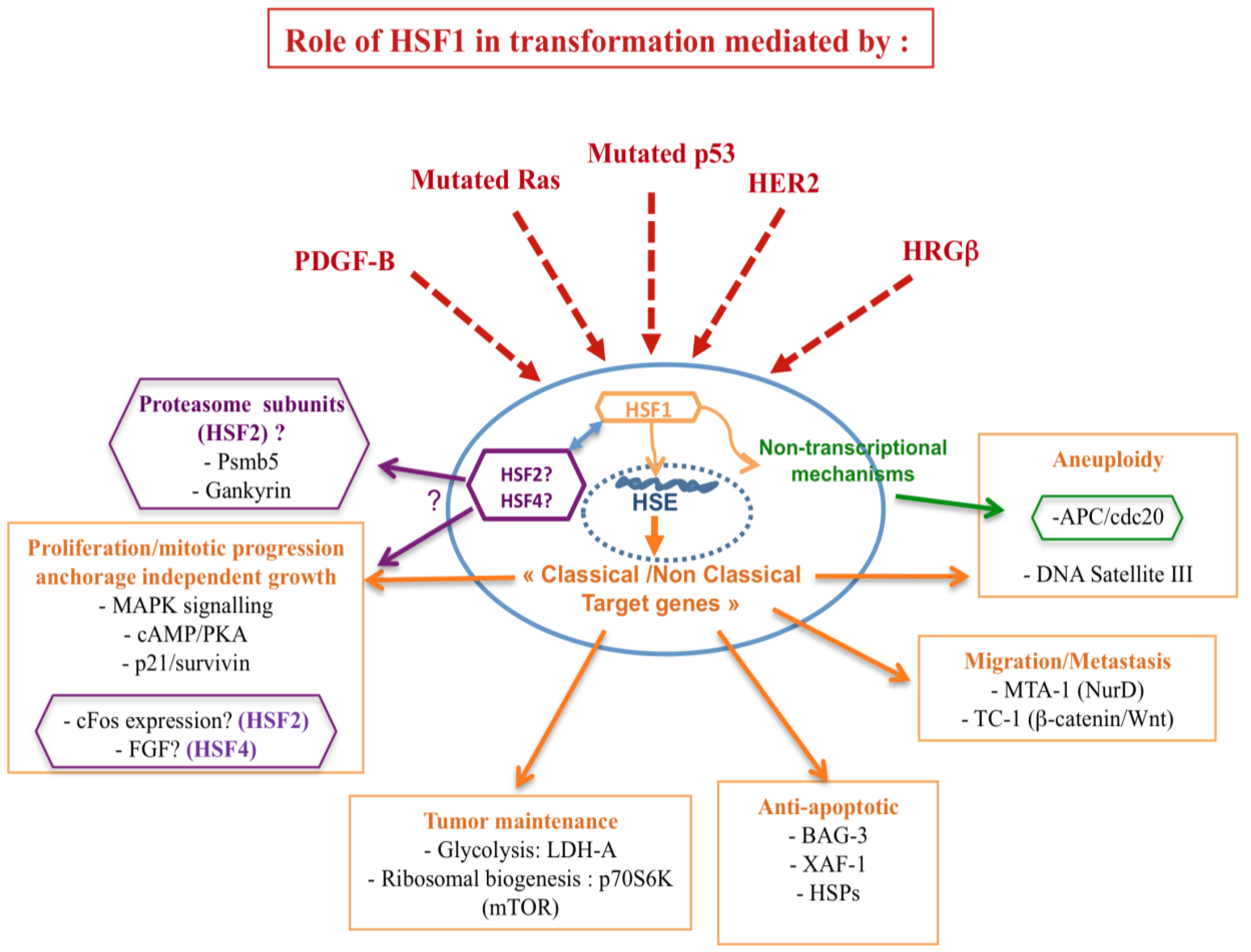
In light of the involvement of HSFs in mediating tumorigenesis, convincing evidence refers to the impact of HSF1 on the tumor suppressor p53 and the oncogene RAS. Herein, p53 and Ras, pivotal integrators of signaling pathways and key regulators of cell fate decisions, are the two most altered genes in human cancers. Moreover, HSF1 seems to participate to the metastatic potential of the cancer cell through cooperation with different metastatic related genes, such as the prometastatic co-repressor gene MTA1. It is noteworthy that HSF1 possesses additional properties in cancer, such as the enhancement of pro-malignant signaling pathways, involving PKA and TOR [<xref ref-type="bibr" rid="b10-cancers-03-01158">10</xref>]. In addition, HSFs act via targets involved in the establishment of genomic instability and epigenetic mechanisms (<xref ref-type="fig" rid="f2-cancers-03-01158">Figure 2</xref>).</p>
<sec>
<label>4.1.</label>
<title>HSF1, Tumor Suppressors, and Oncogenes</title>
<sec>
<label>4.1.1.</label>
<title>Tumor Suppressor p53</title>
<p>The first direct <italic>in vivo</italic> evidence of HSF1 implication in the development of spontaneous tumors arises from <italic>p53</italic><sup>−/−</sup> mice. The rapid tumor evolution observed in mice lacking p53 mainly results in lymphoma, while <italic>p53</italic><sup>−/−</sup><italic>Hsf1</italic><sup>−/−</sup> mice rarely develop lymphomas, but rather succumb to other types of cancers, like testicular carcinoma [<xref ref-type="bibr" rid="b89-cancers-03-01158">89</xref>]. Tumor suppressor p53 is frequently inactivated by genetic mutations in different cancers [<xref ref-type="bibr" rid="b90-cancers-03-01158">90</xref>]. In this context, Dai <italic>et al.</italic> have shown that HSF1 deficiency dramatically reduces spontaneous tumor formation in mice carrying a common, dominant-negative mutation of the <italic>p53</italic> gene, whereas <italic>Hsf1</italic><sup>+/+</sup> and <italic>Hsf1</italic><sup>+/−</sup> mice bearing dominant-negative mutation of p<italic>53</italic> develop a broad spectrum of tumor types (sarcomas, lymphoma, carcinomas; [<xref ref-type="bibr" rid="b10-cancers-03-01158">10</xref>]). This was confirmed by <italic>Hsf1</italic> knock down, using specific siRNAs, in different mouse and human cell systems [<xref ref-type="bibr" rid="b10-cancers-03-01158">10</xref>]. These different results observed in these complementary works [<xref ref-type="bibr" rid="b10-cancers-03-01158">10</xref>,<xref ref-type="bibr" rid="b89-cancers-03-01158">89</xref>] may be explained by differences in the models used, involving distinct mouse genetic background, and the use of clinically relevant <italic>p53</italic> mutation <italic>versus p53</italic> null strategy.</p>
<p>Dysfunction in the proteasome pathway can lead to many disorders including cancers [<xref ref-type="bibr" rid="b91-cancers-03-01158">91</xref>], and proteasome inhibitors are currently used in therapeutical approaches [<xref ref-type="bibr" rid="b92-cancers-03-01158">92</xref>]. Recently, Lecomte <italic>et al.</italic> have reported that some proteasome subunits, such as Psmb5 and Gankyrin, were significantly downregulated in <italic>Hsf2</italic><sup>−/−</sup> MEFs [<xref ref-type="bibr" rid="b6-cancers-03-01158">6</xref>]. Interestingly, Gankyrin is an oncoprotein involved in p53 degradation, thus suggesting a potential role of HSF2 in the regulation of the tumor suppressor p53.</p></sec>
<sec>
<label>4.1.2.</label>
<title>Oncogene RAS and HSF1</title>
<p>Compared with wild-type MEFs, <italic>Hsf1</italic><sup>−/−</sup> MEFs are refractory to proliferation and resistant to focus formation, driven by oncogenic H-RASV12D or by the proto-oncogene PDGF-B, which are both mitogenic signal transducers [<xref ref-type="bibr" rid="b10-cancers-03-01158">10</xref>]. In contrast, expression of c-MYC and LTA (regulator of cell-cycle progression, which are not expected to increase proliferation in already-immortalized cells, but predisposing cells to apoptosis) does not induce proliferation in immortalized <italic>Hsf1</italic><sup>+/+</sup> MEFs. Accordingly, <italic>Hsf1</italic><sup>−/−</sup> MEFs show no enhanced survival in response to RAS and PDGF/B expression, but reduced survival in response to c-MYC and LTA expression [<xref ref-type="bibr" rid="b10-cancers-03-01158">10</xref>].</p></sec>
<sec>
<label>4.1.3.</label>
<title>Oncogene HER2 and HSF1</title>
<p>HSF1 is also required for cell transformation and tumorigenesis induced by the oncogene HER2 (Human Epidermal growth factor Receptor-2), which is responsible for aggressive breast tumors. Indeed, knockdown of <italic>Hsf1</italic> prevents neoplastic transformation (foci formation or tumor growth in xenografts) induced by HER2 expression in untransformed human mammary epithelial MCF-10A cells. This suggests that the proliferation of HER2-expressing cells is critically dependent on HSF1 [<xref ref-type="bibr" rid="b63-cancers-03-01158">63</xref>]. Strikingly, this anti-tumorigenic effect of HSF1 downregulation was associated with HER2-induced upregulation of the cyclin-dependent kinase inhibitor p21, a major regulator of senescence in cancer cells [<xref ref-type="bibr" rid="b93-cancers-03-01158">93</xref>], and accompanied by a decrease in the mitotic regulator survivin, which is also involved in growth arrest and senescence. Survivin is a member of the inhibitors of apoptosis (IAPs) family and a critical regulator of mitosis by modulation of aurora B kinase [<xref ref-type="bibr" rid="b94-cancers-03-01158">94</xref>]. Its decrease is partially controlled by both HSF1 and p21 [<xref ref-type="bibr" rid="b63-cancers-03-01158">63</xref>]. In addition, HSF1-mediated control of breast cancer cell senescence was due, at least partly, to the regulation of HSP70 and HSP27 expression.</p></sec>
<sec>
<label>4.1.4.</label>
<title>Loss of Hormonal Control, Epigenetic and Metastasis-Related Targets of HSF1</title>
<p>The potential role of HSF1 in cancer metastasis was first reported in prostate cancer where HSF1 overexpression was correlated with aggressiveness of the tumors [<xref ref-type="bibr" rid="b70-cancers-03-01158">70</xref>,<xref ref-type="bibr" rid="b71-cancers-03-01158">71</xref>]. Since then, studies have shown that HSF1 is able to mediate tumorigenic effects through the recruitment of the prometastatic co-repressor gene MTA1 (a component of the NuRD complex containing the histone deacetylases HDAC1 and HDAC2) [<xref ref-type="bibr" rid="b95-cancers-03-01158">95</xref>]. MTA1 is expressed in numerous human cancers, including breast, prostate and gastro-intestinal cancers and its expression correlates with tumor aggressiveness and metastasis [<xref ref-type="bibr" rid="b96-cancers-03-01158">96</xref>–<xref ref-type="bibr" rid="b98-cancers-03-01158">98</xref>]. Herein, MTA1 protein expression is higher in hormone-refractory metastatic prostate cancer compared to clinically localized disease and benign prostatic tissues [<xref ref-type="bibr" rid="b97-cancers-03-01158">97</xref>].</p>
<p>Breast cancer cell can also escape from hormonal control, leading to higher metastatic potential. Khaleque <italic>et al.</italic>, have reported that HSF1 binds to the corepressor MTA1 in human cultured breast cancer cells and carcinoma samples. The HSF1-MTA1 complex, strongly induced by the transforming ligand heregulin (a ligand for erbB receptor from the EGF family), assembles on the chromatin of breast cancer cells and mainly binds to the promoter of estrogen responsive genes [<xref ref-type="bibr" rid="b95-cancers-03-01158">95</xref>]. Thus, HSF1 induces repression of estrogen-dependent gene transcription, an effect linked to cancer invasiveness.</p>
<p>Migration properties in correlation with metastasis potential were also shown to be controlled by HSF1. Mouse embryonic cells derived from <italic>Hsf1</italic><sup>−/−</sup> mice are deficient in both basal and EGF-mediated migration. This default in migration, which is partly due to the downregulation of EGF receptor and likely involves Ras, is associated with an impairment of the MAPK signaling pathway [<xref ref-type="bibr" rid="b65-cancers-03-01158">65</xref>].</p></sec>
<sec>
<label>4.1.5.</label>
<title>Cdc20/APC Genomic Instability and HSF1</title>
<p>As described above, HSFs have been described to modulate the structure of chromatin and to establish some epigenetic marks (see introduction). In particular HSF1, in association with HSF2, was shown to steer the transcription of the human DNA satellite III in the centromeric regions, establishing an epigenetic status resembling euchromatin [<xref ref-type="bibr" rid="b14-cancers-03-01158">14</xref>,<xref ref-type="bibr" rid="b99-cancers-03-01158">99</xref>,<xref ref-type="bibr" rid="b100-cancers-03-01158">100</xref>]. The involvement of HSF2 in the expression of satellite III DNA was further confirmed [<xref ref-type="bibr" rid="b27-cancers-03-01158">27</xref>]. Remarkably, transcription of satellite III DNA is associated with aneuploidy in several cancers such as ovarian cancers, melanoma or myeloma [<xref ref-type="bibr" rid="b101-cancers-03-01158">101</xref>–<xref ref-type="bibr" rid="b103-cancers-03-01158">103</xref>] and thus could be related to HSF status.</p>
<p>Accordingly, Lee <italic>et al.</italic> have recently shown that HSF1, via its regulatory domain, when overexpressed in radiation induced-fibrosarcoma cells, interacts with cdc20, the co-activator of anaphase-promoting complex (APC), and thus, inhibits the mitotic exit and the ubiquitination activity of APC on two key anaphase inhibitors, Cyclin B1 and securin. As a consequence, this non-transcriptional HSF1 effect leads to aneuploidy and multinucleated cells associated with micronuclei and genomic alteration [<xref ref-type="bibr" rid="b104-cancers-03-01158">104</xref>]. In prostate carcinoma cells, a dominant negative construct of HSF1 dramatically alters the DNA content of PC3 cells and inhibits aneuploidy and Cyclin B1 stabilization [<xref ref-type="bibr" rid="b73-cancers-03-01158">73</xref>]. Further, HSF1-mediated aneuploidy was facilitated in p53-defective cells. This phenomenon was associated to an increase in HSF1 activity through phosphorylation by a Polo-like kinase, as well as cdc20/HSF1 interaction [<xref ref-type="bibr" rid="b105-cancers-03-01158">105</xref>].</p>
<p>Altogether, HSF1 activity is hijacked in a pleiotropic manner by a large diversity of cancer cells and oncogenes to favor tumor initiation and progression. In sharp contrast, it is noteworthy that HSF1 knockout has a minimal effect on the proliferation/survival of normal primary human cells.</p></sec></sec>
<sec>
<label>4.2.</label>
<title>Potential Tumorigenic Functions for HSF2 and HSF4</title>
<p>In contrast to HSF1, there is little direct evidence on the involvement of HSF2 and HSF4 in tumorigenesis [<xref ref-type="bibr" rid="b106-cancers-03-01158">106</xref>]. Although they can play a role indirectly by their ability to modulate HSF1, several works pointed out their regulatory role in cancer related genes. During mitosis, HSF2 has been shown to bind and activate, not only the HSE promoter of the different <italic>Hsps</italic> (bookmarking), but also the promoter of the proto-oncogene c-Fos. The protein c-Fos is often up-regulated in tumor cells and is well-known for its oncogenic activity [<xref ref-type="bibr" rid="b107-cancers-03-01158">107</xref>]. Thereby, it is tempting to conclude that HSF2 might regulate the oncogenic potential of c-Fos [<xref ref-type="bibr" rid="b108-cancers-03-01158">108</xref>].</p>
<p>In addition, HSF2 is a labile protein whose degradation was recently shown to be regulated by anaphase-promoting complex/cyclosome (APC/C), a ubiquitin E3 ligase, which drives the degradation of cell cycle regulators in cycling cells by associating with the coactivators Cdc20 and Cdh1 [<xref ref-type="bibr" rid="b28-cancers-03-01158">28</xref>]. Therefore, APC/C, by its effect on HSF2, could be involved in a cell cycle-dependent manner in the modulation of HSF1 activity, which could have profound effects on cancer progression.</p>
<p>Finally, the oncomiR miR18 has been shown to regulate the expression of HSF2 during spermatogenesis [<xref ref-type="bibr" rid="b52-cancers-03-01158">52</xref>]. It could be possible that miR18 or other miRNA could regulate HSF2 expression in cancer cells, which, by modulating HSF1, could influence cancer initiation or progression.</p>
<p>HSF4 regulates normal cell proliferation and differentiation during mouse lens development, and belongs to the first group in the hierarchy of genes involved in this process [<xref ref-type="bibr" rid="b5-cancers-03-01158">5</xref>,<xref ref-type="bibr" rid="b59-cancers-03-01158">59</xref>]. Interestingly, HSF4, through its effect repressing the expression of the <italic>Fibroblast Growth factor-7</italic> (<italic>FGF-7</italic>) gene which encodes a specific mitogen for epithelial cells, is also important for governing ocular surface morphogenesis. Upregulation of FGF-7 has been reported to be associated with many human neoplastic tumors of epithelial origin including pancreatic, breast and gastric cancer, thus underlying the oncogenic role of FGF-7 [<xref ref-type="bibr" rid="b109-cancers-03-01158">109</xref>–<xref ref-type="bibr" rid="b111-cancers-03-01158">111</xref>]. Most importantly, Chimaka <italic>et al.</italic>, have demonstrated that overexpression of FGF-7 in Keratin 12-rtTA/tet-O-FGF-7 double transgenic mouse model, in which FGF-7 expression in corneal epithelium is driven by doxycycline treatment, induces epithelial hyperplasia that mimic the human ocular surface squamous neoplasia (OSSN) [<xref ref-type="bibr" rid="b112-cancers-03-01158">112</xref>]. In light of this work and the observation that FGF-7 is upregulated in <italic>Hsf4</italic><sup>−/−</sup> lens epithelial cells, a likely scenario is that HSF4 may interfere with the tumorigenic function of FGF-7 [<xref ref-type="bibr" rid="b4-cancers-03-01158">4</xref>].</p></sec></sec>
<sec>
<label>5.</label>
<title>The Inhibition of HSFs in Cancer Therapy</title>
<sec>
<label>5.1.</label>
<title>Targeting HSF1</title>
<p>HSF1 knockdown experiments in cancer cells demonstrate the interest of blocking this transcription factor in cancer therapy. A study using different human HSF1 targeted shRNAi constructs was performed using a collection of breast cancer cell lines that differed in their p53 status, HER2 expression, estrogen sensitivity and metastatic potential. All cancer cells were strongly affected by the HSF1–inhibitory hairpins [<xref ref-type="bibr" rid="b10-cancers-03-01158">10</xref>], Further, Rossi <italic>et al.</italic>, have determined that the ideal size target for siRNA mediated HSF1 silencing is 322–340 nucleotides. By generating a pSUPER-HSF1 vector, able to potently suppress HSF1 gene, they dramatically increased the sensitivity to hyperthermochemotherapy (combination of a Cisplatin treatment with heat-shock), leading to massive apoptosis of Hela cervical cancer cells [<xref ref-type="bibr" rid="b113-cancers-03-01158">113</xref>]. These approaches, albeit attractive, are still not established for clinical use. In this respect, an expanding array of small, drug-like compounds is currently available, some with potent HSF1-modulating activity in organisms, but only a few of them are in clinical trials (see below).</p></sec>
<sec>
<label>5.2.</label>
<title>Chemical Inhibitors of HSF1</title>
<p>Chemical inhibitors of HSF1 activation have been described including quercetin, genistein and the synthetic benzylidene lactams, KNK437. The natural product Stresgenin B has also been reported to inhibit induction of HSPs, but its mode of action is still unknown [<xref ref-type="bibr" rid="b114-cancers-03-01158">114</xref>]. Quercetin increases the sensitivity of drug-resistant cancer cells to anti-cancer agents [<xref ref-type="bibr" rid="b115-cancers-03-01158">115</xref>], and sensitizes cancer cells to hyperthermia, cisplatin [<xref ref-type="bibr" rid="b116-cancers-03-01158">116</xref>] and tiazofurin [<xref ref-type="bibr" rid="b117-cancers-03-01158">117</xref>]. HSF1 is strongly depleted following quercetin treatment and this effect appeared more marked in neuroblastoma cells. Moreover, a strict correspondence between the quercetin concentrations necessary to cause both HSP inhibition and doxorubicin sensitizing effect was observed, suggesting that neuroblastoma cell' resistance to doxorubicin treatment might be due to high levels of HSPs, under the control of HSF1. As for quercetin treatment, neuroblastoma cells were the most sensitive to HSF1 silencing effect. Knockdown of HSF1 in these cells strongly increased the anti-proliferative and pro-apoptotic effects induced by cisplatin [<xref ref-type="bibr" rid="b118-cancers-03-01158">118</xref>]. Although efficient in cancer experimental models, the clinical use of these inhibitors is limited because of their pleiotropic effects [<xref ref-type="bibr" rid="b119-cancers-03-01158">119</xref>].</p>
<p>To date, the most potent inhibitor of the HSF1 transcriptional function is Triptolide, a diterpene triepoxide from the plant Triptergium wilfordii. Triptolide does not interfere with the early events leading to trimer formation, hyperphosphorylation and DNA binding of HSF1 [<xref ref-type="bibr" rid="b120-cancers-03-01158">120</xref>]. Albeit toxic at high concentrations, a therapeutic dose of Triptolide has been defined to treat pancreatic cancer xenografts [<xref ref-type="bibr" rid="b121-cancers-03-01158">121</xref>]. Nevertheless, the precise mechanism of triptolide action remains to be investigated.</p>
<p>Several cancer treatment approaches, such as HSP90 inhibitors (geldanamycin) and proteasome inhibitors (Bortezomib), induced a proteotoxic stress that activates a pro-survival pathway, which explains the low efficiency of these therapies [<xref ref-type="bibr" rid="b122-cancers-03-01158">122</xref>]. Both HSP90 and proteasome inhibition are known to activate HSF1 and HSF2 [<xref ref-type="bibr" rid="b21-cancers-03-01158">21</xref>,<xref ref-type="bibr" rid="b25-cancers-03-01158">25</xref>,<xref ref-type="bibr" rid="b123-cancers-03-01158">123</xref>–<xref ref-type="bibr" rid="b125-cancers-03-01158">125</xref>]. From a high-throughput screening of molecular products that can interfere with the heat shock response, two new compounds were identified namely, NZ28 and emunin (emetine derivative). These two molecules exhibit little acute toxicity and allowed a strong sensitization of myeloma cells to proteasome and HSP90 inhibitors, as well as of prostate carcinoma cells to proteasome inhibitors [<xref ref-type="bibr" rid="b126-cancers-03-01158">126</xref>]. Their molecular action is not yet known but seems to involve post-transcriptional events downstream HSF1. Similarly, by a high content target based screening, Au <italic>et al.</italic>, have identified another small molecule inhibitor of HSF1 transcriptional activity [<xref ref-type="bibr" rid="b127-cancers-03-01158">127</xref>]. This molecule substantially hampers granules formation in heat-shocked Hela cells and significantly inhibits HSF1 phosphorylation. This effect is associated with a reduction in HSP70 and HSP90 expression. Recently, a malaria drug, quinacrine (QC) has been reported to prevent the induction of HSF1-dependent transcription of <italic>hsp70</italic> gene in a relative selective manner. The combinational treatment with a HSP90 inhibitor (17-DMAG) results in suppression of tumor growth in mouse syngeneic models [<xref ref-type="bibr" rid="b122-cancers-03-01158">122</xref>].</p></sec>
<sec>
<label>5.3.</label>
<title>Viral Approaches</title>
<p>Recently, an interesting anti-cancer approach has been developed based on the fact that HSF1 overexpression enhances the oncolytic effect of a replicative adenovirus. E1B55kD deleted oncolityc adenovirus, Adel55, was designed to achieve cancer specific cytotoxicity. A construct bearing Adel55-cHSF1 for tumor gene therapy, demonstrated that it favored oncolysis and viral replication by increasing its burst in breast cancer cells and SW620 xenografts [<xref ref-type="bibr" rid="b128-cancers-03-01158">128</xref>]. The future will tell of the feasibility of such a viral therapy in cancer patients.</p></sec>
<sec>
<label>5.4.</label>
<title>Targeting other HSFs</title>
<p>It has recently been reported that, in <italic>Hsf2</italic> knockout cells, the proteasome activity is lower compared to normal counterparts. This inhibitory effect is due to the regulatory role of HSF2 on the transcription of some proteasome subunits. Therapeutic use of proteasome inhibitors participates to the selection of the resistant clones, which are associated to the induction of beta5 and beta2 proteasome subunits expression [<xref ref-type="bibr" rid="b129-cancers-03-01158">129</xref>,<xref ref-type="bibr" rid="b130-cancers-03-01158">130</xref>]. Since in <italic>Hsf2</italic><sup>−/−</sup> MEFs one of these two subunits is downregulated, this suggests that inhibition of HSF2 could be beneficial for the sensitization of cancer cells to proteasome inhibitors. More importantly, HSF2 regulates Gankyrin expression that is responsible for p53 tumor suppressor degradation [<xref ref-type="bibr" rid="b6-cancers-03-01158">6</xref>]. Targeting HSF2 could stabilize p53 and favor apoptosis. Therefore, these observations suggest that specific HSF2 inhibitors could reduce the chemoresistance to proteasome inhibition that is so frequently observed in clinical studies. This pioneer study defines a new cancer research axe focused on HSF2 that definitely deserves further investigation.</p></sec></sec>
<sec>
<label>6.</label>
<title>Concluding Remarks</title>
<p>HSF1 expression is likely to be crucial for the “non-oncogene addiction” and the stressed phenotype of cancer cells. These stresses include proteotoxic and oxidative stress, frequent spontaneous DNA damage and aneuploidy [<xref ref-type="bibr" rid="b69-cancers-03-01158">69</xref>]. HSF1 by itself does not act as a classical oncogene or tumor suppressor. Neither enforced overexpression nor knockout directly drives transformation. HSF1 acts as a major multifaceted enhancer of tumorigenesis by regulating diverse core cellular functions that include proliferation, survival, protein synthesis and glucose metabolism, and therefore, is an attractive potential target in cancer therapy. However, some considerations must be taken when using HSF1 inhibitors because, although they seem beneficial in cancer treatment, they might, in parallel, accelerate neurodegenerative processes and aging. Indeed, HSF1, partly through the induction of chaperones that inhibit protein aggregation like HSP27, displays a protective effect against neurodegenerative diseases [<xref ref-type="bibr" rid="b131-cancers-03-01158">131</xref>,<xref ref-type="bibr" rid="b132-cancers-03-01158">132</xref>]. In this respect, therapeutic induction of HSF1-mediated stress response by non-toxic agents, like HSP90 inhibitors and Celastroloids, is currently being explored in Huntington disease [<xref ref-type="bibr" rid="b131-cancers-03-01158">131</xref>,<xref ref-type="bibr" rid="b132-cancers-03-01158">132</xref>]. It should also be mentioned here that hyperthermia, a strategy in cancer that could be considered as the opposite of HSF inhibition, has proved its therapeutical value in clinical trials and is still used in association with chemotherapeutic drugs in some countries (Germany, Italy, Japan, [<xref ref-type="bibr" rid="b133-cancers-03-01158">133</xref>]). Therefore, regarding the development of new tools/potential drugs targeting HSFs, it seems important that compounds do not cross the blood-brain barrier in order to minimize neurodegenerative risks and that they take into account HSF3 multiple post-translational modifications, as well as the formation of heterocomplexes between them. Ideally, rather than to simply activate or inhibit a given HSF, it would be more relevant/innovative to target a specific regulatory status, allowing a restrictive effect on designated signaling pathways.</p></sec></body>
<back>
<sec sec-type="display-objects">
<title>Figures and Tables</title>
<fig id="f1-cancers-03-01158" position="float">
<label>Figure 1.</label>
<caption>
<p>Structure of Heat Shock Factors (HSFs).</p></caption>
<graphic xlink:href="cancers-03-01158f1.gif"/></fig>
<fig id="f2-cancers-03-01158" position="float">
<label>Figure 2.</label>
<caption>
<p>HSF1 potential roles in cancer. Schematic model of the role of HSF1 in cancer transformation: HSF1 activity is hijacked by several oncogenes or mutated tumor suppressors (represented by dashed arrows) allowing activation of a set of genes involved in cancer development, <italic>i.e.</italic>, proliferation and anchorage independent growth, tumor maintenance, anti-apoptotic signaling, migration/metastasis, aneuploidy and probably other mechanisms, including non-transcriptional mechanisms related to the chromatin modification and genomic instability. Notably, there is growing evidence about the role of HSF2 and HSF4 in tumorigenesis since they are directly connected to different cancer related genes (<italic>FGF</italic>, <italic>cFos</italic>, Proteasome subunits). The double arrow represents the crosstalk existing between HSFs.</p></caption>
<graphic xlink:href="cancers-03-01158f2.gif"/></fig>
<table-wrap id="t1-cancers-03-01158" position="float">
<label>Table 1.</label>
<caption>
<p>HSF main characteristics.</p></caption>
<table frame="box" rules="all">
<thead>
<tr>
<th align="left" valign="top"/>
<th align="left" valign="top" content-type="background-color:#666699"><bold>HSF1</bold></th>
<th align="left" valign="top" content-type="background-color:#666699"><bold>HSF2</bold></th>
<th align="left" valign="top" content-type="background-color:#666699"><bold>HSF3</bold></th>
<th align="left" valign="top" content-type="background-color:#666699"><bold>HSF4</bold></th></tr></thead>
<tbody>
<tr>
<td align="left" valign="middle" content-type="background-color:#666699"><bold>Species Conservation</bold></td>
<td align="left" valign="middle" content-type="background-color:#CCFFFF">Human, Mouse, Chicken</td>
<td align="left" valign="middle" content-type="background-color:#CCFFFF">Chicken, Mouse</td>
<td align="left" valign="middle" content-type="background-color:#CCFFFF">Human, Mouse, Chicken</td></tr>
<tr>
<td align="left" valign="middle" content-type="background-color:#666699"><bold>Oligomerization</bold></td>
<td align="left" valign="middle" content-type="background-color:#CCFFFF">NC: Monomer<break/>HS: Trimer</td>
<td align="left" valign="middle" content-type="background-color:#CCFFFF">HS: Dimer-heterotrimers<break/>HSF1/HSF2 Differentiation: Dimer-heterotrimers<break/>HSF1/HSF2 Development: homotrimers/heterotrimers HSF1/HSF2</td>
<td align="left" valign="middle" content-type="background-color:#CCFFFF">NC and HS: Dimer</td>
<td align="left" valign="middle" content-type="background-color:#CCFFFF">NC and HS: Trimer</td></tr>
<tr>
<td align="left" valign="middle" content-type="background-color:#666699"><bold>Target genes in stress responses</bold></td>
<td align="left" valign="middle" content-type="background-color:#CCFFFF"><italic>Hsp</italic> genes (+ non-classical stress genes)</td>
<td align="left" valign="middle" content-type="background-color:#CCFFFF"><italic>Hsp</italic> genes (+ non-classical stress genes) Proteasome subunits</td>
<td align="left" valign="middle" content-type="background-color:#CCFFFF">Mainly <italic>Hsp</italic> genes in chicken Non classical stress genes in mice</td>
<td align="left" valign="middle" content-type="background-color:#CCFFFF">Not heat-responsive Repression of hsp genes in ectopic expression experiments</td></tr>
<tr>
<td align="left" valign="middle" content-type="background-color:#666699"><bold>Developmental role of HSF</bold></td>
<td align="left" valign="middle" content-type="background-color:#CCFFFF">Oogenesis, maternal factor, development and maintenance of germ, ciliated and immune cells</td>
<td align="left" valign="middle" content-type="background-color:#CCFFFF">Brain and gametogenesis development</td>
<td align="left" valign="middle" content-type="background-color:#CCFFFF">ND</td>
<td align="left" valign="middle" content-type="background-color:#CCFFFF">Development and maintenance of sensory organs</td></tr>
<tr>
<td align="left" valign="middle" content-type="background-color:#666699"><bold>Adult tissue localization</bold></td>
<td align="left" valign="middle" content-type="background-color:#CCFFFF">Ubiquitous</td>
<td align="left" valign="middle" content-type="background-color:#CCFFFF">Tissue specific (heart, brain, skeletal muscle, and pancreas)</td></tr></tbody></table>
<table-wrap-foot><fn id="tfn1-cancers-03-01158">
<p>NC: Normal conditions; HS: Heat shock; ND: Not determined.</p></fn></table-wrap-foot></table-wrap>
<table-wrap id="t2-cancers-03-01158" position="float">
<label>Table 2.</label>
<caption>
<p>HSF regulation in cancer.</p></caption>
<table frame="box" rules="all">
<thead>
<tr>
<th align="center" valign="top" content-type="background-color:#666699"><bold>HSF</bold></th>
<th align="center" valign="top" content-type="background-color:#666699"><bold>HSF Positive/Negative Regulators of interest in cancer</bold></th>
<th align="center" valign="top" content-type="background-color:#666699"><bold>Effect on HSFs</bold></th></tr></thead>
<tbody>
<tr>
<td align="center" valign="top" content-type="background-color:#CCFFFF">HSF1</td>
<td align="center" valign="top" content-type="background-color:#CCFFFF">HRGβ</td>
<td align="center" valign="top" content-type="background-color:#CCFFFF">Increase HSF1 expression =&gt; activation of LDH-A (glycolytic enzyme) and the formation of the complex HSF1/MTA1 (prometastasic)</td></tr>
<tr>
<td align="center" valign="top">HSF1</td>
<td align="center" valign="top">TC1 (Wnt Signaling)</td>
<td align="center" valign="top">Increases HSF1 expression in gastro-intestinal cancer =&gt; tumor aggressiveness</td></tr>
<tr>
<td align="center" valign="top" content-type="background-color:#CCFFFF">HSF1</td>
<td align="center" valign="top" content-type="background-color:#CCFFFF">GSK3</td>
<td align="center" valign="top" content-type="background-color:#CCFFFF">Inhibits HSF1 activity-GSK3 is inhibited in breast cancer</td></tr>
<tr>
<td align="center" valign="top">HSF2</td>
<td align="center" valign="top">OncomiR18</td>
<td align="center" valign="top">Inhibits HSF2 expression in spermatogenesis</td></tr>
<tr>
<td align="center" valign="top" content-type="background-color:#CCFFFF">HSF4</td>
<td align="center" valign="top" content-type="background-color:#CCFFFF">DUSP26</td>
<td align="center" valign="top" content-type="background-color:#CCFFFF">Inhibits HSF4 activation induced by MAPK</td></tr>
<tr>
<td align="center" valign="top">HSF3</td>
<td align="center" valign="top">c-Myb</td>
<td align="center" valign="top">Induces HSF3 expression</td></tr></tbody></table></table-wrap></sec>
<ack>
<p>We are very grateful to Ryma Abane, Anne Le Mouël, and Federico Miozzo (UMR7216), and Jessica Gobbo (U866) for helpful comments on the manuscript. Our works are supported by Agence Nationale pour la Recherche (Programme “Neurosciences, Neurologie and Psychiatrie”), ATC Inserm, Association pour la Recherche sur le Cancer (ARC, 3609 and 3997), Institut de Recherche sur les Boissons (IREB), Institut Nationale contre le Cancer (INCa), “Ligue Nationale Contre le Cancer” and its committees in “Nièvre” and “Sâone et Loire” and the ≪Conseil Régional de Bourgogne≫. AdT has an INCa financing. CG's team has the “Label” from the “Ligue Nationale contre le Cancer”.</p></ack>
<ref-list>
<title>References</title>
<ref id="b1-cancers-03-01158"><label>1.</label><citation citation-type="journal"><person-group person-group-type="author"><name><surname>Murray</surname><given-names>J.I.</given-names></name><name><surname>Whitfield</surname><given-names>M.L.</given-names></name><name><surname>Trinklein</surname><given-names>N.D.</given-names></name><name><surname>Myers</surname><given-names>R.M</given-names></name><name><surname>Brown</surname><given-names>P.O.</given-names></name><name><surname>Botstein</surname><given-names>D.</given-names></name></person-group><article-title>Diverse and specific gene expression responses to stresses in cultured human cells</article-title><source>Mol. Biol. Cell</source><year>2004</year><volume>15</volume><fpage>2361</fpage><lpage>2374</lpage><pub-id pub-id-type="doi">10.1091/mbc.E03-11-0799</pub-id><pub-id pub-id-type="pmid">15004229</pub-id></citation></ref>
<ref id="b2-cancers-03-01158"><label>2.</label><citation citation-type="journal"><person-group person-group-type="author"><name><surname>Trinklein</surname><given-names>N.D.</given-names></name><name><surname>Chen</surname><given-names>W.C.</given-names></name><name><surname>Kingston</surname><given-names>R.E.</given-names></name><name><surname>Myers</surname><given-names>R.M.</given-names></name></person-group><article-title>Transcriptional regulation and binding of heat shock factor 1 and heat shock factor 2 to 32 human heat shock genes during thermal stress and differentiation</article-title><source>Cell Stress Chaperones</source><year>2004</year><volume>9</volume><fpage>21</fpage><lpage>28</lpage><pub-id pub-id-type="pmid">15270074</pub-id></citation></ref>
<ref id="b3-cancers-03-01158"><label>3.</label><citation citation-type="journal"><person-group person-group-type="author"><name><surname>Trinklein</surname><given-names>N.D.</given-names></name><name><surname>Murray</surname><given-names>J.I.</given-names></name><name><surname>Hartman</surname><given-names>S.J.</given-names></name><name><surname>Botstein</surname><given-names>D.</given-names></name><name><surname>Myers</surname><given-names>R.M.</given-names></name></person-group><article-title>The role of heat shock transcription factor 1 in the genome-wide regulation of the mammalian heat shock response</article-title><source>Mol. Biol. Cell</source><year>2004</year><volume>15</volume><fpage>1254</fpage><lpage>1261</lpage><pub-id pub-id-type="pmid">14668476</pub-id></citation></ref>
<ref id="b4-cancers-03-01158"><label>4.</label><citation citation-type="journal"><person-group person-group-type="author"><name><surname>Fujimoto</surname><given-names>M.</given-names></name><name><surname>Oshima</surname><given-names>K.</given-names></name><name><surname>Shinkawa</surname><given-names>T.</given-names></name><name><surname>Wang</surname><given-names>B.B.</given-names></name><name><surname>Inouye</surname><given-names>S.</given-names></name><name><surname>Hayashida</surname><given-names>N.</given-names></name><name><surname>Takii</surname><given-names>R.</given-names></name><name><surname>Nakai</surname><given-names>A.</given-names></name></person-group><article-title>Analysis of HSF4 binding regions reveals its necessity for gene regulation during development and heat shock response in mouse lenses</article-title><source>J. Biol. Chem.</source><year>2008</year><volume>283</volume><fpage>29961</fpage><lpage>29970</lpage><pub-id pub-id-type="doi">10.1074/jbc.M804629200</pub-id><pub-id pub-id-type="pmid">18755693</pub-id></citation></ref>
<ref id="b5-cancers-03-01158"><label>5.</label><citation citation-type="journal"><person-group person-group-type="author"><name><surname>Abane</surname><given-names>R.</given-names></name><name><surname>Mezger</surname><given-names>V.</given-names></name></person-group><article-title>Roles of heat shock factors in gametogenesis and development</article-title><source>FEBS J.</source><year>2010</year><volume>277</volume><fpage>4150</fpage><lpage>4172</lpage><pub-id pub-id-type="doi">10.1111/j.1742-4658.2010.07830.x</pub-id><pub-id pub-id-type="pmid">20945531</pub-id></citation></ref>
<ref id="b6-cancers-03-01158"><label>6.</label><citation citation-type="journal"><person-group person-group-type="author"><name><surname>Lecomte</surname><given-names>S.</given-names></name><name><surname>Desmots</surname><given-names>F.</given-names></name><name><surname>Le Masson</surname><given-names>F.</given-names></name><name><surname>Le Goff</surname><given-names>P.</given-names></name><name><surname>Michel</surname><given-names>D.</given-names></name><name><surname>Christians</surname><given-names>E.S.</given-names></name><name><surname>Le Drean</surname><given-names>Y.</given-names></name></person-group><article-title>Roles of heat shock factor 1 and 2 in response to proteasome inhibition: Consequence on p53 stability</article-title><source>Oncogene</source><year>2010</year><volume>29</volume><fpage>4216</fpage><lpage>4224</lpage><pub-id pub-id-type="doi">10.1038/onc.2010.171</pub-id><pub-id pub-id-type="pmid">20498630</pub-id></citation></ref>
<ref id="b7-cancers-03-01158"><label>7.</label><citation citation-type="journal"><person-group person-group-type="author"><name><surname>Westerheide</surname><given-names>S.D.</given-names></name><name><surname>Morimoto</surname><given-names>R.I.</given-names></name></person-group><article-title>Heat shock response modulators as therapeutic tools for diseases of protein conformation</article-title><source>J. Biol. Chem.</source><year>2005</year><volume>280</volume><fpage>33097</fpage><lpage>33100</lpage><pub-id pub-id-type="doi">10.1074/jbc.R500010200</pub-id><pub-id pub-id-type="pmid">16076838</pub-id></citation></ref>
<ref id="b8-cancers-03-01158"><label>8.</label><citation citation-type="journal"><person-group person-group-type="author"><name><surname>Ohtsuka</surname><given-names>H.</given-names></name><name><surname>Azuma</surname><given-names>K.</given-names></name><name><surname>Murakami</surname><given-names>H.</given-names></name><name><surname>Aiba</surname><given-names>H.</given-names></name></person-group><article-title>hsf1 (+) extends chronological lifespan through Ecl1 family genes in fission yeast</article-title><source>Mol. Genet. Genomics</source><year>2010</year><volume>285</volume><fpage>67</fpage><lpage>77</lpage><pub-id pub-id-type="pmid">21072667</pub-id></citation></ref>
<ref id="b9-cancers-03-01158"><label>9.</label><citation citation-type="journal"><person-group person-group-type="author"><name><surname>Fujimoto</surname><given-names>M.</given-names></name><name><surname>Takaki</surname><given-names>E.</given-names></name><name><surname>Hayashi</surname><given-names>T.</given-names></name><name><surname>Kitaura</surname><given-names>Y.</given-names></name><name><surname>Tanaka</surname><given-names>Y.</given-names></name><name><surname>Inouye</surname><given-names>S.</given-names></name><name><surname>Nakai</surname><given-names>A.</given-names></name></person-group><article-title>Active HSF1 significantly suppresses polyglutamine aggregate formation in cellular and mouse models</article-title><source>J. Biol. Chem.</source><year>2005</year><volume>280</volume><fpage>34908</fpage><lpage>34916</lpage><pub-id pub-id-type="doi">10.1074/jbc.M506288200</pub-id><pub-id pub-id-type="pmid">16051598</pub-id></citation></ref>
<ref id="b10-cancers-03-01158"><label>10.</label><citation citation-type="journal"><person-group person-group-type="author"><name><surname>Dai</surname><given-names>C.</given-names></name><name><surname>Whitesell</surname><given-names>L.</given-names></name><name><surname>Rogers</surname><given-names>A.B.</given-names></name><name><surname>Lindquist</surname><given-names>S.</given-names></name></person-group><article-title>Heat shock factor 1 is a powerful multifaceted modifier of carcinogenesis</article-title><source>Cell</source><year>2007</year><volume>130</volume><fpage>1005</fpage><lpage>1018</lpage><pub-id pub-id-type="doi">10.1016/j.cell.2007.07.020</pub-id><pub-id pub-id-type="pmid">17889646</pub-id></citation></ref>
<ref id="b11-cancers-03-01158"><label>11.</label><citation citation-type="journal"><person-group person-group-type="author"><name><surname>Stephanou</surname><given-names>A.</given-names></name><name><surname>Latchman</surname><given-names>D.S.</given-names></name></person-group><article-title>Transcriptional modulation of heat-shock protein gene expression</article-title><source>Biochem. Res. Int.</source><year>2010</year><volume>2011</volume><fpage>238601</fpage><pub-id pub-id-type="pmid">21152185</pub-id></citation></ref>
<ref id="b12-cancers-03-01158"><label>12.</label><citation citation-type="journal"><person-group person-group-type="author"><name><surname>Ciocca</surname><given-names>D.R.</given-names></name><name><surname>Calderwood</surname><given-names>S.K.</given-names></name></person-group><article-title>Heat shock proteins in cancer: diagnostic, prognostic, predictive, and treatment implications</article-title><source>Cell Stress Chaperones</source><year>2005</year><volume>10</volume><fpage>86</fpage><lpage>103</lpage><pub-id pub-id-type="doi">10.1379/CSC-99r.1</pub-id><pub-id pub-id-type="pmid">16038406</pub-id></citation></ref>
<ref id="b13-cancers-03-01158"><label>13.</label><citation citation-type="journal"><person-group person-group-type="author"><name><surname>Calderwood</surname><given-names>S.K.</given-names></name><name><surname>Khaleque</surname><given-names>M.A.</given-names></name><name><surname>Sawyer</surname><given-names>D.B.</given-names></name><name><surname>Ciocca</surname><given-names>D.R.</given-names></name></person-group><article-title>Heat shock proteins in cancer: chaperones of tumorigenesis</article-title><source>Trends Biochem. Sci.</source><year>2006</year><volume>31</volume><fpage>164</fpage><lpage>172</lpage><pub-id pub-id-type="doi">10.1016/j.tibs.2006.01.006</pub-id><pub-id pub-id-type="pmid">16483782</pub-id></citation></ref>
<ref id="b14-cancers-03-01158"><label>14.</label><citation citation-type="journal"><person-group person-group-type="author"><name><surname>Akerfelt</surname><given-names>M.</given-names></name><name><surname>Trouillet</surname><given-names>D.</given-names></name><name><surname>Mezger</surname><given-names>V.</given-names></name><name><surname>Sistonen</surname><given-names>L.</given-names></name></person-group><article-title>Heat shock factors at a crossroad between stress and development</article-title><source>Ann. NY Acad. Sci.</source><year>2007</year><volume>1113</volume><fpage>15</fpage><lpage>27</lpage><pub-id pub-id-type="doi">10.1196/annals.1391.005</pub-id><pub-id pub-id-type="pmid">17483205</pub-id></citation></ref>
<ref id="b15-cancers-03-01158"><label>15.</label><citation citation-type="journal"><person-group person-group-type="author"><name><surname>Akerfelt</surname><given-names>M.</given-names></name><name><surname>Morimoto</surname><given-names>R.I.</given-names></name><name><surname>Sistonen</surname><given-names>L.</given-names></name></person-group><article-title>Heat shock factors: integrators of cell stress, development and lifespan</article-title><source>Nat. Rev. Mol. Cell Biol.</source><year>2010</year><volume>11</volume><fpage>545</fpage><lpage>555</lpage><pub-id pub-id-type="doi">10.1038/nrm2938</pub-id><pub-id pub-id-type="pmid">20628411</pub-id></citation></ref>
<ref id="b16-cancers-03-01158"><label>16.</label><citation citation-type="journal"><person-group person-group-type="author"><name><surname>Anckar</surname><given-names>J.</given-names></name><name><surname>Sistonen</surname><given-names>L.</given-names></name></person-group><article-title>Heat shock factor 1 as a coordinator of stress and developmental pathways</article-title><source>Adv. Exp. Med. Biol.</source><year>2007</year><volume>594</volume><fpage>78</fpage><lpage>88</lpage><pub-id pub-id-type="pmid">17205677</pub-id></citation></ref>
<ref id="b17-cancers-03-01158"><label>17.</label><citation citation-type="journal"><person-group person-group-type="author"><name><surname>Bjork</surname><given-names>J.K.</given-names></name><name><surname>Sistonen</surname><given-names>L.</given-names></name></person-group><article-title>Regulation of the mem- bers of the mammalian heat shock factor family</article-title><source>FEBS J.</source><year>2010</year><volume>277</volume><fpage>4126</fpage><lpage>4139</lpage><pub-id pub-id-type="doi">10.1111/j.1742-4658.2010.07828.x</pub-id><pub-id pub-id-type="pmid">20945529</pub-id></citation></ref>
<ref id="b18-cancers-03-01158"><label>18.</label><citation citation-type="journal"><person-group person-group-type="author"><name><surname>Fujimoto</surname><given-names>M.</given-names></name><name><surname>Nakai</surname><given-names>A.</given-names></name></person-group><article-title>The heat shock factor family and adaptation to proteotoxic stress</article-title><source>FEBS J.</source><year>2010</year><volume>277</volume><fpage>4112</fpage><lpage>4125</lpage><pub-id pub-id-type="doi">10.1111/j.1742-4658.2010.07827.x</pub-id><pub-id pub-id-type="pmid">20945528</pub-id></citation></ref>
<ref id="b19-cancers-03-01158"><label>19.</label><citation citation-type="journal"><person-group person-group-type="author"><name><surname>Wu</surname><given-names>C.</given-names></name></person-group><article-title>Heat shock transcription factors: structure and regulation</article-title><source>Annu. Rev. Cell Dev. Biol.</source><year>1995</year><volume>11</volume><fpage>441</fpage><lpage>469</lpage><pub-id pub-id-type="doi">10.1146/annurev.cb.11.110195.002301</pub-id><pub-id pub-id-type="pmid">8689565</pub-id></citation></ref>
<ref id="b20-cancers-03-01158"><label>20.</label><citation citation-type="journal"><person-group person-group-type="author"><name><surname>Rabindran</surname><given-names>S.K.</given-names></name><name><surname>Haroun</surname><given-names>R.I.</given-names></name><name><surname>Clos</surname><given-names>J.</given-names></name><name><surname>Wisniewski</surname><given-names>J.</given-names></name><name><surname>Wu</surname><given-names>C.</given-names></name></person-group><article-title>Regulation of heat shock factor trimer formation: role of a conserved leucine zipper</article-title><source>Science</source><year>1993</year><volume>259</volume><fpage>230</fpage><lpage>234</lpage><pub-id pub-id-type="doi">10.1126/science.8421783</pub-id><pub-id pub-id-type="pmid">8421783</pub-id></citation></ref>
<ref id="b21-cancers-03-01158"><label>21.</label><citation citation-type="journal"><person-group person-group-type="author"><name><surname>Kawazoe</surname><given-names>Y.</given-names></name><name><surname>Nakai</surname><given-names>A.</given-names></name><name><surname>Tanabe</surname><given-names>M.</given-names></name><name><surname>Nagata</surname><given-names>K.</given-names></name></person-group><article-title>Proteasome inhibition leads to the activation of all members of the heat-shock-factor family</article-title><source>Eur. J. Biochem.</source><year>1998</year><volume>255</volume><fpage>356</fpage><lpage>362</lpage><pub-id pub-id-type="doi">10.1046/j.1432-1327.1998.2550356.x</pub-id><pub-id pub-id-type="pmid">9716376</pub-id></citation></ref>
<ref id="b22-cancers-03-01158"><label>22.</label><citation citation-type="journal"><person-group person-group-type="author"><name><surname>Takemori</surname><given-names>Y.</given-names></name><name><surname>Enoki</surname><given-names>Y.</given-names></name><name><surname>Yamamoto</surname><given-names>N.</given-names></name><name><surname>Fukai</surname><given-names>Y.</given-names></name><name><surname>Adachi</surname><given-names>K.</given-names></name><name><surname>Sakurai</surname><given-names>H.</given-names></name></person-group><article-title>Mutational analysis of human heat-shock transcription factor 1 reveals a regulatory role for oligomerization in DNA-binding specificity</article-title><source>Biochem. J.</source><year>2009</year><volume>424</volume><fpage>253</fpage><lpage>261</lpage><pub-id pub-id-type="doi">10.1042/BJ20090922</pub-id><pub-id pub-id-type="pmid">19758120</pub-id></citation></ref>
<ref id="b23-cancers-03-01158"><label>23.</label><citation citation-type="journal"><person-group person-group-type="author"><name><surname>Sakurai</surname><given-names>H.</given-names></name><name><surname>Enoki</surname><given-names>Y.</given-names></name></person-group><article-title>Novel aspects of heat shock factors: DNA recognition, chromatin modulation and gene expression</article-title><source>FEBS J.</source><year>2010</year><volume>277</volume><fpage>4140</fpage><lpage>4149</lpage><pub-id pub-id-type="doi">10.1111/j.1742-4658.2010.07829.x</pub-id><pub-id pub-id-type="pmid">20945530</pub-id></citation></ref>
<ref id="b24-cancers-03-01158"><label>24.</label><citation citation-type="journal"><person-group person-group-type="author"><name><surname>He</surname><given-names>H.</given-names></name><name><surname>Soncin</surname><given-names>F.</given-names></name><name><surname>Grammatikakis</surname><given-names>N.</given-names></name><name><surname>Li</surname><given-names>Y.</given-names></name><name><surname>Siganou</surname><given-names>A.</given-names></name><name><surname>Gong</surname><given-names>J.</given-names></name><name><surname>Brown</surname><given-names>S.A.</given-names></name><name><surname>Kingston</surname><given-names>R.E.</given-names></name><name><surname>Calderwood</surname><given-names>S.K.</given-names></name></person-group><article-title>Elevated expression of heat shock factor (HSF) 2A stimulates HSF1-induced transcription during stress</article-title><source>J. Biol. Chem.</source><year>2003</year><volume>278</volume><fpage>35465</fpage><lpage>35475</lpage><pub-id pub-id-type="doi">10.1074/jbc.M304663200</pub-id><pub-id pub-id-type="pmid">12813038</pub-id></citation></ref>
<ref id="b25-cancers-03-01158"><label>25.</label><citation citation-type="journal"><person-group person-group-type="author"><name><surname>Loison</surname><given-names>F.</given-names></name><name><surname>Debure</surname><given-names>L.</given-names></name><name><surname>Nizard</surname><given-names>P.</given-names></name><name><surname>le Goff</surname><given-names>P.</given-names></name><name><surname>Michel</surname><given-names>D.</given-names></name><name><surname>le Drean</surname><given-names>Y.</given-names></name></person-group><article-title>Up-regulation of the clusterin gene after proteotoxic stress: implication of HSF1-HSF2 heterocomplexes</article-title><source>Biochem. J.</source><year>2006</year><volume>395</volume><fpage>223</fpage><lpage>231</lpage><pub-id pub-id-type="doi">10.1042/BJ20051190</pub-id><pub-id pub-id-type="pmid">16336210</pub-id></citation></ref>
<ref id="b26-cancers-03-01158"><label>26.</label><citation citation-type="journal"><person-group person-group-type="author"><name><surname>Ostling</surname><given-names>P.</given-names></name><name><surname>Bjork</surname><given-names>J.K.</given-names></name><name><surname>Roos-Mattjus</surname><given-names>P.</given-names></name><name><surname>Mezger</surname><given-names>V.</given-names></name><name><surname>Sistonen</surname><given-names>L.</given-names></name></person-group><article-title>Heat shock factor 2 (HSF2) contributes to inducible expression of hsp genes through interplay with HSF1</article-title><source>J. Biol. Chem.</source><year>2007</year><volume>282</volume><fpage>7077</fpage><lpage>7086</lpage><pub-id pub-id-type="pmid">17213196</pub-id></citation></ref>
<ref id="b27-cancers-03-01158"><label>27.</label><citation citation-type="journal"><person-group person-group-type="author"><name><surname>Sandqvist</surname><given-names>A.</given-names></name><name><surname>Bjork</surname><given-names>J.K.</given-names></name><name><surname>Akerfelt</surname><given-names>M.</given-names></name><name><surname>Chitikova</surname><given-names>Z.</given-names></name><name><surname>Grichine</surname><given-names>A.</given-names></name><name><surname>Vourc'h</surname><given-names>C.</given-names></name><name><surname>Jolly</surname><given-names>C.</given-names></name><name><surname>Salminen</surname><given-names>T.A.</given-names></name><name><surname>Nymalm</surname><given-names>Y.</given-names></name><name><surname>Sistonen</surname><given-names>L.</given-names></name></person-group><article-title>Heterotrimerization of heat-shock factors 1 and 2 provides a transcriptional switch in response to distinct stimuli</article-title><source>Mol. Biol. Cell</source><year>2009</year><volume>20</volume><fpage>1340</fpage><lpage>1347</lpage><pub-id pub-id-type="doi">10.1091/mbc.E08-08-0864</pub-id><pub-id pub-id-type="pmid">19129477</pub-id></citation></ref>
<ref id="b28-cancers-03-01158"><label>28.</label><citation citation-type="journal"><person-group person-group-type="author"><name><surname>Ahlskog</surname><given-names>J.K.</given-names></name><name><surname>Bjork</surname><given-names>J.K.</given-names></name><name><surname>Elsing</surname><given-names>A.N.</given-names></name><name><surname>Aspelin</surname><given-names>C.</given-names></name><name><surname>Kallio</surname><given-names>M.</given-names></name><name><surname>Roos-Mattjus</surname><given-names>P.</given-names></name><name><surname>Sistonen</surname><given-names>L.</given-names></name></person-group><article-title>Anaphase-promoting complex/cyclosome participates in the acute response to protein-damaging stress</article-title><source>Mol. Cell. Biol.</source><year>2010</year><volume>30</volume><fpage>5608</fpage><lpage>5620</lpage><pub-id pub-id-type="doi">10.1128/MCB.01506-09</pub-id><pub-id pub-id-type="pmid">20937767</pub-id></citation></ref>
<ref id="b29-cancers-03-01158"><label>29.</label><citation citation-type="journal"><person-group person-group-type="author"><name><surname>Sullivan</surname><given-names>E.K.</given-names></name><name><surname>Weirich</surname><given-names>C.S.</given-names></name><name><surname>Guyon</surname><given-names>J.R.</given-names></name><name><surname>Sif</surname><given-names>S.</given-names></name><name><surname>Kingston</surname><given-names>R.E.</given-names></name></person-group><article-title>Transcriptional activation domains of human heat shock factor 1 recruit human SWI/SNF</article-title><source>Mol. Cell. Biol.</source><year>2001</year><volume>21</volume><fpage>5826</fpage><lpage>5837</lpage><pub-id pub-id-type="doi">10.1128/MCB.21.17.5826-5837.2001</pub-id><pub-id pub-id-type="pmid">11486022</pub-id></citation></ref>
<ref id="b30-cancers-03-01158"><label>30.</label><citation citation-type="journal"><person-group person-group-type="author"><name><surname>Tu</surname><given-names>N.</given-names></name><name><surname>Hu</surname><given-names>Y.</given-names></name><name><surname>Mivechi</surname><given-names>N.F.</given-names></name></person-group><article-title>Heat shock transcription factor (Hsf)-4b recruits Brg1 during the G1 phase of the cell cycle and regulates the expression of heat shock proteins</article-title><source>J. Cell. Biochem.</source><year>2006</year><volume>98</volume><fpage>1528</fpage><lpage>1542</lpage><pub-id pub-id-type="doi">10.1002/jcb.20865</pub-id><pub-id pub-id-type="pmid">16552721</pub-id></citation></ref>
<ref id="b31-cancers-03-01158"><label>31.</label><citation citation-type="journal"><person-group person-group-type="author"><name><surname>Thomson</surname><given-names>S.</given-names></name><name><surname>Hollis</surname><given-names>A.</given-names></name><name><surname>Hazzalin</surname><given-names>C.A.</given-names></name><name><surname>Mahadevan</surname><given-names>L.C.</given-names></name></person-group><article-title>Distinct stimulus-specific histone modifications at hsp70 chromatin targeted by the transcription factor heat shock factor-1</article-title><source>Mol. Cell.</source><year>2004</year><volume>15</volume><fpage>585</fpage><lpage>594</lpage><pub-id pub-id-type="doi">10.1016/j.molcel.2004.08.002</pub-id><pub-id pub-id-type="pmid">15327774</pub-id></citation></ref>
<ref id="b32-cancers-03-01158"><label>32.</label><citation citation-type="journal"><person-group person-group-type="author"><name><surname>Jolly</surname><given-names>C.</given-names></name><name><surname>Metz</surname><given-names>A.</given-names></name><name><surname>Govin</surname><given-names>J.</given-names></name><name><surname>Vigneron</surname><given-names>M.</given-names></name><name><surname>Turner</surname><given-names>B.M.</given-names></name><name><surname>Khochbin</surname><given-names>S.</given-names></name><name><surname>Vourc'h</surname><given-names>C.</given-names></name></person-group><article-title>Stress-induced transcription of satellite III repeats</article-title><source>J. Cell Biol.</source><year>2004</year><volume>164</volume><fpage>25</fpage><lpage>33</lpage><pub-id pub-id-type="doi">10.1083/jcb.200306104</pub-id><pub-id pub-id-type="pmid">14699086</pub-id></citation></ref>
<ref id="b33-cancers-03-01158"><label>33.</label><citation citation-type="journal"><person-group person-group-type="author"><name><surname>Valgardsdottir</surname><given-names>R.</given-names></name><name><surname>Chiodi</surname><given-names>I.</given-names></name><name><surname>Giordano</surname><given-names>M.</given-names></name><name><surname>Cobianchi</surname><given-names>F.</given-names></name><name><surname>Riva</surname><given-names>S.</given-names></name><name><surname>Biamonti</surname><given-names>G.</given-names></name></person-group><article-title>Structural and functional characterization of noncoding repetitive RNAs transcribed in stressed human cells</article-title><source>Mol. Biol. Cell</source><year>2005</year><volume>16</volume><fpage>2597</fpage><lpage>2604</lpage><pub-id pub-id-type="doi">10.1091/mbc.E04-12-1078</pub-id><pub-id pub-id-type="pmid">15788562</pub-id></citation></ref>
<ref id="b34-cancers-03-01158"><label>34.</label><citation citation-type="journal"><person-group person-group-type="author"><name><surname>Eymery</surname><given-names>A.</given-names></name><name><surname>Souchier</surname><given-names>C.</given-names></name><name><surname>Vourc'h</surname><given-names>C.</given-names></name><name><surname>Jolly</surname><given-names>C.</given-names></name></person-group><article-title>Heat shock factor 1 binds to and transcribes satellite II and III sequences at several pericentromeric regions in heat-shocked cells</article-title><source>Exp. Cell Res.</source><year>2010</year><volume>316</volume><fpage>1845</fpage><lpage>1855</lpage><pub-id pub-id-type="doi">10.1016/j.yexcr.2010.02.002</pub-id><pub-id pub-id-type="pmid">20152833</pub-id></citation></ref>
<ref id="b35-cancers-03-01158"><label>35.</label><citation citation-type="journal"><person-group person-group-type="author"><name><surname>Xing</surname><given-names>H.</given-names></name><name><surname>Wilkerson</surname><given-names>D.C.</given-names></name><name><surname>Mayhew</surname><given-names>C.N.</given-names></name><name><surname>Lubert</surname><given-names>E.J.</given-names></name><name><surname>Skaggs</surname><given-names>H.S.</given-names></name><name><surname>Goodson</surname><given-names>M.L.</given-names></name><name><surname>Hong</surname><given-names>Y.</given-names></name><name><surname>Park-Sarge</surname><given-names>O.K.</given-names></name><name><surname>Sarge</surname><given-names>K.D.</given-names></name></person-group><article-title>Mechanism of hsp70i gene bookmarking</article-title><source>Science</source><year>2005</year><volume>307</volume><fpage>421</fpage><lpage>423</lpage><pub-id pub-id-type="doi">10.1126/science.1106478</pub-id><pub-id pub-id-type="pmid">15662014</pub-id></citation></ref>
<ref id="b36-cancers-03-01158"><label>36.</label><citation citation-type="journal"><person-group person-group-type="author"><name><surname>Christians</surname><given-names>E.</given-names></name><name><surname>Davis</surname><given-names>A.A.</given-names></name><name><surname>Thomas</surname><given-names>S.D.</given-names></name><name><surname>Benjamin</surname><given-names>I.J.</given-names></name></person-group><article-title>Maternal effect of Hsf1 on reproductive success</article-title><source>Nature</source><year>2000</year><volume>407</volume><fpage>693</fpage><lpage>694</lpage><pub-id pub-id-type="doi">10.1038/35037669</pub-id><pub-id pub-id-type="pmid">11048707</pub-id></citation></ref>
<ref id="b37-cancers-03-01158"><label>37.</label><citation citation-type="journal"><person-group person-group-type="author"><name><surname>Metchat</surname><given-names>A</given-names></name><name><surname>Akerfelt</surname><given-names>M.</given-names></name><name><surname>Bierkamp</surname><given-names>C</given-names></name><name><surname>Delsinne</surname><given-names>V.</given-names></name><name><surname>Sistonen</surname><given-names>L.</given-names></name><name><surname>Alexandre</surname><given-names>H.</given-names></name><name><surname>Christians</surname><given-names>E.S.</given-names></name></person-group><article-title>Mammalian heat shock factor 1 is essential for oocyte meiosis and directly regulates Hsp90alpha expression</article-title><source>J. Biol. Chem.</source><year>2009</year><volume>284</volume><fpage>9521</fpage><lpage>9528</lpage><pub-id pub-id-type="doi">10.1074/jbc.M808819200</pub-id><pub-id pub-id-type="pmid">19158073</pub-id></citation></ref>
<ref id="b38-cancers-03-01158"><label>38.</label><citation citation-type="journal"><person-group person-group-type="author"><name><surname>Bierkamp</surname><given-names>C.</given-names></name><name><surname>Luxey</surname><given-names>M.</given-names></name><name><surname>Metchat</surname><given-names>A.</given-names></name><name><surname>Audouard</surname><given-names>C.</given-names></name><name><surname>Dumollard</surname><given-names>R.</given-names></name><name><surname>Christians</surname><given-names>E.</given-names></name></person-group><article-title>Lack of maternal Heat Shock Factor 1 results in multiple cellular and developmental defects, including mitochondrial damage and altered redox homeostasis, and leads to reduced survival of mammalian oocytes and embryos</article-title><source>Dev. Biol.</source><year>2010</year><volume>339</volume><fpage>338</fpage><lpage>353</lpage><pub-id pub-id-type="doi">10.1016/j.ydbio.2009.12.037</pub-id><pub-id pub-id-type="pmid">20045681</pub-id></citation></ref>
<ref id="b39-cancers-03-01158"><label>39.</label><citation citation-type="journal"><person-group person-group-type="author"><name><surname>Santos</surname><given-names>S.D.</given-names></name><name><surname>Saraiva</surname><given-names>M.J.</given-names></name></person-group><article-title>Enlarged ventricles, astrogliosis and neurodegeneration in heat shock factor 1 null mouse brain</article-title><source>Neuroscience</source><year>2004</year><volume>126</volume><fpage>657</fpage><lpage>663</lpage><pub-id pub-id-type="doi">10.1016/j.neuroscience.2004.03.023</pub-id><pub-id pub-id-type="pmid">15183515</pub-id></citation></ref>
<ref id="b40-cancers-03-01158"><label>40.</label><citation citation-type="journal"><person-group person-group-type="author"><name><surname>Homma</surname><given-names>S.</given-names></name><name><surname>Jin</surname><given-names>X.</given-names></name><name><surname>Wang</surname><given-names>G.</given-names></name><name><surname>Tu</surname><given-names>N.</given-names></name><name><surname>Min</surname><given-names>J.</given-names></name><name><surname>Yanasak</surname><given-names>N.</given-names></name><name><surname>Mivechi</surname><given-names>N.F.</given-names></name></person-group><article-title>Demyelination, astrogliosis, and accumulation of ubiquitinated proteins, hallmarks of CNS disease in HSF1-deficient mice</article-title><source>J. Neurosci.</source><year>2007</year><volume>27</volume><fpage>7974</fpage><lpage>7986</lpage><pub-id pub-id-type="doi">10.1523/JNEUROSCI.0006-07.2007</pub-id><pub-id pub-id-type="pmid">17652588</pub-id></citation></ref>
<ref id="b41-cancers-03-01158"><label>41.</label><citation citation-type="journal"><person-group person-group-type="author"><name><surname>Uchida</surname><given-names>S.</given-names></name><name><surname>Har</surname><given-names>K.</given-names></name><name><surname>Kobayashi</surname><given-names>A.</given-names></name><name><surname>Fujimoto</surname><given-names>M.</given-names></name><name><surname>Otsuki</surname><given-names>K.</given-names></name><name><surname>Yamagata</surname><given-names>H.</given-names></name><name><surname>Hobara</surname><given-names>T.</given-names></name><name><surname>Abe</surname><given-names>N.</given-names></name><name><surname>Higuchi</surname><given-names>F.</given-names></name><name><surname>Shibata</surname><given-names>T.</given-names></name><name><surname>Hasegawa</surname><given-names>S.</given-names></name><name><surname>Kida</surname><given-names>S.</given-names></name><name><surname>Nakai</surname><given-names>A.</given-names></name><name><surname>Watanabe</surname><given-names>Y.</given-names></name></person-group><article-title>Impaired hippocampal spinogenesis and neurogenesis and altered affective behavior in mice lacking heat shock factor 1</article-title><source>Proc. Natl. Acad. Sci. USA</source><year>2011</year><volume>108</volume><fpage>1681</fpage><lpage>1686</lpage><pub-id pub-id-type="doi">10.1073/pnas.1016424108</pub-id><pub-id pub-id-type="pmid">21205885</pub-id></citation></ref>
<ref id="b42-cancers-03-01158"><label>42.</label><citation citation-type="journal"><person-group person-group-type="author"><name><surname>Nakai</surname><given-names>A.</given-names></name><name><surname>Suzuki</surname><given-names>M.</given-names></name><name><surname>Tanabe</surname><given-names>M.</given-names></name></person-group><article-title>Arrest of spermatogenesis in mice expressing an active heat shock transcription factor 1</article-title><source>EMBO J.</source><year>2000</year><volume>19</volume><fpage>1545</fpage><lpage>1554</lpage><pub-id pub-id-type="doi">10.1093/emboj/19.7.1545</pub-id><pub-id pub-id-type="pmid">10747023</pub-id></citation></ref>
<ref id="b43-cancers-03-01158"><label>43.</label><citation citation-type="journal"><person-group person-group-type="author"><name><surname>Izu</surname><given-names>H.</given-names></name><name><surname>Inouye</surname><given-names>S.</given-names></name><name><surname>Fujimoto</surname><given-names>M</given-names></name><name><surname>Shiraishi</surname><given-names>K</given-names></name><name><surname>Naito</surname><given-names>K.</given-names></name><name><surname>Nakai</surname><given-names>A.</given-names></name></person-group><article-title>Heat shock transcription factor 1 is involved in quality-control mechanisms in male germ cells</article-title><source>Biol. Reprod.</source><year>2004</year><volume>70</volume><fpage>18</fpage><lpage>24</lpage><pub-id pub-id-type="pmid">12954729</pub-id></citation></ref>
<ref id="b44-cancers-03-01158"><label>44.</label><citation citation-type="journal"><person-group person-group-type="author"><name><surname>Wang</surname><given-names>G</given-names></name><name><surname>Ying</surname><given-names>Z.</given-names></name><name><surname>Jin</surname><given-names>X.</given-names></name><name><surname>Tu</surname><given-names>N.</given-names></name><name><surname>Zhang</surname><given-names>Y.</given-names></name><name><surname>Phillips</surname><given-names>M.</given-names></name><name><surname>Moskophidis</surname><given-names>D.</given-names></name><name><surname>Mivechi</surname><given-names>N.F.</given-names></name></person-group><article-title>Essential requirement for both HSF1 and HSF2 transcriptional activity in spermatogenesis and male fertility</article-title><source>Genesis</source><year>2004</year><volume>38</volume><fpage>66</fpage><lpage>80</lpage><pub-id pub-id-type="doi">10.1002/gene.20005</pub-id><pub-id pub-id-type="pmid">14994269</pub-id></citation></ref>
<ref id="b45-cancers-03-01158"><label>45.</label><citation citation-type="journal"><person-group person-group-type="author"><name><surname>Takaki</surname><given-names>E.</given-names></name><name><surname>Fujimoto</surname><given-names>M.</given-names></name><name><surname>Sugahara</surname><given-names>K.</given-names></name><name><surname>Nakahari</surname><given-names>T.</given-names></name><name><surname>Yonemura</surname><given-names>S.</given-names></name><name><surname>Tanaka</surname><given-names>Y.</given-names></name><name><surname>Hayashida</surname><given-names>N.</given-names></name><name><surname>Inouye</surname><given-names>S.</given-names></name><name><surname>Takemoto</surname><given-names>T.</given-names></name><name><surname>Yamashita</surname><given-names>H.</given-names></name><name><surname>Nakai</surname><given-names>A.</given-names></name></person-group><article-title>Maintenance of olfactory neurogenesis requires HSF1, a major heat shock transcription factor in mice</article-title><source>J. Biol. Chem.</source><year>2006</year><volume>281</volume><fpage>4931</fpage><lpage>4937</lpage><pub-id pub-id-type="pmid">16306048</pub-id></citation></ref>
<ref id="b46-cancers-03-01158"><label>46.</label><citation citation-type="journal"><person-group person-group-type="author"><name><surname>Inouye</surname><given-names>S.</given-names></name><name><surname>Izu</surname><given-names>H.</given-names></name><name><surname>Takaki</surname><given-names>E.</given-names></name><name><surname>Suzuki</surname><given-names>H.</given-names></name><name><surname>Shirai</surname><given-names>M.</given-names></name><name><surname>Yokota</surname><given-names>Y.</given-names></name><name><surname>Ichikawa</surname><given-names>H.</given-names></name><name><surname>Fujimoto</surname><given-names>M.</given-names></name><name><surname>Nakai</surname><given-names>A.</given-names></name></person-group><article-title>Impaired IgG production in mice deficient for heat shock transcription factor 1</article-title><source>J. Biol. Chem.</source><year>2004</year><volume>279</volume><fpage>38701</fpage><lpage>38709</lpage><pub-id pub-id-type="doi">10.1074/jbc.M405986200</pub-id><pub-id pub-id-type="pmid">15226319</pub-id></citation></ref>
<ref id="b47-cancers-03-01158"><label>47.</label><citation citation-type="journal"><person-group person-group-type="author"><name><surname>Zheng</surname><given-names>H.</given-names></name><name><surname>Li</surname><given-names>Z.</given-names></name></person-group><article-title>Cutting edge: cross-presentation of cell-associated antigens to MHC class I molecule is regulated by a major transcription factor for heat shock proteins</article-title><source>J. Immunol.</source><year>2004</year><volume>173</volume><fpage>5929</fpage><lpage>5933</lpage><pub-id pub-id-type="pmid">15528326</pub-id></citation></ref>
<ref id="b48-cancers-03-01158"><label>48.</label><citation citation-type="journal"><person-group person-group-type="author"><name><surname>Kallio</surname><given-names>M.</given-names></name><name><surname>Chang</surname><given-names>Y.</given-names></name><name><surname>Manuel</surname><given-names>M.</given-names></name><name><surname>Alastalo</surname><given-names>T.P</given-names></name><name><surname>Rallu</surname><given-names>M.</given-names></name><name><surname>Gitton</surname><given-names>Y.</given-names></name><name><surname>Pirkkala</surname><given-names>L.</given-names></name><name><surname>Loones</surname><given-names>M.T.</given-names></name><name><surname>Paslaru</surname><given-names>L.</given-names></name><name><surname>Larney</surname><given-names>S.</given-names></name><name><surname>Hiard</surname><given-names>S.</given-names></name><name><surname>Morange</surname><given-names>M.</given-names></name><name><surname>Sistonen</surname><given-names>L.</given-names></name><name><surname>Mezger</surname><given-names>V.</given-names></name></person-group><article-title>Brain abnormalities, defective meiotic chromosome synapsis and female subfertility in HSF2 null mice</article-title><source>EMBO J.</source><year>2002</year><volume>21</volume><fpage>2591</fpage><lpage>2601</lpage><pub-id pub-id-type="doi">10.1093/emboj/21.11.2591</pub-id><pub-id pub-id-type="pmid">12032072</pub-id></citation></ref>
<ref id="b49-cancers-03-01158"><label>49.</label><citation citation-type="journal"><person-group person-group-type="author"><name><surname>Wang</surname><given-names>G.</given-names></name><name><surname>Zhang</surname><given-names>J.</given-names></name><name><surname>Moskophidis</surname><given-names>D.</given-names></name><name><surname>Mivechi</surname><given-names>N.F.</given-names></name></person-group><article-title>Targeted disruption of the heat shock transcription factor (HSF)-2 gene results in increased embryonic lethality, neuronal defects, and reduced spermatogenesis</article-title><source>Genesis</source><year>2003</year><volume>36</volume><fpage>48</fpage><lpage>61</lpage><pub-id pub-id-type="doi">10.1002/gene.10200</pub-id><pub-id pub-id-type="pmid">12748967</pub-id></citation></ref>
<ref id="b50-cancers-03-01158"><label>50.</label><citation citation-type="journal"><person-group person-group-type="author"><name><surname>Chang</surname><given-names>Y.</given-names></name><name><surname>Ostling</surname><given-names>P.</given-names></name><name><surname>Akerfelt</surname><given-names>M.</given-names></name><name><surname>Trouillet</surname><given-names>D.</given-names></name><name><surname>Rallu</surname><given-names>M.</given-names></name><name><surname>Gitton</surname><given-names>Y.</given-names></name><name><surname>El Fatimy</surname><given-names>R.</given-names></name><name><surname>Fardeau</surname><given-names>V.</given-names></name><name><surname>Le Crom</surname><given-names>S.</given-names></name><name><surname>Morange</surname><given-names>M.</given-names></name><name><surname>Sistonen</surname><given-names>L.</given-names></name><name><surname>Mezger</surname><given-names>V.</given-names></name></person-group><article-title>Role of heat-shock factor 2 in cerebral cortex formation and as a regulator of p35 expression</article-title><source>Genes Dev.</source><year>2006</year><volume>20</volume><fpage>836</fpage><lpage>847</lpage><pub-id pub-id-type="doi">10.1101/gad.366906</pub-id><pub-id pub-id-type="pmid">16600913</pub-id></citation></ref>
<ref id="b51-cancers-03-01158"><label>51.</label><citation citation-type="journal"><person-group person-group-type="author"><name><surname>Alastalo</surname><given-names>T.P.</given-names></name><name><surname>Lonnstrom</surname><given-names>M.</given-names></name><name><surname>Leppa</surname><given-names>S.</given-names></name><name><surname>Kaarniranta</surname><given-names>K.</given-names></name><name><surname>Pelto-Huikko</surname><given-names>M.</given-names></name><name><surname>Sistonen</surname><given-names>L.</given-names></name><name><surname>Parvinen</surname><given-names>M.</given-names></name></person-group><article-title>Stage-specific expression and cellular localization of the heat shock factor 2 isoforms in the rat seminiferous epithelium</article-title><source>Exp. Cell Res.</source><year>1998</year><volume>240</volume><fpage>16</fpage><lpage>27</lpage><pub-id pub-id-type="doi">10.1006/excr.1997.3926</pub-id><pub-id pub-id-type="pmid">9570917</pub-id></citation></ref>
<ref id="b52-cancers-03-01158"><label>52.</label><citation citation-type="journal"><person-group person-group-type="author"><name><surname>Bjork</surname><given-names>J.K.</given-names></name><name><surname>Sandqvist</surname><given-names>A.</given-names></name><name><surname>Elsing</surname><given-names>A.N.</given-names></name><name><surname>Kotaja</surname><given-names>N.</given-names></name><name><surname>Sistonen</surname><given-names>L.</given-names></name></person-group><article-title>miR-18, a member of Oncomir-1, targets heat shock transcription factor 2 in spermatogenesis</article-title><source>Development</source><year>2010</year><volume>137</volume><fpage>3177</fpage><lpage>3184</lpage><pub-id pub-id-type="doi">10.1242/dev.050955</pub-id><pub-id pub-id-type="pmid">20724452</pub-id></citation></ref>
<ref id="b53-cancers-03-01158"><label>53.</label><citation citation-type="journal"><person-group person-group-type="author"><name><surname>Akerfelt</surname><given-names>M.</given-names></name><name><surname>Henriksson</surname><given-names>E.</given-names></name><name><surname>Laiho</surname><given-names>A.</given-names></name><name><surname>Vihervaara</surname><given-names>A.</given-names></name><name><surname>Rautoma</surname><given-names>K.</given-names></name><name><surname>Kotaja</surname><given-names>N.</given-names></name><name><surname>Sistonen</surname><given-names>L.</given-names></name></person-group><article-title>Promoter ChIP-chip analysis in mouse testis reveals Y chromosome occupancy by HSF2</article-title><source>Proc. Natl. Acad. Sci. USA</source><year>2008</year><volume>105</volume><fpage>11224</fpage><lpage>11229</lpage><pub-id pub-id-type="doi">10.1073/pnas.0800620105</pub-id><pub-id pub-id-type="pmid">18682557</pub-id></citation></ref>
<ref id="b54-cancers-03-01158"><label>54.</label><citation citation-type="journal"><person-group person-group-type="author"><name><surname>Fujimoto</surname><given-names>M.</given-names></name><name><surname>Hayashida</surname><given-names>N.</given-names></name><name><surname>Katoh</surname><given-names>T.</given-names></name><name><surname>Oshima</surname><given-names>K.</given-names></name><name><surname>Shinkawa</surname><given-names>T.</given-names></name><name><surname>Prakasam</surname><given-names>R.</given-names></name><name><surname>Tan</surname><given-names>K.</given-names></name><name><surname>Inouye</surname><given-names>S.</given-names></name><name><surname>Takii</surname><given-names>R.</given-names></name><name><surname>Nakai</surname><given-names>A.</given-names></name></person-group><article-title>A novel mouse HSF3 has the potential to activate nonclassical heat-shock genes during heat shock</article-title><source>Mol. Biol. Cell</source><year>2010</year><volume>21</volume><fpage>106</fpage><lpage>116</lpage><pub-id pub-id-type="doi">10.1091/mbc.E09-07-0639</pub-id><pub-id pub-id-type="pmid">19864465</pub-id></citation></ref>
<ref id="b55-cancers-03-01158"><label>55.</label><citation citation-type="journal"><person-group person-group-type="author"><name><surname>Tanabe</surname><given-names>M.</given-names></name><name><surname>Nakai</surname><given-names>A.</given-names></name><name><surname>Kawazoe</surname><given-names>Y.</given-names></name><name><surname>Nagata</surname><given-names>K.</given-names></name></person-group><article-title>Different thresholds in the responses of two heat shock transcription factors, HSF1 and HSF3</article-title><source>J. Biol. Chem.</source><year>1997</year><volume>272</volume><fpage>15389</fpage><lpage>15395</lpage><pub-id pub-id-type="doi">10.1074/jbc.272.24.15389</pub-id><pub-id pub-id-type="pmid">9182569</pub-id></citation></ref>
<ref id="b56-cancers-03-01158"><label>56.</label><citation citation-type="journal"><person-group person-group-type="author"><name><surname>Nakai</surname><given-names>A.</given-names></name><name><surname>Tanabe</surname><given-names>M</given-names></name><name><surname>Kawazoe</surname><given-names>Y.</given-names></name><name><surname>Inazawa</surname><given-names>J.</given-names></name><name><surname>Morimoto</surname><given-names>R.I.</given-names></name><name><surname>Nagata</surname><given-names>K.</given-names></name></person-group><article-title>HSF4, a new member of the human heat shock factor family which lacks properties of a transcriptional activator</article-title><source>Mol. Cell. Biol.</source><year>1997</year><volume>17</volume><fpage>469</fpage><lpage>481</lpage><pub-id pub-id-type="pmid">8972228</pub-id></citation></ref>
<ref id="b57-cancers-03-01158"><label>57.</label><citation citation-type="journal"><person-group person-group-type="author"><name><surname>Somasundaram</surname><given-names>T.</given-names></name><name><surname>Bhat</surname><given-names>S.P.</given-names></name></person-group><article-title>Canonical heat shock element in the alpha B-crystallin gene shows tissue-specific and developmentally controlled interactions with heat shock factor</article-title><source>J. Biol. Chem.</source><year>2000</year><volume>275</volume><fpage>17154</fpage><lpage>17159</lpage><pub-id pub-id-type="doi">10.1074/jbc.M000304200</pub-id><pub-id pub-id-type="pmid">10747896</pub-id></citation></ref>
<ref id="b58-cancers-03-01158"><label>58.</label><citation citation-type="journal"><person-group person-group-type="author"><name><surname>Nakai</surname><given-names>A.</given-names></name></person-group><article-title>Heat shock transcription factors and sensory placode development</article-title><source>BMB Rep.</source><year>2009</year><volume>42</volume><fpage>631</fpage><lpage>635</lpage><pub-id pub-id-type="doi">10.5483/BMBRep.2009.42.10.631</pub-id><pub-id pub-id-type="pmid">19874706</pub-id></citation></ref>
<ref id="b59-cancers-03-01158"><label>59.</label><citation citation-type="journal"><person-group person-group-type="author"><name><surname>Fujimoto</surname><given-names>M.</given-names></name><name><surname>Izu</surname><given-names>H.</given-names></name><name><surname>Seki</surname><given-names>K.</given-names></name><name><surname>Fukuda</surname><given-names>K.</given-names></name><name><surname>Nishida</surname><given-names>T.</given-names></name><name><surname>Yamada</surname><given-names>S.</given-names></name><name><surname>Kato</surname><given-names>K.</given-names></name><name><surname>Yonemura</surname><given-names>S.</given-names></name><name><surname>Inouye</surname><given-names>S.</given-names></name><name><surname>Nakai</surname><given-names>A.</given-names></name></person-group><article-title>HSF4 is required for normal cell growth and differentiation during mouse lens development</article-title><source>EMBO J.</source><year>2004</year><volume>23</volume><fpage>4297</fpage><lpage>4306</lpage><pub-id pub-id-type="doi">10.1038/sj.emboj.7600435</pub-id><pub-id pub-id-type="pmid">15483628</pub-id></citation></ref>
<ref id="b60-cancers-03-01158"><label>60.</label><citation citation-type="journal"><person-group person-group-type="author"><name><surname>Takaki</surname><given-names>E.</given-names></name><name><surname>Fujimoto</surname><given-names>M.</given-names></name><name><surname>Nakahari</surname><given-names>T.</given-names></name><name><surname>Yonemura</surname><given-names>S.</given-names></name><name><surname>Miyata</surname><given-names>Y.</given-names></name><name><surname>Hayashida</surname><given-names>N.</given-names></name><name><surname>Yamamoto</surname><given-names>K.</given-names></name><name><surname>Vallee</surname><given-names>R.B.</given-names></name><name><surname>Mikuriya</surname><given-names>T.</given-names></name><name><surname>Sugahara</surname><given-names>K.</given-names></name><name><surname>Yamashita</surname><given-names>H.</given-names></name><name><surname>Inouye</surname><given-names>S.</given-names></name><name><surname>Nakai</surname><given-names>A.</given-names></name></person-group><article-title>Heat shock transcription factor 1 is required for maintenance of ciliary beating in mice</article-title><source>J. Biol. Chem.</source><year>2007</year><volume>282</volume><fpage>37285</fpage><lpage>37292</lpage><pub-id pub-id-type="doi">10.1074/jbc.M704562200</pub-id><pub-id pub-id-type="pmid">17965413</pub-id></citation></ref>
<ref id="b61-cancers-03-01158"><label>61.</label><citation citation-type="journal"><person-group person-group-type="author"><name><surname>Shi</surname><given-names>X.</given-names></name><name><surname>Cui</surname><given-names>B.</given-names></name><name><surname>Wang</surname><given-names>Z.</given-names></name><name><surname>Weng</surname><given-names>L.</given-names></name><name><surname>Xu</surname><given-names>Z.</given-names></name><name><surname>Ma</surname><given-names>J.</given-names></name><name><surname>Xu</surname><given-names>G.</given-names></name><name><surname>Kong</surname><given-names>X.</given-names></name><name><surname>Hu</surname><given-names>L.</given-names></name></person-group><article-title>Removal of Hsf4 leads to cataract development in mice through down-regulation of gamma S-crystallin and Bfsp expression</article-title><source>BMC Mol. Biol.</source><year>2009</year><volume>10</volume><fpage>10</fpage><pub-id pub-id-type="doi">10.1186/1471-2199-10-10</pub-id><pub-id pub-id-type="pmid">19224648</pub-id></citation></ref>
<ref id="b62-cancers-03-01158"><label>62.</label><citation citation-type="journal"><person-group person-group-type="author"><name><surname>Akerfelt</surname><given-names>M.</given-names></name><name><surname>Vihervaara</surname><given-names>A.</given-names></name><name><surname>Laiho</surname><given-names>A.</given-names></name><name><surname>Conter</surname><given-names>A.</given-names></name><name><surname>Christians</surname><given-names>E.S.</given-names></name><name><surname>Sistonen</surname><given-names>L.</given-names></name><name><surname>Henriksson</surname><given-names>E.</given-names></name></person-group><article-title>Heat shock transcription factor 1 localizes to sex chromatin during meiotic repression</article-title><source>J. Biol. Chem.</source><year>2010</year><volume>285</volume><fpage>34469</fpage><lpage>34476</lpage><pub-id pub-id-type="doi">10.1074/jbc.M110.157552</pub-id><pub-id pub-id-type="pmid">20802198</pub-id></citation></ref>
<ref id="b63-cancers-03-01158"><label>63.</label><citation citation-type="journal"><person-group person-group-type="author"><name><surname>Meng</surname><given-names>L.</given-names></name><name><surname>Gabai</surname><given-names>V.L.</given-names></name><name><surname>Sherman</surname><given-names>M.Y.</given-names></name></person-group><article-title>Heat-shock transcription factor HSF1 has a critical role in human epidermal growth factor receptor-2-induced cellular transformation and tumorigenesis</article-title><source>Oncogene</source><year>2010</year><volume>29</volume><fpage>5204</fpage><lpage>5213</lpage><pub-id pub-id-type="doi">10.1038/onc.2010.277</pub-id><pub-id pub-id-type="pmid">20622894</pub-id></citation></ref>
<ref id="b64-cancers-03-01158"><label>64.</label><citation citation-type="journal"><person-group person-group-type="author"><name><surname>Elenbaas</surname><given-names>B.</given-names></name><name><surname>Spirio</surname><given-names>L.</given-names></name><name><surname>Koerner</surname><given-names>F.</given-names></name><name><surname>Fleming</surname><given-names>M.D.</given-names></name><name><surname>Zimonjic</surname><given-names>D.B.</given-names></name><name><surname>Donaher</surname><given-names>J.L.</given-names></name><name><surname>Popescu</surname><given-names>N.C.</given-names></name><name><surname>Hahn</surname><given-names>W.C.</given-names></name><name><surname>Weinberg</surname><given-names>R.A.</given-names></name></person-group><article-title>Human breast cancer cells generated by oncogenic transformation of primary mammary epithelial cells</article-title><source>Genes Dev.</source><year>2001</year><volume>15</volume><fpage>50</fpage><lpage>65</lpage><pub-id pub-id-type="doi">10.1101/gad.828901</pub-id><pub-id pub-id-type="pmid">11156605</pub-id></citation></ref>
<ref id="b65-cancers-03-01158"><label>65.</label><citation citation-type="journal"><person-group person-group-type="author"><name><surname>O'Callaghan-Sunol</surname><given-names>C.</given-names></name><name><surname>Sherman</surname><given-names>M.Y.</given-names></name></person-group><article-title>Heat shock transcription factor (HSF1) plays a critical role in cell migration via maintaining MAP kinase signaling</article-title><source>Cell Cycle</source><year>2006</year><volume>5</volume><fpage>1431</fpage><lpage>1437</lpage><pub-id pub-id-type="doi">10.4161/cc.5.13.2915</pub-id><pub-id pub-id-type="pmid">16855393</pub-id></citation></ref>
<ref id="b66-cancers-03-01158"><label>66.</label><citation citation-type="journal"><person-group person-group-type="author"><name><surname>Gallo</surname><given-names>G.J.</given-names></name><name><surname>Prentice</surname><given-names>H.</given-names></name><name><surname>Kingston</surname><given-names>R.E.</given-names></name></person-group><article-title>Heat shock factor is required for growth at normal temperatures in the fission yeast Schizosaccharomyces pombe</article-title><source>Mol. Cell. Biol.</source><year>1993</year><volume>13</volume><fpage>749</fpage><lpage>761</lpage><pub-id pub-id-type="pmid">8423799</pub-id></citation></ref>
<ref id="b67-cancers-03-01158"><label>67.</label><citation citation-type="journal"><person-group person-group-type="author"><name><surname>Hahn</surname><given-names>J.S.</given-names></name><name><surname>Hu</surname><given-names>Z.</given-names></name><name><surname>Thiele</surname><given-names>D.J.</given-names></name><name><surname>Iyer</surname><given-names>V.R.</given-names></name></person-group><article-title>Genome-wide analysis of the biology of stress responses through heat shock transcription factor</article-title><source>Mol. Cell. Biol.</source><year>2004</year><volume>24</volume><fpage>5249</fpage><lpage>5256</lpage><pub-id pub-id-type="doi">10.1128/MCB.24.12.5249-5256.2004</pub-id><pub-id pub-id-type="pmid">15169889</pub-id></citation></ref>
<ref id="b68-cancers-03-01158"><label>68.</label><citation citation-type="journal"><person-group person-group-type="author"><name><surname>Xiao</surname><given-names>X.</given-names></name><name><surname>Zuo</surname><given-names>X.</given-names></name><name><surname>Davis</surname><given-names>A.A.</given-names></name><name><surname>McMillan</surname><given-names>D.R.</given-names></name><name><surname>Curry</surname><given-names>B.B.</given-names></name><name><surname>Richardson</surname><given-names>J.A.</given-names></name><name><surname>Benjamin</surname><given-names>I.J.</given-names></name></person-group><article-title>HSF1 is required for extra-embryonic development, postnatal growth and protection during inflammatory responses in mice</article-title><source>EMBO J.</source><year>1999</year><volume>18</volume><fpage>5943</fpage><lpage>5952</lpage><pub-id pub-id-type="doi">10.1093/emboj/18.21.5943</pub-id><pub-id pub-id-type="pmid">10545106</pub-id></citation></ref>
<ref id="b69-cancers-03-01158"><label>69.</label><citation citation-type="journal"><person-group person-group-type="author"><name><surname>Whitesell</surname><given-names>L.</given-names></name><name><surname>Lindquist</surname><given-names>S.</given-names></name></person-group><article-title>Inhibiting the transcription factor HSF1 as an anticancer strategy</article-title><source>Exp. Opin. Ther. Targets</source><year>2009</year><volume>13</volume><fpage>469</fpage><lpage>478</lpage><pub-id pub-id-type="doi">10.1517/14728220902832697</pub-id></citation></ref>
<ref id="b70-cancers-03-01158"><label>70.</label><citation citation-type="journal"><person-group person-group-type="author"><name><surname>Tang</surname><given-names>D.</given-names></name><name><surname>Khaleque</surname><given-names>M.A.</given-names></name><name><surname>Jones</surname><given-names>E.L.</given-names></name><name><surname>Theriault</surname><given-names>J.R.</given-names></name><name><surname>Li</surname><given-names>C.</given-names></name><name><surname>Wong</surname><given-names>W.H.</given-names></name><name><surname>Stevenson</surname><given-names>M.A.</given-names></name><name><surname>Calderwood</surname><given-names>S.K.</given-names></name></person-group><article-title>Expression of heat shock proteins and heat shock protein messenger ribonucleic acid in human prostate carcinoma <italic>in vitro</italic> and in tumors <italic>in vivo</italic></article-title><source>Cell Stress Chaperones</source><year>2005</year><volume>10</volume><fpage>46</fpage><lpage>58</lpage><pub-id pub-id-type="doi">10.1379/CSC-44R.1</pub-id><pub-id pub-id-type="pmid">15832947</pub-id></citation></ref>
<ref id="b71-cancers-03-01158"><label>71.</label><citation citation-type="journal"><person-group person-group-type="author"><name><surname>Hoang</surname><given-names>A.T.</given-names></name><name><surname>Huang</surname><given-names>J.</given-names></name><name><surname>Rudra-Ganguly</surname><given-names>N.</given-names></name><name><surname>Zheng</surname><given-names>J.</given-names></name><name><surname>Powell</surname><given-names>W.C.</given-names></name><name><surname>Rabindran</surname><given-names>S.K.</given-names></name><name><surname>Wu</surname><given-names>C.</given-names></name><name><surname>Roy-Burman</surname><given-names>P.</given-names></name></person-group><article-title>A novel association between the human heat shock transcription factor 1 (HSF1) and prostate adenocarcinoma</article-title><source>Am. J. Pathol.</source><year>2000</year><volume>156</volume><fpage>857</fpage><lpage>864</lpage><pub-id pub-id-type="doi">10.1016/S0002-9440(10)64954-1</pub-id><pub-id pub-id-type="pmid">10702402</pub-id></citation></ref>
<ref id="b72-cancers-03-01158"><label>72.</label><citation citation-type="journal"><person-group person-group-type="author"><name><surname>Kurahashi</surname><given-names>T.</given-names></name><name><surname>Miyake</surname><given-names>H.</given-names></name><name><surname>Hara</surname><given-names>I.</given-names></name><name><surname>Fujisawa</surname><given-names>M.</given-names></name></person-group><article-title>Expression of major heat shock proteins in prostate cancer: correlation with clinicopathological outcomes in patients undergoing radical prostatectomy</article-title><source>J. Urol.</source><year>2007</year><volume>177</volume><fpage>757</fpage><lpage>761</lpage><pub-id pub-id-type="doi">10.1016/j.juro.2006.09.073</pub-id><pub-id pub-id-type="pmid">17222676</pub-id></citation></ref>
<ref id="b73-cancers-03-01158"><label>73.</label><citation citation-type="journal"><person-group person-group-type="author"><name><surname>Wang</surname><given-names>Y.</given-names></name><name><surname>Theriault</surname><given-names>J.R.</given-names></name><name><surname>He</surname><given-names>H.</given-names></name><name><surname>Gong</surname><given-names>J.</given-names></name><name><surname>Calderwood</surname><given-names>S.K.</given-names></name></person-group><article-title>Expression of a dominant negative heat shock factor-1 construct inhibits aneuploidy in prostate carcinoma cells</article-title><source>J. Biol. Chem.</source><year>2004</year><volume>279</volume><fpage>32651</fpage><lpage>32659</lpage><pub-id pub-id-type="doi">10.1074/jbc.M401475200</pub-id><pub-id pub-id-type="pmid">15152009</pub-id></citation></ref>
<ref id="b74-cancers-03-01158"><label>74.</label><citation citation-type="journal"><person-group person-group-type="author"><name><surname>Khaleque</surname><given-names>M.A.</given-names></name><name><surname>Bharti</surname><given-names>A.</given-names></name><name><surname>Sawyer</surname><given-names>D.</given-names></name><name><surname>Gong</surname><given-names>J.</given-names></name><name><surname>Benjamin</surname><given-names>I.J.</given-names></name><name><surname>Stevenson</surname><given-names>M.A.</given-names></name><name><surname>Calderwood</surname><given-names>S.K.</given-names></name></person-group><article-title>Induction of heat shock proteins by heregulin beta1 leads to protection from apoptosis and anchorage-independent growth</article-title><source>Oncogene</source><year>2005</year><volume>24</volume><fpage>6564</fpage><lpage>6573</lpage><pub-id pub-id-type="pmid">16007186</pub-id></citation></ref>
<ref id="b75-cancers-03-01158"><label>75.</label><citation citation-type="journal"><person-group person-group-type="author"><name><surname>Fanelli</surname><given-names>M.A.</given-names></name><name><surname>Montt-Guevara</surname><given-names>M.</given-names></name><name><surname>Diblasi</surname><given-names>A.M.</given-names></name><name><surname>Gago</surname><given-names>F.E.</given-names></name><name><surname>Tello</surname><given-names>O.</given-names></name><name><surname>Cuello-Carrion</surname><given-names>F.D.</given-names></name><name><surname>Callegari</surname><given-names>E.</given-names></name><name><surname>Bausero</surname><given-names>M.A.</given-names></name><name><surname>Ciocca</surname><given-names>D.R.</given-names></name></person-group><article-title>P-cadherin and beta-catenin are useful prognostic markers in breast cancer patients; beta-catenin interacts with heat shock protein Hsp27</article-title><source>Cell Stress Chaperones</source><year>2008</year><volume>13</volume><fpage>207</fpage><lpage>220</lpage><pub-id pub-id-type="doi">10.1007/s12192-007-0007-z</pub-id><pub-id pub-id-type="pmid">18320359</pub-id></citation></ref>
<ref id="b76-cancers-03-01158"><label>76.</label><citation citation-type="journal"><person-group person-group-type="author"><name><surname>Zhao</surname><given-names>Y.H.</given-names></name><name><surname>Zhou</surname><given-names>M.</given-names></name><name><surname>Liu</surname><given-names>H.</given-names></name><name><surname>Ding</surname><given-names>Y.</given-names></name><name><surname>Khong</surname><given-names>H.T.</given-names></name><name><surname>Yu</surname><given-names>D.</given-names></name><name><surname>Fodstad</surname><given-names>O.</given-names></name><name><surname>Tan</surname><given-names>M.</given-names></name></person-group><article-title>Upregulation of lactate dehydrogenase A by ErbB2 through heat shock factor 1 promotes breast cancer cell glycolysis and growth</article-title><source>Oncogene</source><year>2009</year><volume>28</volume><fpage>3689</fpage><lpage>3701</lpage><pub-id pub-id-type="doi">10.1038/onc.2009.229</pub-id><pub-id pub-id-type="pmid">19668225</pub-id></citation></ref>
<ref id="b77-cancers-03-01158"><label>77.</label><citation citation-type="journal"><person-group person-group-type="author"><name><surname>Cen</surname><given-names>H.</given-names></name><name><surname>Zheng</surname><given-names>S.</given-names></name><name><surname>Fang</surname><given-names>Y.M.</given-names></name><name><surname>Tang</surname><given-names>X.P.</given-names></name><name><surname>Dong</surname><given-names>Q.</given-names></name></person-group><article-title>Induction of HSF1 expression is associated with sporadic colorectal cancer</article-title><source>World J. Gastroenterol.</source><year>2004</year><volume>10</volume><fpage>3122</fpage><lpage>3126</lpage><pub-id pub-id-type="pmid">15457556</pub-id></citation></ref>
<ref id="b78-cancers-03-01158"><label>78.</label><citation citation-type="journal"><person-group person-group-type="author"><name><surname>Jacobs</surname><given-names>A.T.</given-names></name><name><surname>Marnett</surname><given-names>L.J.</given-names></name></person-group><article-title>HSF1-mediated BAG3 expression attenuates apoptosis in 4-hydroxynonenal-treated colon cancer cells via stabilization of anti-apoptotic Bcl-2 proteins</article-title><source>J. Biol. Chem.</source><year>2009</year><volume>284</volume><fpage>9176</fpage><lpage>9183</lpage><pub-id pub-id-type="doi">10.1074/jbc.M808656200</pub-id><pub-id pub-id-type="pmid">19179333</pub-id></citation></ref>
<ref id="b79-cancers-03-01158"><label>79.</label><citation citation-type="journal"><person-group person-group-type="author"><name><surname>Potter</surname><given-names>J.D.</given-names></name><name><surname>Bigler</surname><given-names>J.</given-names></name><name><surname>Fosdick</surname><given-names>L.</given-names></name><name><surname>Bostick</surname><given-names>R.M.</given-names></name><name><surname>Kampman</surname><given-names>E.</given-names></name><name><surname>Chen</surname><given-names>C.</given-names></name><name><surname>Louis</surname><given-names>T.A.</given-names></name><name><surname>Grambsch</surname><given-names>P.</given-names></name></person-group><article-title>Colorectal adenomatous and hyperplastic polyps: smoking and <italic>N</italic>-acetyltransferase 2 polymorphisms</article-title><source>Cancer Epidemiol. Biomarker. Prev.</source><year>1999</year><volume>8</volume><fpage>69</fpage><lpage>75</lpage></citation></ref>
<ref id="b80-cancers-03-01158"><label>80.</label><citation citation-type="journal"><person-group person-group-type="author"><name><surname>Tejpar</surname><given-names>S.</given-names></name><name><surname>Li</surname><given-names>C.</given-names></name><name><surname>Yu</surname><given-names>C.</given-names></name><name><surname>Poon</surname><given-names>R.</given-names></name><name><surname>Denys</surname><given-names>H.</given-names></name><name><surname>Sciot</surname><given-names>R.</given-names></name><name><surname>Van Cutsem</surname><given-names>E.</given-names></name><name><surname>Cassiman</surname><given-names>J.J.</given-names></name><name><surname>Alman</surname><given-names>B.A.</given-names></name></person-group><article-title>Tcf-3 expression and beta-catenin mediated transcriptional activation in aggressive fibromatosis (desmoid tumour)</article-title><source>Br. J. Cancer</source><year>2001</year><volume>85</volume><fpage>98</fpage><lpage>101</lpage><pub-id pub-id-type="doi">10.1054/bjoc.2001.1857</pub-id><pub-id pub-id-type="pmid">11437409</pub-id></citation></ref>
<ref id="b81-cancers-03-01158"><label>81.</label><citation citation-type="journal"><person-group person-group-type="author"><name><surname>Ehrenfried</surname><given-names>J.A.</given-names></name><name><surname>Herron</surname><given-names>B.E.</given-names></name><name><surname>Townsend</surname><given-names>C.M.</given-names><suffix>Jr.</suffix></name><name><surname>Evers</surname><given-names>B.M.</given-names></name></person-group><article-title>Heat shock proteins are differentially expressed in human gastrointestinal cancers</article-title><source>Surg. Oncol.</source><year>1995</year><volume>4</volume><fpage>197</fpage><lpage>203</lpage><pub-id pub-id-type="doi">10.1016/S0960-7404(10)80036-2</pub-id><pub-id pub-id-type="pmid">8528482</pub-id></citation></ref>
<ref id="b82-cancers-03-01158"><label>82.</label><citation citation-type="journal"><person-group person-group-type="author"><name><surname>Wang</surname><given-names>J.</given-names></name><name><surname>He</surname><given-names>H.</given-names></name><name><surname>Yu</surname><given-names>L.</given-names></name><name><surname>Xia</surname><given-names>H.H.</given-names></name><name><surname>Lin</surname><given-names>M.C.</given-names></name><name><surname>Gu</surname><given-names>Q.</given-names></name><name><surname>Li</surname><given-names>M.</given-names></name><name><surname>Zou</surname><given-names>B.</given-names></name><name><surname>An</surname><given-names>X.</given-names></name><name><surname>Jiang</surname><given-names>B.</given-names></name><name><surname>Kung</surname><given-names>H.F.</given-names></name><name><surname>Wong</surname><given-names>B.C.</given-names></name></person-group><article-title>HSF1 down-regulates XAF1 through transcriptional regulation</article-title><source>J. Biol. Chem.</source><year>2006</year><volume>281</volume><fpage>2451</fpage><lpage>2459</lpage><pub-id pub-id-type="pmid">16303760</pub-id></citation></ref>
<ref id="b83-cancers-03-01158"><label>83.</label><citation citation-type="journal"><person-group person-group-type="author"><name><surname>Park</surname><given-names>J.</given-names></name><name><surname>Jung</surname><given-names>Y</given-names></name><name><surname>Kim</surname><given-names>J.</given-names></name><name><surname>Kim</surname><given-names>K.Y.</given-names></name><name><surname>Ahn</surname><given-names>S.G.</given-names></name><name><surname>Song</surname><given-names>K.</given-names></name><name><surname>Lee</surname><given-names>I.</given-names></name></person-group><article-title>TC1 (C8orf4) is upregulated by cellular stress and mediates heat shock response</article-title><source>Biochem. Biophys. Res. Commun.</source><year>2007</year><volume>360</volume><fpage>447</fpage><lpage>452</lpage><pub-id pub-id-type="doi">10.1016/j.bbrc.2007.06.077</pub-id><pub-id pub-id-type="pmid">17603013</pub-id></citation></ref>
<ref id="b84-cancers-03-01158"><label>84.</label><citation citation-type="journal"><person-group person-group-type="author"><name><surname>Kim</surname><given-names>B.</given-names></name><name><surname>Koo</surname><given-names>H.</given-names></name><name><surname>Yang</surname><given-names>S.</given-names></name><name><surname>Bang</surname><given-names>S.</given-names></name><name><surname>Jung</surname><given-names>Y.</given-names></name><name><surname>Kim</surname><given-names>Y.</given-names></name><name><surname>Kim</surname><given-names>J.</given-names></name><name><surname>Park</surname><given-names>J.</given-names></name><name><surname>Moon</surname><given-names>R.T.</given-names></name><name><surname>Song</surname><given-names>K.</given-names></name><name><surname>Lee</surname><given-names>I.</given-names></name></person-group><article-title>TC1(C8orf4) correlates with Wnt/beta-catenin target genes and aggressive biological behavior in gastric cancer</article-title><source>Clin. Cancer Res.</source><year>2006</year><volume>12</volume><fpage>3541</fpage><lpage>3548</lpage><pub-id pub-id-type="doi">10.1158/1078-0432.CCR-05-2440</pub-id><pub-id pub-id-type="pmid">16740781</pub-id></citation></ref>
<ref id="b85-cancers-03-01158"><label>85.</label><citation citation-type="journal"><person-group person-group-type="author"><name><surname>Strik</surname><given-names>H.M.</given-names></name><name><surname>Weller</surname><given-names>M.</given-names></name><name><surname>Frank</surname><given-names>B.</given-names></name><name><surname>Hermisson</surname><given-names>M.</given-names></name><name><surname>Deininger</surname><given-names>M.H.</given-names></name><name><surname>Dichgans</surname><given-names>J.</given-names></name><name><surname>Meyermann</surname><given-names>R.</given-names></name></person-group><article-title>Heat shock protein expression in human gliomas</article-title><source>Anticancer Res.</source><year>2000</year><volume>20</volume><fpage>4457</fpage><lpage>4462</lpage><pub-id pub-id-type="pmid">11205288</pub-id></citation></ref>
<ref id="b86-cancers-03-01158"><label>86.</label><citation citation-type="journal"><person-group person-group-type="author"><name><surname>Mustafa</surname><given-names>D.A.</given-names></name><name><surname>Sieuwerts</surname><given-names>A.M.</given-names></name><name><surname>Zheng</surname><given-names>P.P.</given-names></name><name><surname>Kros</surname><given-names>J.M.</given-names></name></person-group><article-title>Overexpression of colligin 2 in glioma vasculature is associated with overexpression of heat shock factor 2</article-title><source>Gene Regul. Syst. Biol.</source><year>2010</year><volume>4</volume><fpage>103</fpage><lpage>107</lpage></citation></ref>
<ref id="b87-cancers-03-01158"><label>87.</label><citation citation-type="journal"><person-group person-group-type="author"><name><surname>Hu</surname><given-names>Y.</given-names></name><name><surname>Mivechi</surname><given-names>N.F.</given-names></name></person-group><article-title>Association and regulation of heat shock transcription factor 4b with both extracellular signal-regulated kinase mitogen-activated protein kinase and dual-specificity tyrosine phosphatase DUSP26</article-title><source>Mol. Cell. Biol.</source><year>2006</year><volume>26</volume><fpage>3282</fpage><lpage>3294</lpage><pub-id pub-id-type="doi">10.1128/MCB.26.8.3282-3294.2006</pub-id><pub-id pub-id-type="pmid">16581800</pub-id></citation></ref>
<ref id="b88-cancers-03-01158"><label>88.</label><citation citation-type="journal"><person-group person-group-type="author"><name><surname>Tanikawa</surname><given-names>J.</given-names></name><name><surname>Ichikawa-Iwata</surname><given-names>E.</given-names></name><name><surname>Kanei-Ishii</surname><given-names>C.</given-names></name><name><surname>Nakai</surname><given-names>A.</given-names></name><name><surname>Matsuzawa</surname><given-names>S.</given-names></name><name><surname>Reed</surname><given-names>J.C.</given-names></name><name><surname>Ishii</surname><given-names>S.</given-names></name></person-group><article-title>p53 suppresses the c-Myb-induced activation of heat shock transcription factor 3</article-title><source>J. Biol. Chem.</source><year>2000</year><volume>275</volume><fpage>15578</fpage><lpage>15585</lpage><pub-id pub-id-type="doi">10.1074/jbc.M000372200</pub-id><pub-id pub-id-type="pmid">10747903</pub-id></citation></ref>
<ref id="b89-cancers-03-01158"><label>89.</label><citation citation-type="journal"><person-group person-group-type="author"><name><surname>Min</surname><given-names>J.N.</given-names></name><name><surname>Huang</surname><given-names>L.</given-names></name><name><surname>Zimonjic</surname><given-names>D.B.</given-names></name><name><surname>Moskophidis</surname><given-names>D.</given-names></name><name><surname>Mivechi</surname><given-names>N.F.</given-names></name></person-group><article-title>Selective suppression of lymphomas by functional loss of Hsf1 in a p53-deficient mouse model for spontaneous tumors</article-title><source>Oncogene</source><year>2007</year><volume>26</volume><fpage>5086</fpage><lpage>5097</lpage><pub-id pub-id-type="doi">10.1038/sj.onc.1210317</pub-id><pub-id pub-id-type="pmid">17310987</pub-id></citation></ref>
<ref id="b90-cancers-03-01158"><label>90.</label><citation citation-type="journal"><person-group person-group-type="author"><name><surname>Oren</surname><given-names>M.</given-names></name><name><surname>Rotter</surname><given-names>V.</given-names></name></person-group><article-title>Mutant p53 gain-of-function in cancer</article-title><source>Cold Spring Harb. Perspect. Biol.</source><year>2010</year><volume>2</volume><fpage>a001107</fpage><pub-id pub-id-type="doi">10.1101/cshperspect.a001107</pub-id><pub-id pub-id-type="pmid">20182618</pub-id></citation></ref>
<ref id="b91-cancers-03-01158"><label>91.</label><citation citation-type="journal"><person-group person-group-type="author"><name><surname>Paul</surname><given-names>S.</given-names></name></person-group><article-title>Dysfunction of the ubiquitin-proteasome system in multiple disease conditions: therapeutic approaches</article-title><source>Bioessays</source><year>2008</year><volume>30</volume><fpage>1172</fpage><lpage>1184</lpage><pub-id pub-id-type="doi">10.1002/bies.20852</pub-id><pub-id pub-id-type="pmid">18937370</pub-id></citation></ref>
<ref id="b92-cancers-03-01158"><label>92.</label><citation citation-type="journal"><person-group person-group-type="author"><name><surname>Borissenko</surname><given-names>L.</given-names></name><name><surname>Groll</surname><given-names>M.</given-names></name></person-group><article-title>20S proteasome and its inhibitors: crystallographic knowledge for drug development</article-title><source>Chem. Rev.</source><year>2007</year><volume>107</volume><fpage>687</fpage><lpage>717</lpage><pub-id pub-id-type="doi">10.1021/cr0502504</pub-id><pub-id pub-id-type="pmid">17316053</pub-id></citation></ref>
<ref id="b93-cancers-03-01158"><label>93.</label><citation citation-type="journal"><person-group person-group-type="author"><name><surname>Roninson</surname><given-names>I.B.</given-names></name></person-group><article-title>Tumor cell senescence in cancer treatment</article-title><source>Cancer Res.</source><year>2003</year><volume>63</volume><fpage>2705</fpage><lpage>2715</lpage><pub-id pub-id-type="pmid">12782571</pub-id></citation></ref>
<ref id="b94-cancers-03-01158"><label>94.</label><citation citation-type="journal"><person-group person-group-type="author"><name><surname>Altieri</surname><given-names>D.C.</given-names></name></person-group><article-title>Survivin, versatile modulation of cell division and apoptosis in cancer</article-title><source>Oncogene</source><year>2003</year><volume>22</volume><fpage>8581</fpage><lpage>8589</lpage><pub-id pub-id-type="doi">10.1038/sj.onc.1207113</pub-id><pub-id pub-id-type="pmid">14634620</pub-id></citation></ref>
<ref id="b95-cancers-03-01158"><label>95.</label><citation citation-type="journal"><person-group person-group-type="author"><name><surname>Khaleque</surname><given-names>M.A.</given-names></name><name><surname>Bharti</surname><given-names>A.</given-names></name><name><surname>Gong</surname><given-names>J.</given-names></name><name><surname>Gray</surname><given-names>P.J.</given-names></name><name><surname>Sachdev</surname><given-names>V.</given-names></name><name><surname>Ciocca</surname><given-names>D.R.</given-names></name><name><surname>Stati</surname><given-names>A.</given-names></name><name><surname>Fanelli</surname><given-names>M.</given-names></name><name><surname>Calderwood</surname><given-names>S.K.</given-names></name></person-group><article-title>Heat shock factor 1 represses estrogen-dependent transcription through association with MTA1</article-title><source>Oncogene</source><year>2008</year><volume>27</volume><fpage>1886</fpage><lpage>1893</lpage><pub-id pub-id-type="doi">10.1038/sj.onc.1210834</pub-id><pub-id pub-id-type="pmid">17922035</pub-id></citation></ref>
<ref id="b96-cancers-03-01158"><label>96.</label><citation citation-type="journal"><person-group person-group-type="author"><name><surname>Martin</surname><given-names>M.D.</given-names></name><name><surname>Hilsenbeck</surname><given-names>S.G.</given-names></name><name><surname>Mohsin</surname><given-names>S.K.</given-names></name><name><surname>Hopp</surname><given-names>T.A.</given-names></name><name><surname>Clark</surname><given-names>G.M.</given-names></name><name><surname>Osborne</surname><given-names>C.K.</given-names></name><name><surname>Allred</surname><given-names>D.C.</given-names></name><name><surname>O'Connell</surname><given-names>P.</given-names></name></person-group><article-title>Breast tumors that overexpress nuclear metastasis-associated 1 (MTA1) protein have high recurrence risks but enhanced responses to systemic therapies</article-title><source>Breast Cancer Res. Treat.</source><year>2006</year><volume>95</volume><fpage>7</fpage><lpage>12</lpage><pub-id pub-id-type="doi">10.1007/s10549-005-9016-8</pub-id><pub-id pub-id-type="pmid">16244788</pub-id></citation></ref>
<ref id="b97-cancers-03-01158"><label>97.</label><citation citation-type="journal"><person-group person-group-type="author"><name><surname>Hofer</surname><given-names>M.D.</given-names></name><name><surname>Kuefer</surname><given-names>R.</given-names></name><name><surname>Varambally</surname><given-names>S.</given-names></name><name><surname>Li</surname><given-names>H.</given-names></name><name><surname>Ma</surname><given-names>J.</given-names></name><name><surname>Shapiro</surname><given-names>G.I.</given-names></name><name><surname>Gschwend</surname><given-names>J.E.</given-names></name><name><surname>Hautmann</surname><given-names>R.E.</given-names></name><name><surname>Sanda</surname><given-names>M.G.</given-names></name><name><surname>Giehl</surname><given-names>K.</given-names></name><name><surname>Menke</surname><given-names>A.</given-names></name><name><surname>Chinnaiyan</surname><given-names>A.M.</given-names></name><name><surname>Rubin</surname><given-names>M.A.</given-names></name></person-group><article-title>The role of metastasis-associated protein 1 in prostate cancer progression</article-title><source>Cancer Res.</source><year>2004</year><volume>64</volume><fpage>825</fpage><lpage>829</lpage><pub-id pub-id-type="doi">10.1158/0008-5472.CAN-03-2755</pub-id><pub-id pub-id-type="pmid">14871807</pub-id></citation></ref>
<ref id="b98-cancers-03-01158"><label>98.</label><citation citation-type="journal"><person-group person-group-type="author"><name><surname>Toh</surname><given-names>Y.</given-names></name><name><surname>Oki</surname><given-names>E.</given-names></name><name><surname>Oda</surname><given-names>S.</given-names></name><name><surname>Tokunaga</surname><given-names>E.</given-names></name><name><surname>Ohno</surname><given-names>S.</given-names></name><name><surname>Maehara</surname><given-names>Y.</given-names></name><name><surname>Nicolson</surname><given-names>G.L.</given-names></name><name><surname>Sugimachi</surname><given-names>K.</given-names></name></person-group><article-title>Overexpression of the MTA1 gene in gastrointestinal carcinomas: correlation with invasion and metastasis</article-title><source>Int. J. Cancer</source><year>1997</year><volume>74</volume><fpage>459</fpage><lpage>463</lpage><pub-id pub-id-type="doi">10.1002/(SICI)1097-0215(19970822)74:4&lt;459::AID-IJC18&gt;3.0.CO;2-4</pub-id><pub-id pub-id-type="pmid">9291440</pub-id></citation></ref>
<ref id="b99-cancers-03-01158"><label>99.</label><citation citation-type="journal"><person-group person-group-type="author"><name><surname>Biamonti</surname><given-names>G.</given-names></name></person-group><article-title>Nuclear stress bodies: a heterochromatin affair?</article-title><source>Nat. Rev. Mol. Cell Biol.</source><year>2004</year><volume>5</volume><fpage>493</fpage><lpage>498</lpage><pub-id pub-id-type="doi">10.1038/nrm1405</pub-id><pub-id pub-id-type="pmid">15173828</pub-id></citation></ref>
<ref id="b100-cancers-03-01158"><label>100.</label><citation citation-type="journal"><person-group person-group-type="author"><name><surname>Eymery</surname><given-names>A.</given-names></name><name><surname>Callanan</surname><given-names>M.</given-names></name><name><surname>Vourc'h</surname><given-names>C.</given-names></name></person-group><article-title>The secret message of heterochromatin: new insights into the mechanisms and function of centromeric and pericentric repeat sequence transcription</article-title><source>Int. J. Dev. Biol.</source><year>2009</year><volume>53</volume><fpage>259</fpage><lpage>268</lpage><pub-id pub-id-type="doi">10.1387/ijdb.082673ae</pub-id><pub-id pub-id-type="pmid">19412885</pub-id></citation></ref>
<ref id="b101-cancers-03-01158"><label>101.</label><citation citation-type="journal"><person-group person-group-type="author"><name><surname>King</surname><given-names>B.L.</given-names></name><name><surname>Carcangiu</surname><given-names>M.L.</given-names></name><name><surname>Carter</surname><given-names>D.</given-names></name><name><surname>Kiechle</surname><given-names>M.</given-names></name><name><surname>Pfisterer</surname><given-names>J.</given-names></name><name><surname>Pfleiderer</surname><given-names>A.</given-names></name><name><surname>Kacinski</surname><given-names>B.M.</given-names></name></person-group><article-title>Microsatellite instability in ovarian neoplasms</article-title><source>Br. J. Cancer</source><year>1995</year><volume>72</volume><fpage>376</fpage><lpage>382</lpage><pub-id pub-id-type="doi">10.1038/bjc.1995.341</pub-id><pub-id pub-id-type="pmid">7640221</pub-id></citation></ref>
<ref id="b102-cancers-03-01158"><label>102.</label><citation citation-type="journal"><person-group person-group-type="author"><name><surname>Lee</surname><given-names>J.D.</given-names></name><name><surname>Unger</surname><given-names>E.R.</given-names></name><name><surname>Gittenger</surname><given-names>C.</given-names></name><name><surname>Lee</surname><given-names>D.R.</given-names></name><name><surname>Hebert</surname><given-names>R.</given-names></name><name><surname>Maize</surname><given-names>J.C.</given-names></name></person-group><article-title>Interphase cytogenetic analysis of 1q12 satellite III DNA in melanocytic lesions: increased aneuploidy with malignant histology</article-title><source>Am. J. Dermatopathol.</source><year>2001</year><volume>23</volume><fpage>176</fpage><lpage>180</lpage><pub-id pub-id-type="doi">10.1097/00000372-200106000-00002</pub-id><pub-id pub-id-type="pmid">11391095</pub-id></citation></ref>
<ref id="b103-cancers-03-01158"><label>103.</label><citation citation-type="journal"><person-group person-group-type="author"><name><surname>Flactif</surname><given-names>M.</given-names></name><name><surname>Zandecki</surname><given-names>M.</given-names></name><name><surname>Lai</surname><given-names>J.L.</given-names></name><name><surname>Bernardi</surname><given-names>F.</given-names></name><name><surname>Obein</surname><given-names>V.</given-names></name><name><surname>Bauters</surname><given-names>F.</given-names></name><name><surname>Facon</surname><given-names>T.</given-names></name></person-group><article-title>Interphase fluorescence in situ hybridization (FISH) as a powerful tool for the detection of aneuploidy in multiple myeloma</article-title><source>Leukemia</source><year>1995</year><volume>9</volume><fpage>2109</fpage><lpage>2114</lpage><pub-id pub-id-type="pmid">8609724</pub-id></citation></ref>
<ref id="b104-cancers-03-01158"><label>104.</label><citation citation-type="journal"><person-group person-group-type="author"><name><surname>Lee</surname><given-names>Y.J.</given-names></name><name><surname>Lee</surname><given-names>H.J.</given-names></name><name><surname>Lee</surname><given-names>J.S.</given-names></name><name><surname>Jeoung</surname><given-names>D.</given-names></name><name><surname>Kang</surname><given-names>C.M.</given-names></name><name><surname>Bae</surname><given-names>S.</given-names></name><name><surname>Lee</surname><given-names>S.J.</given-names></name><name><surname>Kwon</surname><given-names>S.H.</given-names></name><name><surname>Kang</surname><given-names>D.</given-names></name><name><surname>Lee</surname><given-names>Y.S.</given-names></name></person-group><article-title>A novel function for HSF1-induced mitotic exit failure and genomic instability through direct interaction between HSF1 and Cdc20</article-title><source>Oncogene</source><year>2008</year><volume>27</volume><fpage>2999</fpage><lpage>3009</lpage><pub-id pub-id-type="doi">10.1038/sj.onc.1210966</pub-id><pub-id pub-id-type="pmid">18059335</pub-id></citation></ref>
<ref id="b105-cancers-03-01158"><label>105.</label><citation citation-type="journal"><person-group person-group-type="author"><name><surname>Lee</surname><given-names>Y.J.</given-names></name><name><surname>Kim</surname><given-names>E.H.</given-names></name><name><surname>Lee</surname><given-names>J.S.</given-names></name><name><surname>Jeoung</surname><given-names>D.</given-names></name><name><surname>Bae</surname><given-names>S.</given-names></name><name><surname>Kwon</surname><given-names>S.H.</given-names></name><name><surname>Lee</surname><given-names>Y.S.</given-names></name></person-group><article-title>HSF1 as a mitotic regulator: Phosphorylation of HSF1 by Plk1 is essential for mitotic progression</article-title><source>Cancer Res.</source><year>2008</year><volume>68</volume><fpage>7550</fpage><lpage>7560</lpage><pub-id pub-id-type="doi">10.1158/0008-5472.CAN-08-0129</pub-id><pub-id pub-id-type="pmid">18794143</pub-id></citation></ref>
<ref id="b106-cancers-03-01158"><label>106.</label><citation citation-type="journal"><person-group person-group-type="author"><name><surname>Kavak</surname><given-names>E.</given-names></name><name><surname>Najafov</surname><given-names>A.</given-names></name><name><surname>Ozturk</surname><given-names>N.</given-names></name><name><surname>Seker</surname><given-names>T.</given-names></name><name><surname>Cavusoglu</surname><given-names>K.</given-names></name><name><surname>Aslan</surname><given-names>T.</given-names></name><name><surname>Duru</surname><given-names>A.D.</given-names></name><name><surname>Saygili</surname><given-names>T.</given-names></name><name><surname>Hoxhaj</surname><given-names>G.</given-names></name><name><surname>Hiz</surname><given-names>M.C.</given-names></name><name><surname>Unal</surname><given-names>D.O.</given-names></name><name><surname>Birgül-Iyison</surname><given-names>N.</given-names></name><name><surname>Ozturk</surname><given-names>M.</given-names></name><name><surname>Koman</surname><given-names>A.</given-names></name></person-group><article-title>Analysis of the Wnt/B-catenin/TCF4 pathway using SAGE, genome-wide microarray and promoter analysis: Identification of BRI3 and HSF2 as novel targets</article-title><source>Cell Signal.</source><year>2010</year><volume>22</volume><fpage>1523</fpage><lpage>1535</lpage><pub-id pub-id-type="doi">10.1016/j.cellsig.2010.05.021</pub-id><pub-id pub-id-type="pmid">20538055</pub-id></citation></ref>
<ref id="b107-cancers-03-01158"><label>107.</label><citation citation-type="journal"><person-group person-group-type="author"><name><surname>Schutte</surname><given-names>J.</given-names></name><name><surname>Viallet</surname><given-names>J.</given-names></name><name><surname>Nau</surname><given-names>M.</given-names></name><name><surname>Segal</surname><given-names>S.</given-names></name><name><surname>Fedorko</surname><given-names>J.</given-names></name><name><surname>Minna</surname><given-names>J.</given-names></name></person-group><article-title>jun-B inhibits and c-fos stimulates the transforming and trans-activating activities of c-jun</article-title><source>Cell</source><year>1989</year><volume>59</volume><fpage>987</fpage><lpage>997</lpage><pub-id pub-id-type="doi">10.1016/0092-8674(89)90755-1</pub-id><pub-id pub-id-type="pmid">2513129</pub-id></citation></ref>
<ref id="b108-cancers-03-01158"><label>108.</label><citation citation-type="journal"><person-group person-group-type="author"><name><surname>Wilkerson</surname><given-names>D.C.</given-names></name><name><surname>Skaggs</surname><given-names>H.S.</given-names></name><name><surname>Sarge</surname><given-names>K.D.</given-names></name></person-group><article-title>HSF2 binds to the Hsp90, Hsp27, and c-Fos promoters constitutively and modulates their expression</article-title><source>Cell Stress Chaperones</source><year>2007</year><volume>12</volume><fpage>283</fpage><lpage>290</lpage><pub-id pub-id-type="doi">10.1379/CSC-250.1</pub-id><pub-id pub-id-type="pmid">17915561</pub-id></citation></ref>
<ref id="b109-cancers-03-01158"><label>109.</label><citation citation-type="journal"><person-group person-group-type="author"><name><surname>Cho</surname><given-names>K.</given-names></name><name><surname>Ishiwata</surname><given-names>T.</given-names></name><name><surname>Uchida</surname><given-names>E</given-names></name><name><surname>Nakazawa</surname><given-names>N.</given-names></name><name><surname>Korc</surname><given-names>M.</given-names></name><name><surname>Naito</surname><given-names>Z.</given-names></name><name><surname>Tajiri</surname><given-names>T.</given-names></name></person-group><article-title>Enhanced expression of keratinocyte growth factor and its receptor correlates with venous invasion in pancreatic cancer</article-title><source>Am. J. Pathol.</source><year>2007</year><volume>170</volume><fpage>1964</fpage><lpage>1974</lpage><pub-id pub-id-type="doi">10.2353/ajpath.2007.060935</pub-id><pub-id pub-id-type="pmid">17525264</pub-id></citation></ref>
<ref id="b110-cancers-03-01158"><label>110.</label><citation citation-type="journal"><person-group person-group-type="author"><name><surname>Hishikawa</surname><given-names>Y.</given-names></name><name><surname>Tamaru</surname><given-names>N.</given-names></name><name><surname>Ejima</surname><given-names>K.</given-names></name><name><surname>Hayashi</surname><given-names>T.</given-names></name><name><surname>Koji</surname><given-names>T.</given-names></name></person-group><article-title>Expression of keratinocyte growth factor and its receptor in human breast cancer: its inhibitory role in the induction of apoptosis possibly through the overexpression of Bcl-2</article-title><source>Arch. Histol. Cytol.</source><year>2004</year><volume>67</volume><fpage>455</fpage><lpage>464</lpage><pub-id pub-id-type="doi">10.1679/aohc.67.455</pub-id><pub-id pub-id-type="pmid">15781986</pub-id></citation></ref>
<ref id="b111-cancers-03-01158"><label>111.</label><citation citation-type="journal"><person-group person-group-type="author"><name><surname>Shaoul</surname><given-names>R.</given-names></name><name><surname>Eliahu</surname><given-names>L.</given-names></name><name><surname>Sher</surname><given-names>I.</given-names></name><name><surname>Hamlet</surname><given-names>Y.</given-names></name><name><surname>Miselevich</surname><given-names>I.</given-names></name><name><surname>Goldshmidt</surname><given-names>O.</given-names></name><name><surname>Ron</surname><given-names>D.</given-names></name></person-group><article-title>Elevated expression of FGF7 protein is common in human gastric diseases</article-title><source>Biochem. Biophys. Res. Commun.</source><year>2006</year><volume>350</volume><fpage>825</fpage><lpage>833</lpage><pub-id pub-id-type="doi">10.1016/j.bbrc.2006.08.198</pub-id><pub-id pub-id-type="pmid">17049492</pub-id></citation></ref>
<ref id="b112-cancers-03-01158"><label>112.</label><citation citation-type="journal"><person-group person-group-type="author"><name><surname>Chikama</surname><given-names>T.</given-names></name><name><surname>Liu</surname><given-names>C.Y.</given-names></name><name><surname>Meij</surname><given-names>J.T.</given-names></name><name><surname>Hayashi</surname><given-names>Y.</given-names></name><name><surname>Wang</surname><given-names>I.J.</given-names></name><name><surname>Yang</surname><given-names>L.</given-names></name><name><surname>Nishida</surname><given-names>T.</given-names></name><name><surname>Kao</surname><given-names>W.W.</given-names></name></person-group><article-title>Excess FGF-7 in corneal epithelium causes corneal intraepithelial neoplasia in young mice and epithelium hyperplasia in adult mice</article-title><source>Am. J. Pathol.</source><year>2008</year><volume>172</volume><fpage>638</fpage><lpage>649</lpage><pub-id pub-id-type="doi">10.2353/ajpath.2008.070897</pub-id><pub-id pub-id-type="pmid">18276784</pub-id></citation></ref>
<ref id="b113-cancers-03-01158"><label>113.</label><citation citation-type="journal"><person-group person-group-type="author"><name><surname>Rossi</surname><given-names>A.</given-names></name><name><surname>Ciafre</surname><given-names>S.</given-names></name><name><surname>Balsamo</surname><given-names>M.</given-names></name><name><surname>Pierimarchi</surname><given-names>P.</given-names></name><name><surname>Santoro</surname><given-names>M.G.</given-names></name></person-group><article-title>Targeting the heat shock factor 1 by RNA interference: a potent tool to enhance hyperthermochemotherapy efficacy in cervical cancer</article-title><source>Cancer Res.</source><year>2006</year><volume>66</volume><fpage>7678</fpage><lpage>7685</lpage><pub-id pub-id-type="doi">10.1158/0008-5472.CAN-05-4282</pub-id><pub-id pub-id-type="pmid">16885369</pub-id></citation></ref>
<ref id="b114-cancers-03-01158"><label>114.</label><citation citation-type="journal"><person-group person-group-type="author"><name><surname>Akagawa</surname><given-names>H.</given-names></name><name><surname>Takano</surname><given-names>Y.</given-names></name><name><surname>Ishii</surname><given-names>A.</given-names></name><name><surname>Mizuno</surname><given-names>S.</given-names></name><name><surname>Izui</surname><given-names>R.</given-names></name><name><surname>Sameshima</surname><given-names>T.</given-names></name><name><surname>Kawamura</surname><given-names>N.</given-names></name><name><surname>Dobashi</surname><given-names>K.</given-names></name><name><surname>Yoshioka</surname><given-names>T.</given-names></name></person-group><article-title>Stresgenin B, an inhibitor of heat-induced heat shock protein gene expression, produced by Streptomyces sp. AS-9</article-title><source>J. Antibiot. (Tokyo)</source><year>1999</year><volume>52</volume><fpage>960</fpage><lpage>970</lpage><pub-id pub-id-type="doi">10.7164/antibiotics.52.960</pub-id></citation></ref>
<ref id="b115-cancers-03-01158"><label>115.</label><citation citation-type="journal"><person-group person-group-type="author"><name><surname>Limtrakul</surname><given-names>P.</given-names></name><name><surname>Khantamat</surname><given-names>O.</given-names></name><name><surname>Pintha</surname><given-names>K.</given-names></name></person-group><article-title>Inhibition of P-glycoprotein function and expression by kaempferol and quercetin</article-title><source>J. Chemother.</source><year>2005</year><volume>17</volume><fpage>86</fpage><lpage>95</lpage><pub-id pub-id-type="pmid">15828450</pub-id></citation></ref>
<ref id="b116-cancers-03-01158"><label>116.</label><citation citation-type="journal"><person-group person-group-type="author"><name><surname>Jakubowicz-Gil</surname><given-names>J.</given-names></name><name><surname>Paduch</surname><given-names>R.</given-names></name><name><surname>Piersiak</surname><given-names>T.</given-names></name><name><surname>Glowniak</surname><given-names>K.</given-names></name><name><surname>Gawron</surname><given-names>A.</given-names></name><name><surname>Kandefer-Szerszen</surname><given-names>M.</given-names></name></person-group><article-title>The effect of quercetin on pro-apoptotic activity of cisplatin in HeLa cells</article-title><source>Biochem. Pharmacol.</source><year>2005</year><volume>69</volume><fpage>1343</fpage><lpage>1350</lpage><pub-id pub-id-type="doi">10.1016/j.bcp.2005.01.022</pub-id><pub-id pub-id-type="pmid">15826605</pub-id></citation></ref>
<ref id="b117-cancers-03-01158"><label>117.</label><citation citation-type="journal"><person-group person-group-type="author"><name><surname>Shen</surname><given-names>F.</given-names></name><name><surname>Herenyiova</surname><given-names>M.</given-names></name><name><surname>Weber</surname><given-names>G.</given-names></name></person-group><article-title>Synergistic down-regulation of signal transduction and cytotoxicity by tiazofurin and quercetin in human ovarian carcinoma cells</article-title><source>Life Sci.</source><year>1999</year><volume>64</volume><fpage>1869</fpage><lpage>1876</lpage><pub-id pub-id-type="doi">10.1016/S0024-3205(99)00133-2</pub-id><pub-id pub-id-type="pmid">10353585</pub-id></citation></ref>
<ref id="b118-cancers-03-01158"><label>118.</label><citation citation-type="journal"><person-group person-group-type="author"><name><surname>Zanini</surname><given-names>C.</given-names></name><name><surname>Giribaldi</surname><given-names>G.</given-names></name><name><surname>Mandili</surname><given-names>G.</given-names></name><name><surname>Carta</surname><given-names>F.</given-names></name><name><surname>Crescenzio</surname><given-names>N.</given-names></name><name><surname>Bisaro</surname><given-names>B.</given-names></name><name><surname>Doria</surname><given-names>A.</given-names></name><name><surname>Foglia</surname><given-names>L.</given-names></name><name><surname>di Montezemolo</surname><given-names>L.C.</given-names></name><name><surname>Timeus</surname><given-names>F.</given-names></name><name><surname>Turrini</surname><given-names>F.</given-names></name></person-group><article-title>Inhibition of heat shock proteins (HSP) expression by quercetin and differential doxorubicin sensitization in neuroblastoma and Ewing's sarcoma cell lines</article-title><source>J. Neurochem.</source><year>2007</year><volume>103</volume><fpage>1344</fpage><lpage>1354</lpage><pub-id pub-id-type="doi">10.1111/j.1471-4159.2007.04835.x</pub-id><pub-id pub-id-type="pmid">17680992</pub-id></citation></ref>
<ref id="b119-cancers-03-01158"><label>119.</label><citation citation-type="journal"><person-group person-group-type="author"><name><surname>Asea</surname><given-names>A.</given-names></name><name><surname>Ara</surname><given-names>G.</given-names></name><name><surname>Teicher</surname><given-names>B.A.</given-names></name><name><surname>Stevenson</surname><given-names>M.A.</given-names></name><name><surname>Calderwood</surname><given-names>S.K.</given-names></name></person-group><article-title>Effects of the flavonoid drug quercetin on the response of human prostate tumours to hyperthermia <italic>in vitro</italic> and <italic>in vivo</italic></article-title><source>Int. J. Hyperther.</source><year>2001</year><volume>17</volume><fpage>347</fpage><lpage>356</lpage></citation></ref>
<ref id="b120-cancers-03-01158"><label>120.</label><citation citation-type="journal"><person-group person-group-type="author"><name><surname>Westerheide</surname><given-names>S.D.</given-names></name><name><surname>Kawahara</surname><given-names>T.L.</given-names></name><name><surname>Orton</surname><given-names>K.</given-names></name><name><surname>Morimoto</surname><given-names>R.I.</given-names></name></person-group><article-title>Triptolide, an inhibitor of the human heat shock response that enhances stress-induced cell death</article-title><source>J. Biol. Chem.</source><year>2006</year><volume>281</volume><fpage>9616</fpage><lpage>9622</lpage><pub-id pub-id-type="pmid">16469748</pub-id></citation></ref>
<ref id="b121-cancers-03-01158"><label>121.</label><citation citation-type="journal"><person-group person-group-type="author"><name><surname>Phillips</surname><given-names>P.A.</given-names></name><name><surname>Dudeja</surname><given-names>V.</given-names></name><name><surname>McCarroll</surname><given-names>J.A.</given-names></name><name><surname>Borja-Cacho</surname><given-names>D.</given-names></name><name><surname>Dawra</surname><given-names>R.K.</given-names></name><name><surname>Grizzle</surname><given-names>W.E.</given-names></name><name><surname>Vickers</surname><given-names>S.M.</given-names></name><name><surname>Saluja</surname><given-names>A.K.</given-names></name></person-group><article-title>Triptolide induces pancreatic cancer cell death via inhibition of heat shock protein 70</article-title><source>Cancer Res.</source><year>2007</year><volume>67</volume><fpage>9407</fpage><lpage>9416</lpage><pub-id pub-id-type="doi">10.1158/0008-5472.CAN-07-1077</pub-id><pub-id pub-id-type="pmid">17909050</pub-id></citation></ref>
<ref id="b122-cancers-03-01158"><label>122.</label><citation citation-type="journal"><person-group person-group-type="author"><name><surname>Neznanov</surname><given-names>N.</given-names></name><name><surname>Gorbachev</surname><given-names>A.V.</given-names></name><name><surname>Neznanova</surname><given-names>L.</given-names></name><name><surname>Komarov</surname><given-names>A.P.</given-names></name><name><surname>Gurova</surname><given-names>K.V.</given-names></name><name><surname>Gasparian</surname><given-names>A.V.</given-names></name><name><surname>Banerjee</surname><given-names>A.K.</given-names></name><name><surname>Almasan</surname><given-names>A.</given-names></name><name><surname>Fairchild</surname><given-names>R.L.</given-names></name><name><surname>Gudkov</surname><given-names>A.V.</given-names></name></person-group><article-title>Anti-malaria drug blocks proteotoxic stress response: anti-cancer implications</article-title><source>Cell Cycle</source><year>2009</year><volume>8</volume><fpage>3960</fpage><lpage>3970</lpage><pub-id pub-id-type="doi">10.4161/cc.8.23.10179</pub-id><pub-id pub-id-type="pmid">19901558</pub-id></citation></ref>
<ref id="b123-cancers-03-01158"><label>123.</label><citation citation-type="journal"><person-group person-group-type="author"><name><surname>Mathew</surname><given-names>A.</given-names></name><name><surname>Mathur</surname><given-names>S.K.</given-names></name><name><surname>Morimoto</surname><given-names>R.I.</given-names></name></person-group><article-title>Heat shock response and protein degradation: regulation of HSF2 by the ubiquitin-proteasome pathway</article-title><source>Mol. Cell. Biol.</source><year>1998</year><volume>18</volume><fpage>5091</fpage><lpage>5098</lpage><pub-id pub-id-type="pmid">9710593</pub-id></citation></ref>
<ref id="b124-cancers-03-01158"><label>124.</label><citation citation-type="journal"><person-group person-group-type="author"><name><surname>Boyault</surname><given-names>C.</given-names></name><name><surname>Zhang</surname><given-names>Y.</given-names></name><name><surname>Fritah</surname><given-names>S.</given-names></name><name><surname>Caron</surname><given-names>C.</given-names></name><name><surname>Gilquin</surname><given-names>B.</given-names></name><name><surname>Kwon</surname><given-names>S.H.</given-names></name><name><surname>Garrido</surname><given-names>C.</given-names></name><name><surname>Yao</surname><given-names>T.P.</given-names></name><name><surname>Vourc'h</surname><given-names>C.</given-names></name><name><surname>Matthias</surname><given-names>P.</given-names></name><name><surname>Khochbin</surname><given-names>S.</given-names></name></person-group><article-title>HDAC6 controls major cell response pathways to cytotoxic accumulation of protein aggregates</article-title><source>Genes Dev.</source><year>2007</year><volume>21</volume><fpage>2172</fpage><lpage>2181</lpage><pub-id pub-id-type="doi">10.1101/gad.436407</pub-id><pub-id pub-id-type="pmid">17785525</pub-id></citation></ref>
<ref id="b125-cancers-03-01158"><label>125.</label><citation citation-type="journal"><person-group person-group-type="author"><name><surname>Conde</surname><given-names>R.</given-names></name><name><surname>Belak</surname><given-names>Z.R.</given-names></name><name><surname>Nair</surname><given-names>M.</given-names></name><name><surname>O'Carroll</surname><given-names>R.F.</given-names></name><name><surname>Ovsenek</surname><given-names>N.</given-names></name></person-group><article-title>Modulation of Hsf1 activity by novobiocin and geldanamycin</article-title><source>Biochem. Cell Biol.</source><year>2009</year><volume>87</volume><fpage>845</fpage><lpage>851</lpage><pub-id pub-id-type="doi">10.1139/O09-049</pub-id><pub-id pub-id-type="pmid">19935870</pub-id></citation></ref>
<ref id="b126-cancers-03-01158"><label>126.</label><citation citation-type="journal"><person-group person-group-type="author"><name><surname>Zaarur</surname><given-names>N.</given-names></name><name><surname>Gabai</surname><given-names>V.L.</given-names></name><name><surname>Porco</surname><given-names>J.A.</given-names><suffix>Jr.</suffix></name><name><surname>Calderwood</surname><given-names>S.</given-names></name><name><surname>Sherman</surname><given-names>M.Y.</given-names></name></person-group><article-title>Targeting heat shock response to sensitize cancer cells to proteasome and Hsp90 inhibitors</article-title><source>Cancer Res.</source><year>2006</year><volume>66</volume><fpage>1783</fpage><lpage>1791</lpage><pub-id pub-id-type="doi">10.1158/0008-5472.CAN-05-3692</pub-id><pub-id pub-id-type="pmid">16452239</pub-id></citation></ref>
<ref id="b127-cancers-03-01158"><label>127.</label><citation citation-type="journal"><person-group person-group-type="author"><name><surname>Au</surname><given-names>Q.</given-names></name><name><surname>Zhang</surname><given-names>Y.</given-names></name><name><surname>Barber</surname><given-names>J.R.</given-names></name><name><surname>Ng</surname><given-names>S.C.</given-names></name><name><surname>Zhang</surname><given-names>B.</given-names></name></person-group><article-title>Identification of inhibitors of HSF1 functional activity by high-content target-based screening</article-title><source>J. Biomol. Screen.</source><year>2009</year><volume>14</volume><fpage>1165</fpage><lpage>1175</lpage><pub-id pub-id-type="doi">10.1177/1087057109347472</pub-id><pub-id pub-id-type="pmid">19820069</pub-id></citation></ref>
<ref id="b128-cancers-03-01158"><label>128.</label><citation citation-type="journal"><person-group person-group-type="author"><name><surname>Wang</surname><given-names>C.</given-names></name><name><surname>Dai</surname><given-names>Z.</given-names></name><name><surname>Fan</surname><given-names>R.</given-names></name><name><surname>Deng</surname><given-names>Y.</given-names></name><name><surname>Lv</surname><given-names>G.</given-names></name><name><surname>Lu</surname><given-names>G.</given-names></name></person-group><article-title>HSF1 overexpression enhances oncolytic effect of replicative adenovirus</article-title><source>J. Transl. Med.</source><year>2010</year><volume>8</volume><fpage>44</fpage><pub-id pub-id-type="doi">10.1186/1479-5876-8-44</pub-id><pub-id pub-id-type="pmid">20459615</pub-id></citation></ref>
<ref id="b129-cancers-03-01158"><label>129.</label><citation citation-type="journal"><person-group person-group-type="author"><name><surname>Oerlemans</surname><given-names>R.</given-names></name><name><surname>Franke</surname><given-names>N.E.</given-names></name><name><surname>Assaraf</surname><given-names>Y.G.</given-names></name><name><surname>Cloos</surname><given-names>J.</given-names></name><name><surname>van Zantwijk</surname><given-names>I.</given-names></name><name><surname>Berkers</surname><given-names>C.R.</given-names></name><name><surname>Scheffer</surname><given-names>G.L.</given-names></name><name><surname>Debipersad</surname><given-names>K.</given-names></name><name><surname>Vojtekova</surname><given-names>K.</given-names></name><name><surname>Lemos</surname><given-names>C.</given-names></name><name><surname>van der Heijden</surname><given-names>J.W.</given-names></name><name><surname>Ylstra</surname><given-names>B.</given-names></name><name><surname>Peters</surname><given-names>G.J.</given-names></name><name><surname>Kaspers</surname><given-names>G.L.</given-names></name><name><surname>Dijkmans</surname><given-names>B.A.</given-names></name><name><surname>Scheper</surname><given-names>R.J.</given-names></name><name><surname>Jansen</surname><given-names>G.</given-names></name></person-group><article-title>Molecular basis of bortezomib resistance: proteasome subunit beta5 (PSMB5) gene mutation and overexpression of PSMB5 protein</article-title><source>Blood</source><year>2008</year><volume>112</volume><fpage>2489</fpage><lpage>2499</lpage><pub-id pub-id-type="doi">10.1182/blood-2007-08-104950</pub-id><pub-id pub-id-type="pmid">18565852</pub-id></citation></ref>
<ref id="b130-cancers-03-01158"><label>130.</label><citation citation-type="journal"><person-group person-group-type="author"><name><surname>Ruckrich</surname><given-names>T.</given-names></name><name><surname>Kraus</surname><given-names>M.</given-names></name><name><surname>Gogel</surname><given-names>J.</given-names></name><name><surname>Beck</surname><given-names>A.</given-names></name><name><surname>Ovaa</surname><given-names>H.</given-names></name><name><surname>Verdoes</surname><given-names>M.</given-names></name><name><surname>Overkleeft</surname><given-names>H.S.</given-names></name><name><surname>Kalbacher</surname><given-names>H.</given-names></name><name><surname>Driessen</surname><given-names>C.</given-names></name></person-group><article-title>Characterization of the ubiquitin-proteasome system in bortezomib-adapted cells</article-title><source>Leukemia</source><year>2009</year><volume>23</volume><fpage>1098</fpage><lpage>1105</lpage><pub-id pub-id-type="doi">10.1038/leu.2009.8</pub-id><pub-id pub-id-type="pmid">19225532</pub-id></citation></ref>
<ref id="b131-cancers-03-01158"><label>131.</label><citation citation-type="journal"><person-group person-group-type="author"><name><surname>Lu</surname><given-names>A.</given-names></name><name><surname>Ran</surname><given-names>R.</given-names></name><name><surname>Parmentier-Batteur</surname><given-names>S.</given-names></name><name><surname>Nee</surname><given-names>A.</given-names></name><name><surname>Sharp</surname><given-names>F.R.</given-names></name></person-group><article-title>Geldanamycin induces heat shock proteins in brain and protects against focal cerebral ischemia</article-title><source>J. Neurochem.</source><year>2002</year><volume>81</volume><fpage>355</fpage><lpage>364</lpage><pub-id pub-id-type="pmid">12064483</pub-id></citation></ref>
<ref id="b132-cancers-03-01158"><label>132.</label><citation citation-type="journal"><person-group person-group-type="author"><name><surname>Wang</surname><given-names>J.</given-names></name><name><surname>Gines</surname><given-names>S.</given-names></name><name><surname>MacDonald</surname><given-names>M.E.</given-names></name><name><surname>Gusella</surname><given-names>J.F.</given-names></name></person-group><article-title>Reversal of a full-length mutant huntingtin neuronal cell phenotype by chemical inhibitors of polyglutamine-mediated aggregation</article-title><source>BMC Neurosci.</source><year>2005</year><volume>6</volume><fpage>1</fpage><pub-id pub-id-type="doi">10.1186/1471-2202-6-1</pub-id><pub-id pub-id-type="pmid">15649316</pub-id></citation></ref>
<ref id="b133-cancers-03-01158"><label>133.</label><citation citation-type="journal"><person-group person-group-type="author"><name><surname>Ciocca</surname><given-names>D.R.</given-names></name><name><surname>Fanelli</surname><given-names>M.A.</given-names></name><name><surname>Cuello-Carrion</surname><given-names>F.D.</given-names></name><name><surname>Castro</surname><given-names>G.N.</given-names></name></person-group><article-title>Heat shock proteins in prostate cancer: from tumorigenesis to the clinic</article-title><source>Int. J. Hyperther.</source><year>2010</year><volume>26</volume><fpage>737</fpage><lpage>747</lpage><pub-id pub-id-type="doi">10.3109/02656731003776968</pub-id></citation></ref></ref-list></back></article>
