Detection of Ricin Contamination in Ground Beef by Electrochemiluminescence Immunosorbent Assay

Abstract
:1. Introduction
2. Materials and Methods
2.1. Homogenizer
2.2. Samples
2.3. Toxins
2.4. Assay Plates
2.5. Antibodies and Conjugated Antibodies
2.6. ELISA Conditions
2.7. ECL Assay Conditions
2.8. ECL Assay Using mAb-Coated Assay Plates
2.9. Data Analysis
3. Results
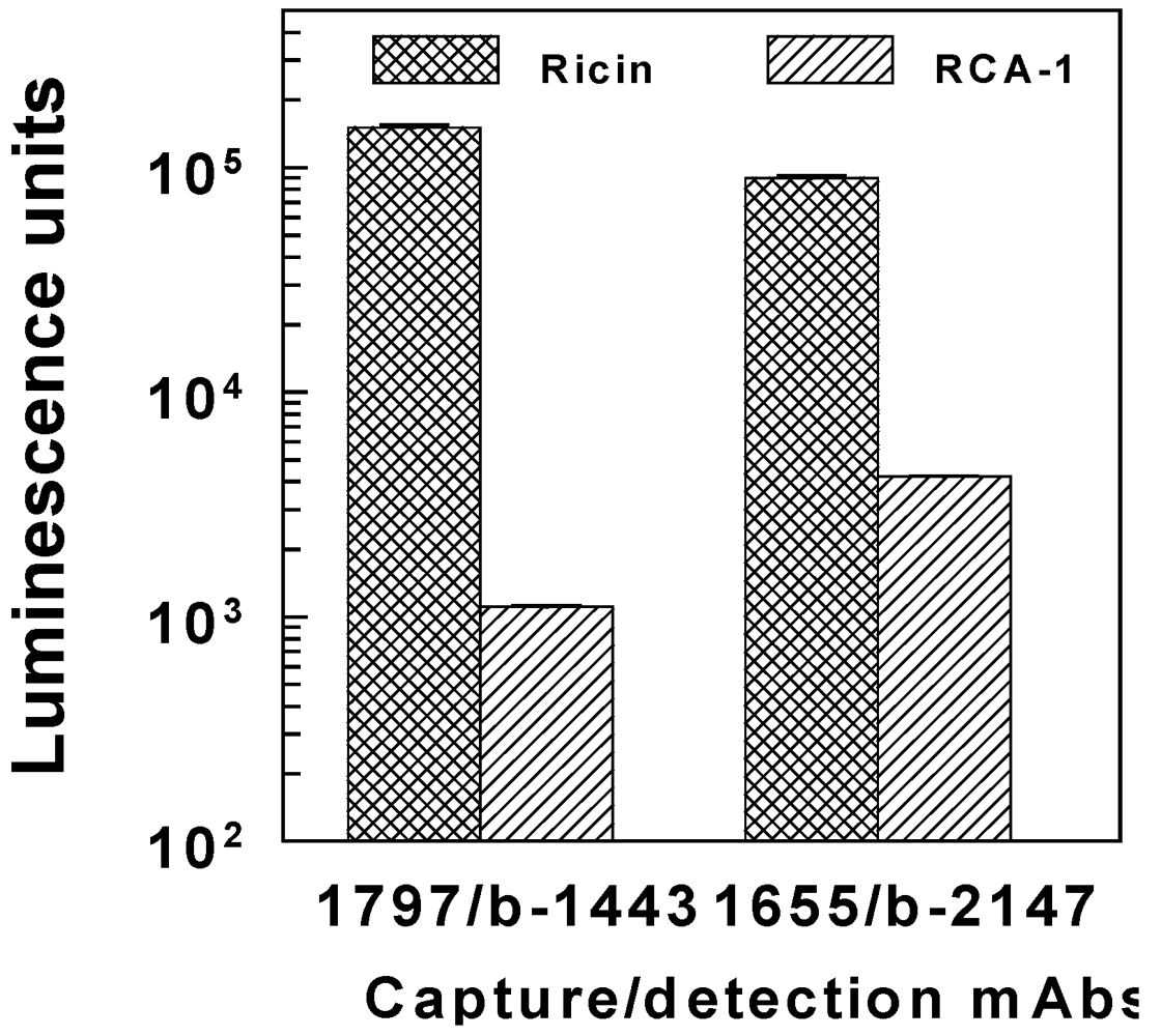
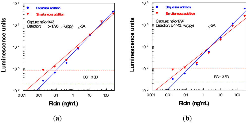
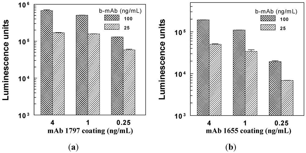
3.1. Capture and Detection Antibody Concentrations


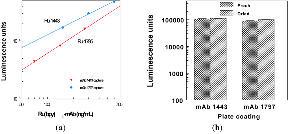
3.2. Directly Labeled mAbs for Detection

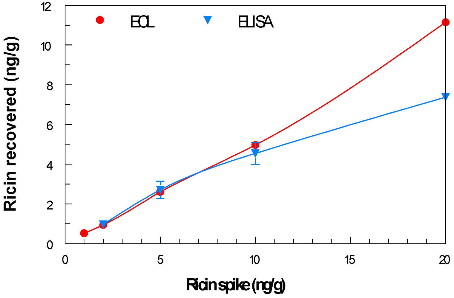
3.3. Recovery of Ricin from Ground Beef
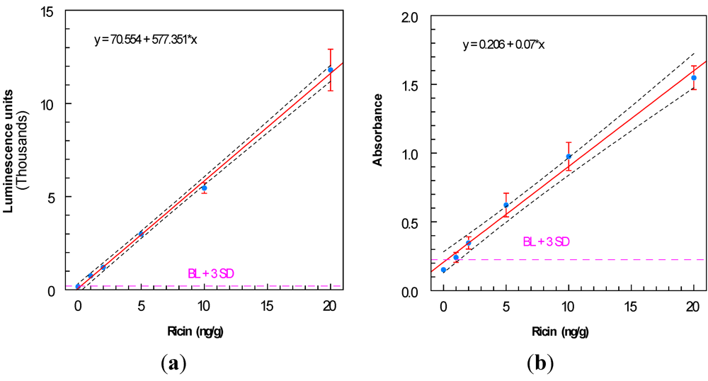
3.4. Purified Ricin

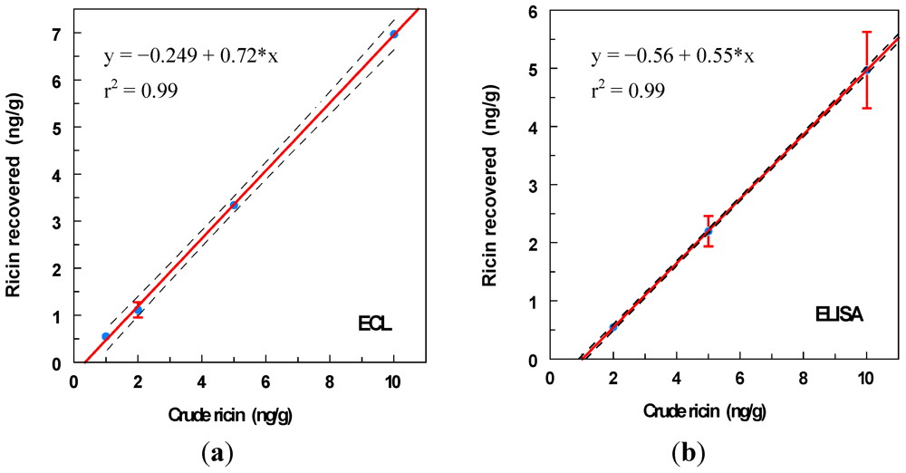
3.5. Crude Ricin
4. Discussion
Acknowledgments
References
- Doan, L.G. Ricin: mechanism of toxicity, clinical manifestations, and vaccine development. A review. J. Toxicol. Clin. Toxicol. 2004, 42, 201–208. [Google Scholar] [CrossRef] [PubMed]
- Lord, J.M.; Roberts, L.M. Ricin: Structure, synthesis, and mode of action. In Microbial Protein Toxins; Schmitt, M.J., Schaffrath, R., Eds.; Springer-Verlag: Berlin, Germany, 2005; pp. 215–233. [Google Scholar]
- Madsen, J.M. Toxins as weapons of mass destruction. A comparison and contrast with biological-warfare and chemical-warfare agents. Clin. Lab. Med. 2001, 21, 593–605. [Google Scholar] [PubMed]
- CDC. Investigation of a ricin-containing envelope at a postal facility—South Carolina, 2003. MMWR Morb. Mortal. Wkly. Rep. 2003, 52, 1129–1131. [PubMed]
- Ishiguro, M.; Tanabe, S.; Matori, Y.; Sakakibara, R. Biochemical studies on oral toxicity of ricin. IV. A fate of orally administered ricin in rats. J. Pharmacobiodyn. 1992, 15, 147–156. [Google Scholar] [PubMed]
- He, X.; Lu, S.; Cheng, L.W.; Rasooly, R.; Carter, J.M. Effect of food matrices on the biological activity of ricin. J. Food Prot. 2008, 71, 2053–2058. [Google Scholar] [PubMed]
- He, X.; McMahon, S.; McKeon, T.A.; Brandon, D.L. Development of a novel immuno-PCR assay for detection of ricin in ground beef, liquid chicken egg and milk. J. Food Prot. 2010, 73, 695–700. [Google Scholar] [PubMed]
- Poli, M.A.; Rivera, V.R.; Hewetson, J.F.; Merrill, G.A. Detection of ricin by colorimetric and chemiluminescence ELISA. Toxicon 1994, 32, 1371–1377. [Google Scholar]
- Shyu, H.F.; Chiao, D.J.; Liu, H.W.; Tang, S.S. Monoclonal antibody-based enzyme immunoassay for detection of ricin. Hybrid. Hybridomics 2002, 21, 69–73. [Google Scholar] [CrossRef] [PubMed]
- Fulton, R.E.; Thompson, H.G. Fluorogenic hand-held immunoassay for the identification of ricin: rapid analyte measurement platform. J. Immunoassay Immunochem. 2007, 28, 227–241. [Google Scholar] [CrossRef] [PubMed]
- Brandon, D.L.; Hernlem, B.J. Development of monoclonal antibodies specific for Ricinus agglutinins. Food Agric. Immunol. 2009, 20, 11–22. [Google Scholar] [CrossRef]
- Hale, M.L. Microtiter-based assay for evaluating the biological activity of ribosome-inactivating proteins. Pharmacol. Toxicol. 2001, 88, 255–260. [Google Scholar] [CrossRef] [PubMed]
- Shyu, R.H.; Shyu, H.F.; Liu, H.W.; Tang, S.S. Colloidal gold-based immunochromatographic assay for detection of ricin. Toxicon 2002, 40, 255–258. [Google Scholar] [CrossRef] [PubMed]
- Huelseweh, B.; Ehricht, R.; Marschall, H. A simple and rapid protein array based method for the simultaneous detection of biowarfare agents. Proteomics 2006, 6, 2972–2981. [Google Scholar] [CrossRef] [PubMed]
- Stine, R.; Pishko, M.V.; Schengrund, C.L. Comparison of glycosphingolipids and antibodies as receptor molecules for ricin detection. Anal. Chem. 2005, 77, 2882–2888. [Google Scholar] [CrossRef] [PubMed]
- Feltis, B.N.; Sexton, B.A.; Glenn, F.L.; Best, M.J.; Wilkins, M.; Davis, T.J. A hand-held surface plasmon resonance biosensor for the detection of ricin and other biological agents. Biosens. Bioelectron. 2008, 23, 1131–1136. [Google Scholar] [CrossRef] [PubMed]
- He, X.; Brandon, D.L.; Chen, G.Q.; McKeon, T.A.; Carter, J.M. Detection of castor contamination by real-time polymerase chain reaction. J. Agric. Food Chem. 2007, 55, 545–550. [Google Scholar] [PubMed]
- He, X.; Carter, J.M.; Brandon, D.L.; Cheng, L.W.; McKeon, T.A. Application of a real time polymerase chain reaction method to detect castor toxin contamination in fluid milk and eggs. J. Agric. Food Chem. 2007, 55, 6897–6902. [Google Scholar] [CrossRef] [PubMed]
- Darby, S.M.; Miller, M.L.; Allen, R.O. Forensic determination of ricin and the alkaloid marker ricinine from castor bean extracts. J. Forensic Sci. 2001, 46, 1033–1042. [Google Scholar] [PubMed]
- Guglielmo-Viret, V.; Thullier, P. Comparison of an electrochemiluminescence assay in plate format over a colorimetric ELISA, for the detection of ricin B chain (RCA-B). J. Immunol. Methods 2007, 328, 70–78. [Google Scholar] [CrossRef] [PubMed]
- Garber, E.A.E.; O’Brien, T.W. Detection of ricin in food using electrochemiluminescence-based technology. J. AOAC Int. 2008, 91, 376–382. [Google Scholar] [PubMed]
- Cho, C.Y.; Keener, W.K.; Garber, E.A.E. Application of deadenylase electrochemiluminescence assay for ricin to foods in a plate format. J. Food Prot. 2009, 72, 903–906. [Google Scholar] [PubMed]
- Garber, E.A.E.; Eppley, R.M.; Stack, M.E.; McLaughlin, M.A.; Park, D.L. Feasibility of immunodiagnostic devices for the detection of ricin, amanitin, and T-2 toxin in food. J. Food Prot. 2005, 68, 1294–1301. [Google Scholar] [PubMed]
- Smith, P.K.; Krohn, R.I.; Hermanson, G.T.; Mallia, A.K.; Gartner, F.H.; Provenzano, M.D.; Fujimoto, E.K.; Goeke, N.M.; Olson, B.J.; Klenk, D.C. Measurement of protein using bicinchoninic acid. Anal. Biochem. 1985, 150, 76–85. [Google Scholar] [PubMed]
- Bradberry, S.M.; Dickers, K.J.; Rice, P.; Griffiths, G.D.; Vale, J.A. Ricin poisoning. Toxicol. Rev. 2003, 22, 65–70. [Google Scholar] [PubMed]
- Audi, J.; Belson, M.; Patel, M.; Schier, J.; Osterloh, J. Ricin poisoning: A comprehensive review. JAMA 2005, 294, 2342–2351. [Google Scholar] [CrossRef] [PubMed]
- Bennett, R.W. An antibody modified automated enzyme-linked immunosorbent assay-based method for detection of staphylococcal enterotoxin. J. Rapid Methods Autom. Microbiol. 2008, 16, 320–329. [Google Scholar] [CrossRef]
- Anderson, G.P.; Bernstein, R.D.; Swain, M.D.; Zabetakis, D.; Goldman, E.R. Binding kinetics of antiricin single domain antibodies and improved detection using a B chain specific binder. Anal. Chem. 2010, 82, 7202–7207. [Google Scholar] [PubMed]
© 2011 by the authors; licensee MDPI, Basel, Switzerland. This article is an open-access article distributed under the terms and conditions of the Creative Commons Attribution license (http://creativecommons.org/licenses/by/3.0/).
Share and Cite
Brandon, D.L. Detection of Ricin Contamination in Ground Beef by Electrochemiluminescence Immunosorbent Assay. Toxins 2011, 3, 398-408. https://doi.org/10.3390/toxins3040398
Brandon DL. Detection of Ricin Contamination in Ground Beef by Electrochemiluminescence Immunosorbent Assay. Toxins. 2011; 3(4):398-408. https://doi.org/10.3390/toxins3040398
Chicago/Turabian StyleBrandon, David L. 2011. "Detection of Ricin Contamination in Ground Beef by Electrochemiluminescence Immunosorbent Assay" Toxins 3, no. 4: 398-408. https://doi.org/10.3390/toxins3040398