1. Introduction
In Shiga-toxin producing
Escherichia coli (STEC) infections, a broad spectrum of central nervous system (CNS) symptoms occurs (abbreviations used in this article are listed in
Table 1). Those symptoms include cortical blindness, poor fine-motor coordination, seizures and coma [
1,
2,
3,
4,
5,
6,
7,
8,
9,
10,
11,
12,
13]. Globotriaosylceramide (Gb
3) is a known receptor of Shiga toxin (Stx), which is central to the intoxication and disease process [
14]. It has been shown that a Gb
3 knockout mouse is resistant to Stx [
15]. To understand target components within the CNS, determining which cell types express Gb
3 is essential. Previously, we reported that in the mouse CNS, Shiga toxin-2 acts on spinal cord neurons which express Gb
3, and leads to hindlimb paralysis [
16]. Other mouse CNS cell types expressing Gb
3 have not been described in detail.
Table 1.
Abbreviations used in this manuscript.
Table 1.
Abbreviations used in this manuscript.
| Abbreviation | Abbreviated Term |
|---|
| AP | Area postrema (e) |
| ARH | Arcuate nucleus of hypothalamus (c) |
| BBB | Blood-brain-barrier |
| BLA | Basolateral nucleus of the amygdala (c) |
| CNS | Central nervous system |
| CP | Caudate-putamen (a) |
| CSF | Cerebro-spinal fluid |
| CVO | Circumventricular organs |
| DMX | Dorsal motor nucleus of the vagus (e) |
| ec | External capsule (c) |
| MD | Mediodorsal nucleus of the thalamus (d) |
| ME | Median eminence (c) |
| MH | Medial habenula (d) |
| NTS | Nucleus of the solitary tract (e) |
| OVLT | Organum vasculosum of the lamina terminalis (b) |
| PVT | Paraventricular nucleus of the thalamus (d) |
| SCO | Subcommissural organ |
| SFO | Subfornical organ (d) |
| V3 | Third ventricle |
| V4 | Fourth ventricle |
| VL | Lateral ventricle |
| XII | Hypoglossal nucleus (e) |
The trafficking route of Stx into the CNS is as important as determining its target. In human STEC patients’ brain magnetic resonance imaging (MRI), regions as the basal ganglia and also thalamus, cerebellum and brain stem, are found positive for increased permeability of fluid [
17,
18,
19,
20]. In a rabbit model, MRI showed enhanced permeability in the area surrounding V3 after Stx injections [
21]. However, precise Stx trafficking routes and the mechanisms involved are still in question. Circumventricluar organs (CVO) are known to be devoid of the blood-brain-barrier (BBB), thus exchange of substances between the plasma and the CNS parenchyma is relatively easy [
22]. The CVO is situated around the V3 (OVLT, SFO, ME, posterior pituitary, pineal gland and SCO) as well as the V4 (AP). Also, the choroid plexus located at both V3 and V4, is sometimes considered as the CVO. If the vessels at the CVO are expressing Gb
3, it may increase the chance of being the primary target in the CNS. In this article, Gb
3 expression in the CVO is addressed. Ependymal cells form a lining of the ventricle, which separates cerebro-spinal fluid (CSF) and parenchyma. As the choroid plexus makes CSF from serum and secretes it into the ventricles, there is a possibility of Stx2 in serum being transferred to the ventricle. If ependymal cells express Gb
3, this also could be an entry point of Stx into the CNS parenchyma.
3. Results and Discussion
Anti-Gb
3-Ab reactive neurons were seen throughout the mouse CNS. In the olfactory bulb, where Mitral cells accept input from nasal epithelium cells to sense smell, and interneurons such as periglomerular cells and granule cells reside, all neurons were Gb
3 positive (
Figure 1). In the cerebrum, neurons in the cortex, including motor cortex were also Gb
3 positive (
Figure 2a). Astrocytes in the corpus callosum which is close to the cerebral cortex were Gb
3 negative (
Figure 2b,c). Hippocampus neurons, CA1, 2, 3 and dentate gyrus, were Gb
3 positive (
Figure 2e). Also, the neurons in the striatum (
Figure 2b), amygdala (
Figure 4i), thalamus (
Figure 4e) and hypothalamus were Gb
3 positive. The hypothalamic nuclei (ARH) neurons are shown to be Gb
3 positive in
Figure 4c. In the cerebellum, Perkinje cells as well as granule cells were also Gb
3 positive (
Figure 3). Neurons throughout the mouse CNS appeared to express Gb
3, hence, depending on the CNS entry site of Stx, all these neurons could become targets of the toxin.
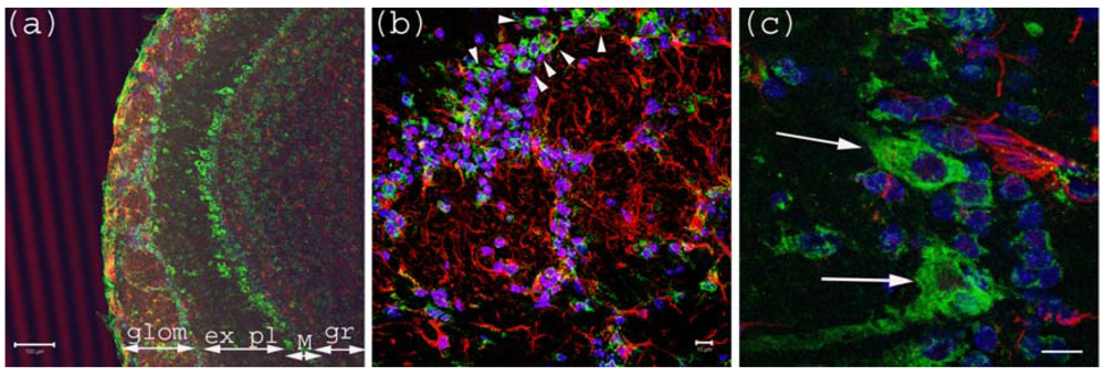
Figure 1.
Anti-Gb3-Ab staining of Olfactory bulb. (a) Olfactory bulb sections stained with anti-Gb3-Ab (Green), and anti-GFAP-Ab (Red) and DAPI (Blue). Glomerular layer (glom), external plexiform (ex pl), Mitral cell layer (M) and granular layer (gr) are visible as neurons are stained anti-Gb3. Bar indicates 100 μm. (b) High magnification of glomerular layer. Arrowheads point to anti-Gb3-Ab reactive periglomerular neurons. Bar indicates 10 μm. (c) High magnification of Mitral cell layer. Arrows show anti-Gb3-Ab positive Mitral cells. Bar indicates 10 μm.
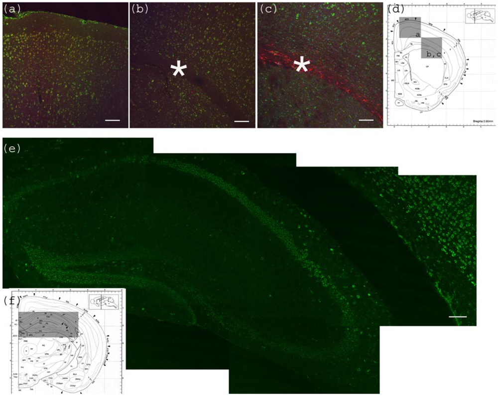
Figure 2.
Anti-Gb
3-Ab staining of cerebrum. (
a) Motor cortex region showing anti-Gb
3 stain (Green) in 2-6 layers of neurons, which are also positive for neuron specific anti-NeuN-Ab (Red). Position of the image corresponding to the Bregma 0.90 mm is shown in (d) as a square. (
b) Corpus callosum region of the same section is shown. Astrocyte rich corpus callosum (*) is anti-Gb
3 and anti-NeuN negative. Lowerleft area to corpus callosum is striatum (CP). Note that neurons of this area are also positive for anti-Gb
3 Ab. Bar indicates 100 μm. (
c) Corpus callosum region is stained with anti-Gb
3-Ab (Green) and anti-GFAP-Ab (an astrocytic marker, Red). Contrast of GFAP (Red) positive layer versus cortex neurons (Green) is clear. Bar indicates 100 m. (
d) Coronal architectural positions at Bregma 0.90 mm is shown with indication of images (a), (b) and (c) in squares. (
e) Hippocampus region is shown with anti-Gb
3-Ab (Green) stain. Neuronal layers of CA1, 2, 3 and dentate gyras are anti-Gb
3 positive. Area in (e) is shown as a rectangle in (f). Bar indicates 100 m. (
f) Coronal section architecture at Bregma −2.40 mm is shown. (d) and (f) are modified from [
23].
![Toxins 02 01997 g002]()
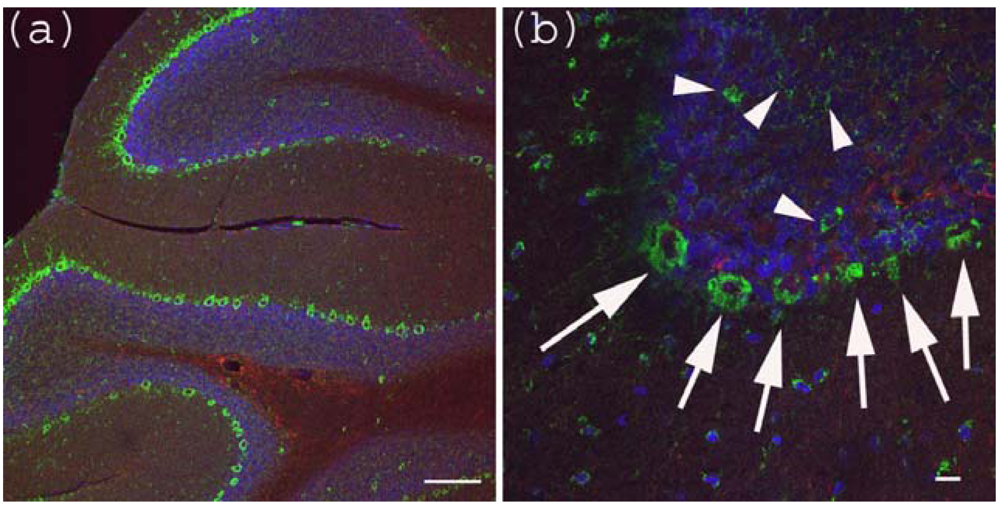
Figure 3.
Anti-Gb3-Ab staining of cerebellum. (a) Cerebellum sections were stained with anti-Gb3 Ab (Green), anti-GFAP-Ab (Red) and DAPI (Blue). Perkinje layer is shown reactive to anti-Gb3-Ab. Bar indicates 100 μm. (b) Higher magnification of (a). Arrows indicate anti-Gb3-Ab positive Perkinje cells, while arrowheads indicate anti-Gb3-Ab positive neurons in the granule layer. Bar indicates 10 μm.
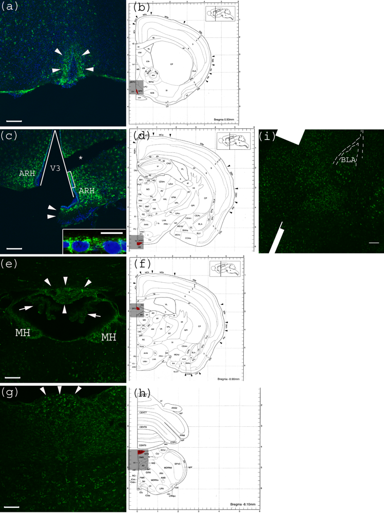
CVO areas in the mouse CNS were also tested for anti-Gb
3-Ab reactivity. Within the tested CVO areas, none appeared to have a Gb
3 expressing vessel structure. In contrast, neurons of these areas were positive for Gb
3. In the OVLT, neurons which have characteristic large nuclei, were shown to be Gb
3 positive (
Figure 4a). In
Figure 4c, the lining of V3 consisting of ependymal cells appears positive for Gb
3. In
Figure 4e, neurons in the SFO, as well as MH are Gb
3 positive, while the choroid plexus is Gb
3 negative. In
Figure 4g, neurons in the medulla oblongata are Gb
3 positive including the AP area. Within this image, a central canal positioned in the center, appears very dim.
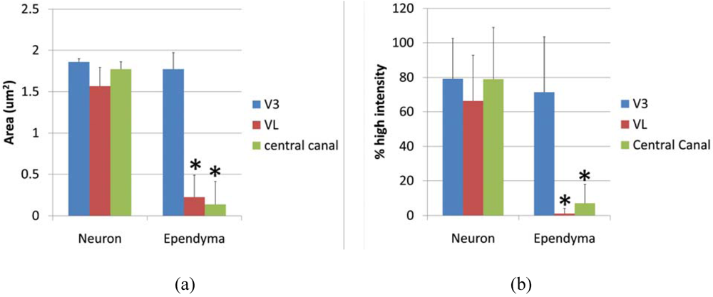
Ependymal cells were tested as a Gb
3 expressing cell type and intensity analysis was performed comparing high intensity pixels in neuron and ependyma in the ventricle area as V3, VL and central canal. A square shaped region of interest (ROI) was used to detect the pixel area which is higher than background intensity. The average area (μm
2) of neurons
vs. ependymal cells in V3, VL and central canal was; 1.857 ± 0.039
vs. 1.773 ± 0.197 (p = 0.275), 1.567 ± 0.085
vs. 0.226 ± 0.262 (p = 0.0097), 1.7739 ± 0.085
vs. 0.137 ± 0.275 (p = 0.0014), respectively.
P values less than 0.01 were considered to be statistically significant, and shown as (*) in
Figure 5a. The line profile intensity analysis revealed a similar result that averages of high intensity percent of neurons
vs. ependyma in V3, VL and central canal were; 79.23 ± 23.29
vs. 71.41 ± 32.03 (p = 0.289), 66.31 ± 26.46
vs. 1.156 ± 2.997 (p = 4.029E-10), 77.73 ± 30.39
vs. 4.346 ± 4.954 (p = 1.042E-08). The results are graphed in
Figure 5b. In both analyses, the intensity level of Gb
3 positive neurons and ependymal cells in the V3 region was similar, thus ependymal cells in this area were Gb
3 positive. In contrast, VL and central canal ependymal cells were Gb
3 negative.
As the CVO is naturally leaky, serum Stx2 might travel through this route where vessels do not express Gb3. As ependymal cells at V3 express Gb3, this could serve as an entry point of Stx, if Stx enters into CSF in the mouse CNS. Trafficking of Stx in the CNS needs to be further investigated.
Figure 4.
Anti-Gb
3-Ab staining of CVO areas (
a) Anti-Gb
3-Ab stain (Green) of OVLT region, which is depicted in (b) as Red. Arrowheads indicate the OVLT area; (
b) Architectural structure of mouse Bregma 0.50 mm; (
c) Anti-Gb
3-Ab stain (Green) of a region containing ME, which is depicted in (d) as Red. Arrowheads indicate the ME area. Neurons in ARH, which are adjacent to ME are anti-Gb
3-Ab reactive. The V3 is shown as a triangular area. A part of ependymal layer (square region) is shown in inset with higher magnification. (*) indicates a broken tissue space; (
d) Architectural structure of mouse Bregma −1.30 mm; (
e) Anti-Gb
3-Ab stain (Green) of a region containing SFO, which is depicted in (f) as Red. Arrowheads indicate the SFO area, while arrows show anti-Gb
3-Ab negative choroid plexus. Neurons of MH are also anti-Gb
3-Ab reactive. Lower area of this field includes thalamic nuclei as PVT and MD, in which neurons are positive for anti-Gb
3-Ab. (
f) Architectural structure of mouse Bregma -0.90 mm; (
g) Anti-Gb
3-Ab stain (Green) of a region containing AP, which is depicted in (h) as Red. Arrowheads indicate AP area. Neurons of several different nuclei in the medulla oblongata such as NTS, DMX and XII are also anti-Gb
3-Ab positive; (
h) Architectural structure of mouse Bregma −8.10 mm; (
i) Neurons in the amygdala are also anti-Gb
3-Ab positive. BLA is indicated in the picture. Dotted line outlines external capsule (ec). See architectural structure in (d). All Bars indicate 100 m, except an inset in (c) which indicates 10 m. Blue color in (a) and (c) indicates nuclei staining using DAPI. (b), (d), (f) and (h) are modified from [
23]. See abbreviations in
Table 1.
![Toxins 02 01997 g004]()
Figure 5.
Intesnsity of anti-Gb3-Ab staining in neurons vs. ependyma. (a) High intensity area comparison of neurons and ependymal cells from V3, VL or central canal. Values from each region (neuron vs. ependyma) are compared, and the statistically significant difference is shown as (*). (b) High intensity percentage from Line profile analysis comparing neurons and ependymal cells from V3, VL or central canal. Values from each region (neuron vs. ependyma) are compared, and statistically significant differences are shown as (*). * indicates p < 0.01 in 2-tailed, paired t-test.

