Article

PepGMV Rep-Protein Expression in Mammalian Cells

1
CA Ingeniería de Biosistemas, División de Investigación y Posgrado, Facultad de Ingeniería, Universidad Autónoma de Querétaro, Cerro de las Campanas s/n, Santiago de Querétaro, Qro., 76010, México
2
División de Investigación y Posgrado, Facultad de Ciencias Naturales, Universidad Autónoma de Querétaro, Avenida de las Ciencias s/n, Juriquilla, Qro., 76230, México
*
Author to whom correspondence should be addressed.
Viruses 2012, 4(9), 1792-1801; https://doi.org/10.3390/v4091792
Submission received: 27 July 2012 / Revised: 6 September 2012 / Accepted: 6 September 2012 / Published: 24 September 2012
(This article belongs to the Special Issue Plant Viruses)

Abstract

:
The Geminiviruses genome is a small, single strand DNA that replicates in the plant cell nucleus. Analogous to animal DNA viruses, Geminiviruses depend on the host replication machinery to amplify their genomes and only supply the factors required to initiate their replication. Consequently, Geminiviruses remove the cell-cycle arrest and induce the host replication machinery using an endocycle process. They encode proteins, such as the conserved replication-associated proteins (Rep) that interact with retinoblastoma-like proteins in plants and alter the cell division cycle in yeasts. Therefore, the aim of this work is to analyze the impact of Pepper Golden Mosaic Virus (PepGMV) Rep protein in mammalian cells. Results indicate that the pTracer-SV40:Rep construction obtained in this work can be used to analyze the Rep protein effect in mammalian cells in order to compare the cell cycle regulation mechanisms in plants and animals.

1. Introduction

Geminiviruses are plant pathogens which have small, single strand DNA genomes. They are characterized by their unique geminate capsids formed by two incomplete icosahedra [1,2]. Geminiviruses constitute a large family that is divided into the Begomovirus, Mastrevirus, Curtovirus, and Topocuvirus genera based on genome arrangement, insect vector, and host range [3,4]. The Begomovirus genus is the largest and includes whitefly-transmited, and bipartite (genome divided into two components) geminivirus [5]. Pepper Golden Mosaic Virus (PepGMV) belongs to Begomovirus genus and naturally infects pepper, tomato, tobacco, and other solanaceous crops. The typical symptoms induced by PepGMV in pepper plants, are yellow mosaic and wrinkled leaf and stunting of the plant [6].
Geminiviruses replicate in the cell nucleus via a rolling circle mechanism. They only supply the factors required to initiate their replication, and depend on the host replication machinery to amplify their genomes. Mammalian DNA-tumor viruses also employ this mechanism [1]. Many Geminiviruses replicate in differentiated cells that no longer contain detectable levels of DNA polymerases and associated factors [1,3]. Therefore, geminiviruses removed the cell-cycle arrest and thereby induce the host replication machinery in order to replicate their genomes [3,7,8].
Mammalian DNA-tumor viruses such as polyoma, papilloma and adeno viruses encode proteins that modify animal cell-cycle controls by binding to the retinoblastoma protein (pRB) or the related‑family members, p107 and p130 [9]. pRB-family members negatively regulate cell cycle progression and promote differentiation, in part, through their interactions with E2F transcription factors [10,11,12].
Analogous to animal DNA viruses, Geminiviruses encode proteins that interact with pRB proteins in plants. They encode for a conserved replication-associated protein (Rep) which is a multifunctional protein involved in viral replication, auto-regulation of its own gene transcription, and inactivation and recruitment of host-encoded proteins related to host DNA synthesis [4,13,14,15,16,17]. Rep is the only viral protein that is essential for viral replication. It initiates and terminates viral DNA synthesis and induces the accumulation of host-replication factors in infected cells [4,12]. Moreover, Rep protein interacts with host regulatory factors, including the proliferating cell nuclear antigen (PCNA), and the pRB, which modulates the plant cell cycle and differentiation [18,19]. Kittelmann et al. [2] found that African Cassava Mosaic Virus (ACMV) Rep protein expression in fission yeast (Schizosaccharomyces pombe) caused cell elongation that resembles cdc (cell division cycle) phenotypes. Cells expressing Rep protein increased DNA contents, suggesting that ACMV Rep protein promotes reinitiation of nuclear DNA replication during the fission yeast cell cycle [2].
Therefore, the objective of this work is to analyze the impact of Pepper Golden Mosaic Virus (PepGMV) Rep-protein expression in mammalian cell cycle regulation. To do this, the Rep open reading frame (ORF) was cloned into the expression vector pTracer-SV40 and 3T3L1 mouse fibroblast cells were transfected with the pTracer-SV40:Rep. Rep expression was analyzed by RT-PCR.

2. Results and Discussion

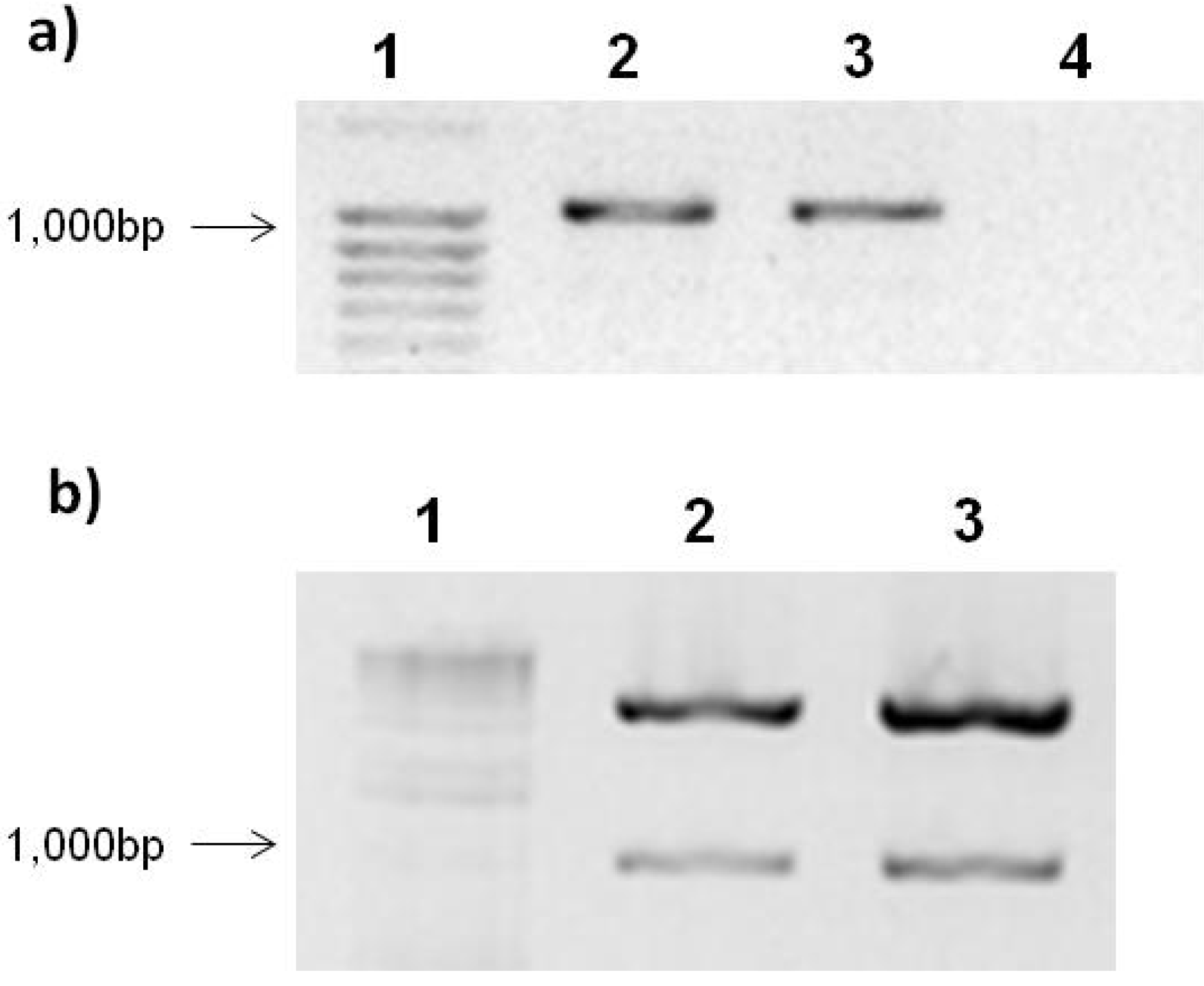
2.1. Expression Vector pTracer-SV40 and Ligation with PepGMV Rep-Protein Gene

The Rep gene open reading frame (ORF) (GenBank: U57457.1) was amplified by PCR as shown in Figure 1a. Lines 2–4 (Figure 2a) show a band of approximately 1,047 bp corresponding to PepGMV Rep protein gene. After PCR amplification, Rep was cloned into the expression vector pTracer-SV40. Ligation was confirmed by double digestion with SpeI and KpnI enzymes. Figure 1b shows the gel electrophoresis of the double digestion product. As expected, two bands were observed in each of the samples. One corresponds to the linearized pTracerSV40 vector (Figure 1b, lines 2–4) which has a size of 4.2 kb and a second band corresponding to Rep (Figure 1b, lines 2–4).
These results confirmed the ligation of PepGMV-Rep gene into the pTracer-SV40 expression vector, obtaining the following constructions: pTracer-SV40:Rep and pTracer-SV40:RepATG-.
Figure 1. Agarose gel electrophoresis of Rep gene PCR amplification and construction double digestion.(a) PCR amplification of Rep gene. Lanes: 1, size molecular marker; 2, Rep; 3, RepATG; 4, negative control. (b) Construction digestion with SpeI and KpnI. Lanes: 1, size molecular marker; 2, pTracerSV40:Rep; 3, pTracerSV40:RepATG. The samples were loaded in a 1.2% agarose gel stained with EtBr.
Viruses 04 01792 g001

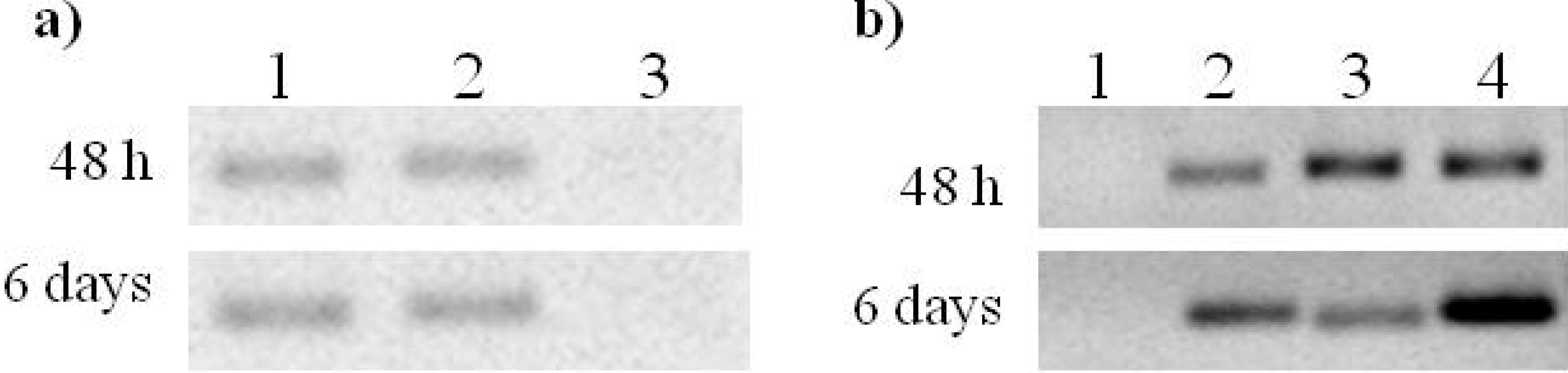
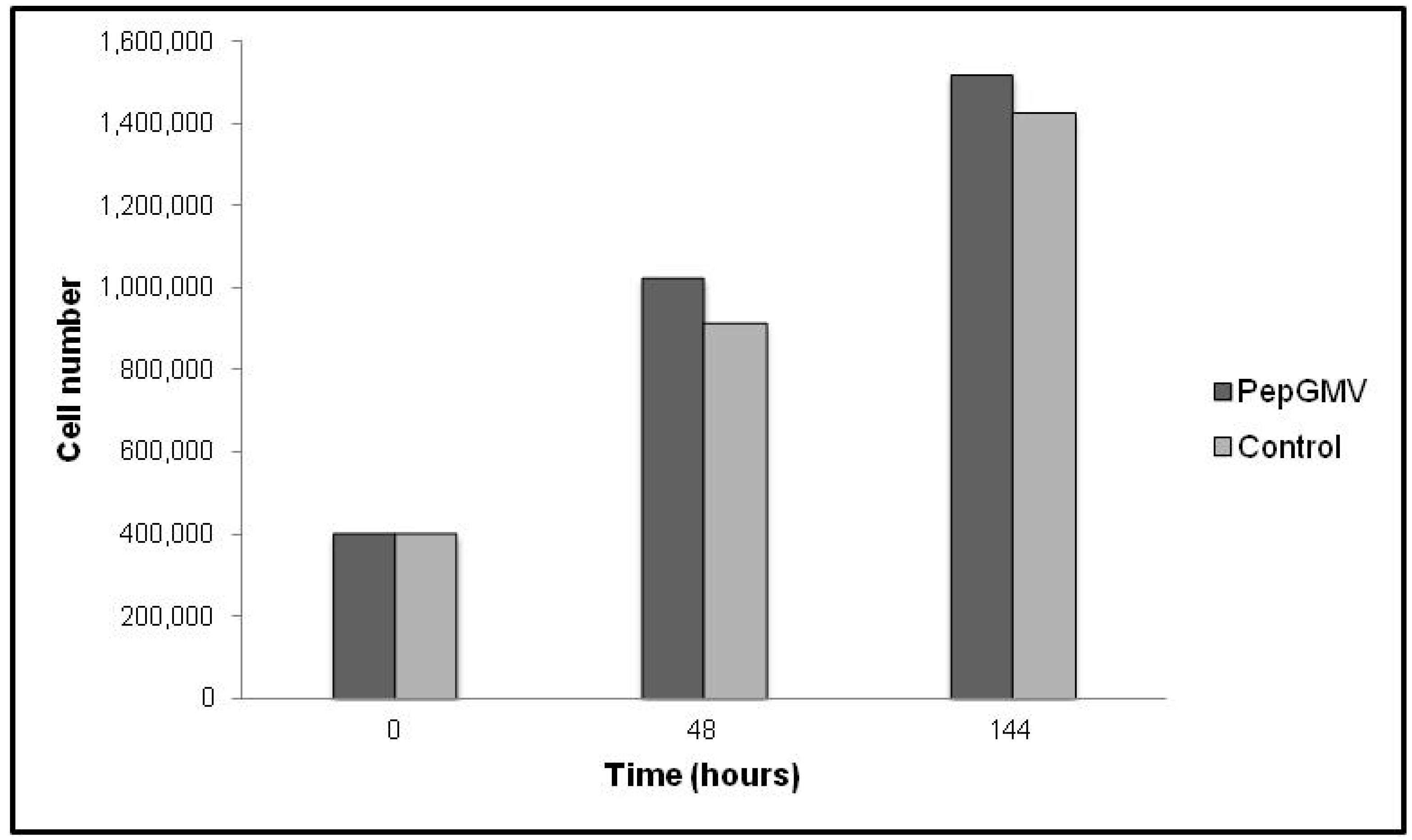
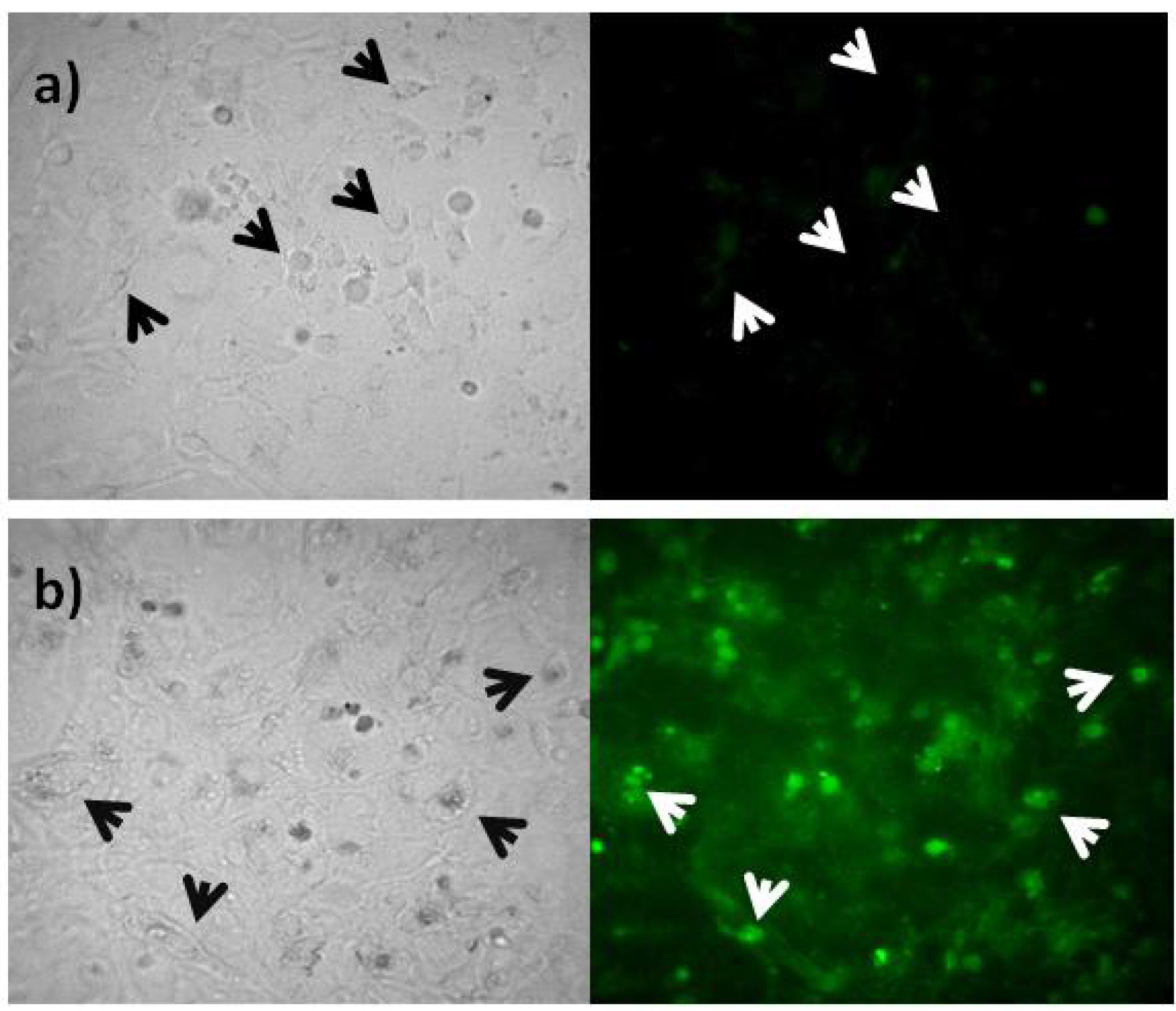
2.2. PepGMV Cell Transfection

In order to indicate if Geminivirus PepGMV is able to replicate in mammalian cells, the PepGMV A component was transfected into 3T3L1 fibroblast cells (as described in the Experimental section). PepGMV A DNA presence was confirmed by PCR amplification using the primers for the Rep gene and the JM23 and JM24 primers (Table 1). Figure 2 shows the PCR amplification of PepGMV A DNA-transfected cells. Again, a band of 1,047 bp corresponding to Rep is observed in samples taken at 24 and 144 h post transfection. Additionally, a band of 280 bp corresponding to the common region of PepGMV is observed, which confirms the presence of Geminivirus DNA inside the cells. In addition, RT-PCR was performed in order to verify Rep gene transcription in transfected cells (Figure 3a). The level of Rep transcription was too low to be clearly seen. Thus a re-amplification was made to corroborate the presence of the Rep transcript (Figure 3b). A band corresponding to Rep gene was observed after re-amplification, indicating that Rep transcription was taking place in transfected cells but with a low efficiency. Therefore geminivirus PepGMV was recognized by mammalian cell machinery and Rep transcription occurred.
Table 1. Oligonucleotides used for Rep gene PCR amplification, PepGMV A component detection and RT-PCR. Translation initiation codon is shown in bold letters. The underlined sequence corresponds to the Kozak sequence that was added in order to improve translation of Rep in mammalian cells.
Oligonuceotide nameForwardReverseFragment size
RepPKGGTACCACCATGCCACTACCACCAAAATCACTAGTTTAGCTATCCTGTGCTGTGCT1,047 bp
RepPAGGTACCACCActGCACTACCACCAAAATCACTAGTTTAGCTATCCTGTGCTGTGCT1,047 bp
JM23TGGTGTAGGACTCCAGCAGAGTC 288 bp
JM24TAGGCCCACACCTTGGTGACCAA
β actinAGGTATCCTGACCCTGAAGTACCCCGGCCACACGCAGCTCATTGTA106 bp
GAPDHATTGTTGCCATCAACGACCCCCAAGCTTCCCATTCTCGGCC116 bp
RepPepGMVCAAAGCTGGTGATCCGAAAACGGTTAAACGAGGATAATGGATAAGG120 bp
Figure 2. PCR amplification of PepGMV A component in transfected cells. (a) 48 h post-transfection. Lines: 1, size molecular marker; 2, Rep primers; 3, Rep primers positive control; 4, JM23 and JM24 primers; 5, JM23 and JM24 primers positive control; 6, negative control. (b) 6 days after transfection. Lines: 1, size molecular marker; 2, Rep primers; 3, Rep primers positive control; 4, JM23 and JM24 primers; 5, JM23 and JM24 primers positive control; 6, negative control. PepGMV A plasmid DNA was used as positive control. The samples were loaded in a 1.2% agarose gel stained with EtBr.
Viruses 04 01792 g002
Figure 3. RT-PCR of PepGMV A transfected cells. (a) RT-PCR. Lines: 1, positive control; 2, GAPDH; 3, Rep. (b) RT-PCR re-amplification. Lines: 1, negative control; 2, GAPDH; 3, Rep; 4, positive control. The expected fragment size was 120 bp. Samples were loaded in a 2% agarose gel stained with EtBr.
Viruses 04 01792 g003
To determine if the Geminivirus affects the cell growth, transfected cells with PepGMV A component were counted. Figure 4 shows the number of cells at 0, 48 and 144 h after transfection. A difference in the cell number was observed in cells transfected with the PepGMV in contrast with control cells, indicating that the presence of PepGMV is affecting cell growth.
Figure 4. Transfected cells growth. Cells were counted using a hematocytometer after 48 and 144 h post transfection with PepGMV A component. The counting was performed in duplicate.
Viruses 04 01792 g004

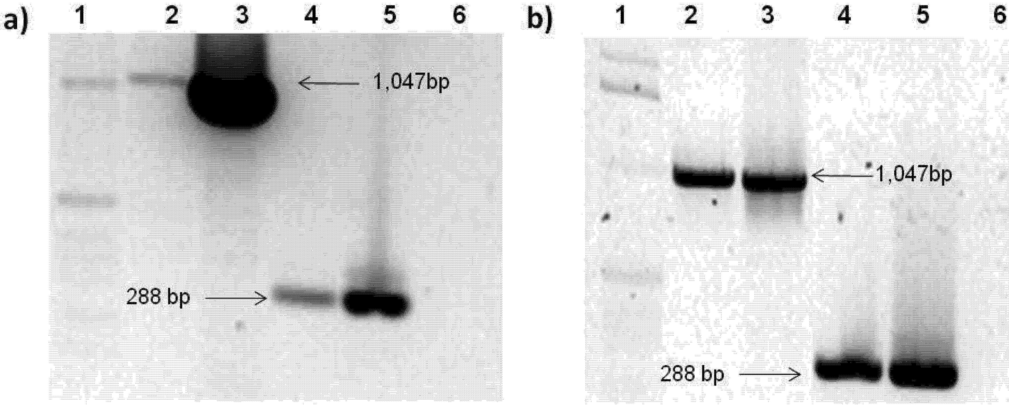
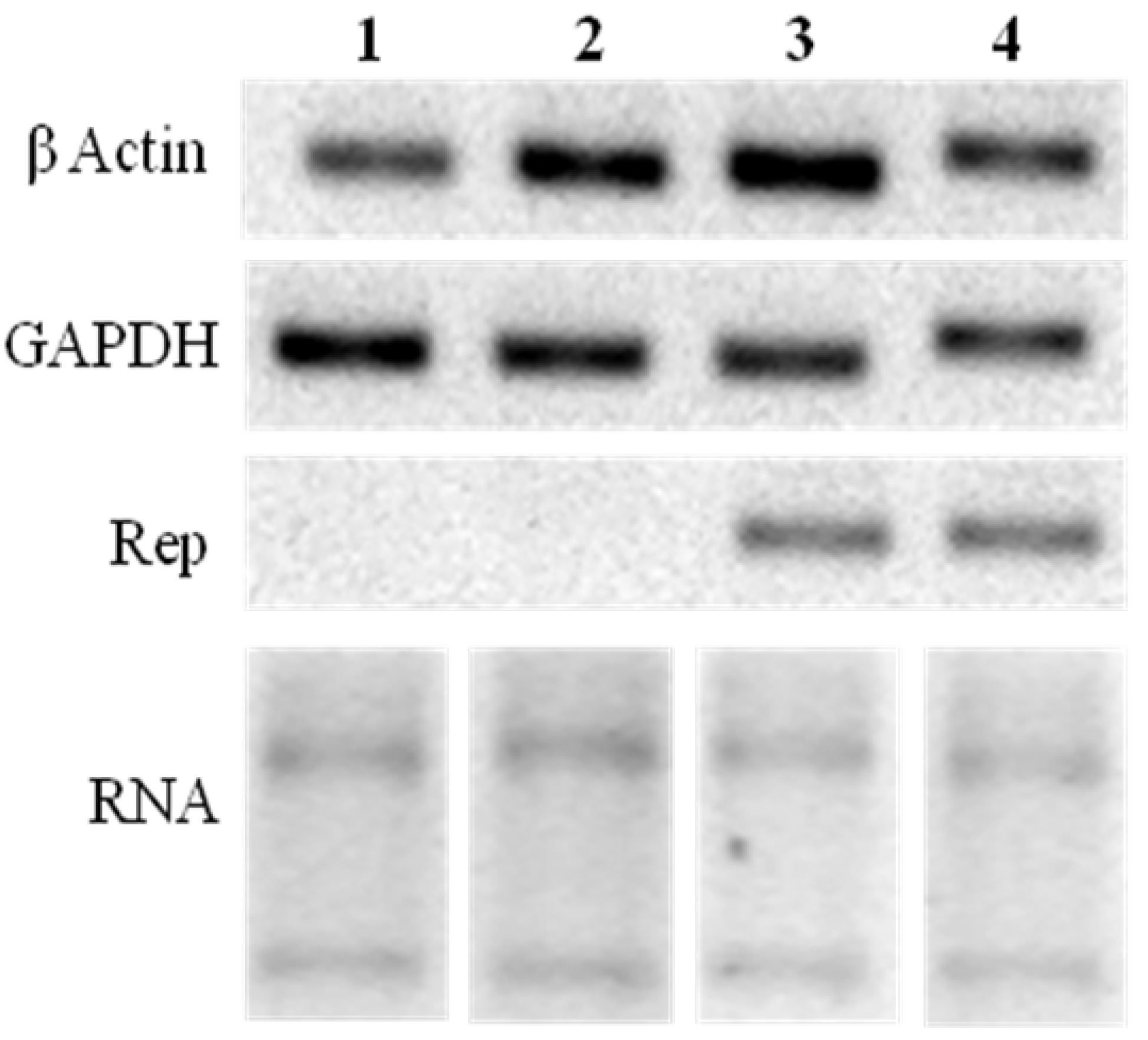
2.3. Rep-Gene Expression

Expression of green fluorescent protein (GFP) was detected in order to verify the construction transfection into cells (Figure 5). Cells transfected with the pTracer-SV40:Rep (Figure 5a) reacted and fluoresced in contrast with control cells in which no fluorescence was observed (Figure 5b). This indicates the insertion of the construction and the expression of the GFP correlates with Rep expression.
Figure 5. Transfected cells image under fluorescent microscopy. (a) Control cells. (b) Cells transfected with pTracer-SV40:Rep. Left: white light. Right: fluorescence. Arrows indicates cell position.
Viruses 04 01792 g005
Additionally, Rep expression was confirmed by RT-PCR. Figure 6 shows the RT-PCR analysis of transfected cells at 72 h post transfection. A band corresponding to Rep gene was observed in cells transfected with both pTracer-SV40:Rep and pTracer-SV40:RepATG constructs. In contrast, with control and vector-transfected cells (Figure 6). This result indicates that Rep gene is being transcribed in transfected cells (Figure 6).
Figure 6. RT-PCR of transfected cells. Lines: 1, control cells; 2, cells transfected with the empty vector; 3, pTracer-SV40:Rep transfected cells; 4, pTracer-SV40:RepATG transfected cells. Samples were loaded in a 2% agarose gel stained with EtBr.
Viruses 04 01792 g006

3. Experimental Section

3.1. Viral Clone and Rep-Gene PCR Amplification

The dimeric clone of Pepper Golden Mosaic Virus (PepGMV) (Tamaulipas isolate) was used in this work. PepGMV genome has been previously described [6].
The Rep gene open reading frame (ORF) was amplified by PCR using the oligonucleotides shown in Table 1. The PepGMV Rep sequence used was obtained from the National Center for Biotechnology Information (NCBI) database (GenBank: U57457.1). To clone the Rep gene in the expression vector pTracer™-SV40, the sites for SpeI (extreme 3') and KpnI (extreme 5') restriction enzymes were used. PCR amplification conditions were as follows: 95 °C for 1 min, 30 cycles of 95 °C for 30 s and 68 °C for 2 min, and a final extension of 68 °C for 2 min. In addition, a negative translation control was generated by changing the Rep-gene initiation codon sequence (ATG) for the sequence ACT.

3.2. Construction of pTracer™-SV40:Rep Expression Vector

After PCR amplification, the 1,047 bp gene was cleaned with the Wizard® PCR Preps DNA Purification System (Promega) and cloned in the intermediate pGem®-T Easy vector (Promega) using the manufacturer’s procedures. The pTracer™-SV40 vector (Invitrogen) was double digested with SpeI and KpnI enzymes (Fermentas) in order to linearize it and clone the Rep gene. Insert and vector purification was performed using the Silica Bead DNA Gel Extraction kit (Fermentas) and ligation reaction was done using the ligase T4 (Invitrogen) and a 4:1 insert/vector molar ratio. The insert presence was confirmed by double digestion with SpeI and KpnI enzymes (Fermentas).Gene correct orientation was confirmed by sequencing (Genetic Analyzer 3130, Applied Biosystems).

3.3. Cell Culture and Transfection

3T3L1 mice fibroblast cells (ATCC No. CL-173) were grown in Dubelco’s Modified Eagle Medium (DMEM, Gibco) supplemented with penicillin (62.1 mg/L; Sigma), streptomycin (100 mg/L; Sigma), anphotericin (250 µg/L; Gibco), and 5% of calf serum (Biowest). The cells grew in an incubator at 37 °C and a 5% CO2 atmosphere.
Cell transfection was performed by calcium-phosphate precipitation method [20]. Twenty four hours before transfection, cells were seeded into 60 mm plates at a concentration of 1 × 10⁵ cells/mL [20]. The culture medium was replaced with fresh medium one hour before transfection. The phosphate-DNA precipitation was performed as follows: 25 µg of plasmid DNA were mix with 100 µL of 2.5 M CaCl2. This solution was diluted with 1/10 TE buffer (1 mM Tris-HCl, 0.1 mM EDTA, pH 7.6) to a final volume of 1 mL. One volume of this 2× Ca/DNA solution was added drop by drop to an equal volume of 2× HEPES buffered saline solution. The resulting solution was incubated at room temperature for one minute prior to adding to the cells medium. One hundred microliters of precipitate per milliliter of culture medium were added to the cells Cells were exposed to the precipitate for 6 h at 37 °C and 5% CO2 atmosphere. After this time, the culture medium was replaced with fresh medium and the cells were incubated for 6 days at 37 °C and 5% CO2 atmosphere. Cells were transfected with pTracerSV40:Rep and pTracerSV40:RepATG-constructions. Additionally, the PepGMV A component was transfected in order to probe if PepGMV is able to replicate in mammalian cells.

3.4. Rep Expression

3T3L1 cells transfection with pTracerSV40:Rep and pTracerSV40:RepATG was verified by detection of green fluorescent protein (GFP) by fluorescent microscopy. Additionally, Rep gene expression was verified by RT-PCR for treatments72 h after transfection. Total RNA was extracted from transfected cells using the SV total RNA isolation system (Promega).DNA first strand was generated by the First strand cDNA synthesis kit (Fermentas). The same treatment was applied to cells transfected with PepGMV A component. cDNA PCR amplification was performed with the oligonucleotides shown in Table 1 and the housekeeping genes glyceraldehyde-3-phosphate dehydrogenase (GADPH) and β actin were used as expression controls [21,22].

4. Conclusions

The Pepper Golden Mosaic Virus Rep gene was expressed in mammalian cells. This gene was cloned and transfected into mammalian cells. The expression level was at the same level as the housekeeping genes. Results indicated that the pTracer-SV40:Rep construction developed in this work can be used to analyze the Rep-protein expression in mammalian cells. Moreover, PepGMV, as Geminivirus, was recognized by mammalian-cell machinery and Rep transcription occurred. This trend indicates that this model can be used to study mammalian cell cycle transcriptome regulation using a Geminivirus member. The next step is to determine the effect of the Rep-expressed protein in the expression of mammalian cell cycle regulation genes.

Acknowledgments

We thank Rafael Rivera Bustamante for kindly providing the PepGMV clone and CONACYT México for supporting this work.

Conflict of Interest

The authors declare no conflict of interest.

References and Notes

  1. Gutierrez, C.; Ramirez-Parra, E.; Castellano, M.M.; Sanz-Burgos, A.P.; Luque, A.; Missich, R. Geminivirus DNA replication and cell cycle interactions. Vet. Microbiol. 2004, 98, 111–119. [Google Scholar] [CrossRef]
  2. Kittelmann, K.; Rau, P.; Gronenborn, B.; Jeske, H. Plant geminivirus Rep protein induces rereplication in fission yeast. J. Virol. 2009, 83, 6769–6778. [Google Scholar] [CrossRef]
  3. Hanley-Bowdoin, L.; Settlage, S.B.; Robertson, D. Reprogramming plant gene expression: A prerequisite to geminivirus DNA replication. Mol. Plant Pathol. 2004, 5, 149–156. [Google Scholar] [CrossRef]
  4. Nash, T.E.; Dallas, M.B.; Reyes, M.I.; Buhrman, G.K.; Ascencio-Ibañez, J.T.; Hanley-Bowdoin, L. Functional analysis of a novel motif conserved across geminivirus Rep proteins. J. Virol. 2011, 85, 1182–1192. [Google Scholar]
  5. Yadava, P.; Suyal, G.; Mukherjee, S.K. Begomovirus DNA replication and pathogenicity. Curr. Sci. 2010, 98, 360–368. [Google Scholar]
  6. Carrillo-Tripp, J.; Lozoya-Gloria, E.; Rivera-Bustamante, R.F. Symptom remission and specific resistance of pepper plants after infection by Pepper golden mosaic virus. Virology 2007, 97, 51–59. [Google Scholar]
  7. Ascencio-Ibáñez, J.T.; Sozzani, R.; Lee, T.J.; Chu, T.M.; Wolfinger, R.D.; Cella, R.; Hanley Bowdoin, L. Global analysis of Arabidopsis gene expression uncovers a complex array of changes impacting pathogen response and cell cycle during geminivirus infection. Plant Physiol. 2008, 148, 436–454. [Google Scholar] [CrossRef]
  8. Torres-Pacheco, I.; Guevara-González, R.G.; Ruíz-Medrano, R.; Rivera-Bustamante, R.F. Los geminivirus como modelos de estudio de ciclo celular y de la apoptosis en plantas (in Spanish). Revista Mexicana de Fitolpatología 1996, 14, 88–96. [Google Scholar]
  9. Herwing, S.; Strauss, M. The retinoblastoma protein: A master regulator of cell cycle, differentiation and apoptosis. Eur. J. Biochem. 1997, 246, 581–601. [Google Scholar]
  10. Sidle, A.; Palaty, C.; Dirks, P.; Wiggan, O.; Kiess, M.; Gill, R.M.; Wong, A.K.; Hamel, P.A. Activity of the retinoblastoma family proteins, pRB, p107, and p130, during cellular proliferation and differentiation. Crit. Rev. Biochem. Molec. Biol. 1996, 31, 237–271. [Google Scholar] [CrossRef]
  11. Burkhart, D.L.; Sage, J. Cellular mechanisms of tumour suppression by the retinoblastoma gene. Nature Rev. Cancer 2008, 8, 671–682. [Google Scholar] [CrossRef]
  12. Desvoyes, B.; Ramirez-Parra, E.; Xie, Q.; Chua, N.H.; Gutierrez, C. Cell type-specific role of the retinoblastoma/E2F pathway during Arabidopsis leaf development. Plant Physiol. 2006, 140, 67–80. [Google Scholar]
  13. Nagar, S.; Pedersen, T.J.; Carrick, K.M.; Hanley-Bowdoin, L.; Robertson, D. A geminivirus induces expression of a host DNA synthesis protein in terminally differentiated plant cells. Plant Cell 1995, 7, 705–719. [Google Scholar]
  14. Martin, D.P.; Biagini, P.; Lefeuvre, P.; Golden, M.; Roumagnac, P.; Varsani, A. Recombination in eukaryotic single stranded DNA viruses. Viruses 2011, 3, 1699–1738. [Google Scholar] [CrossRef] [Green Version]
  15. Laufs, J.; Jupin, I.; David, C.; Schumacher, S.; Heyraud-Nitschke, F.; Gronenbom, B. Geminivirus replication: Genetic and biochemical characterization of Rep protein function, a review. Biochimie 1995, 77, 765–773. [Google Scholar] [CrossRef]
  16. Chapa-Oliver, A.; Guevara-Gonzalez, R.G.; González-Chavira, M.M.; Ocampo-Velazquez, A.A.; Feregrino-Pérez, A.A.; Mejía-Teniente, L.; Herrera-Ruíz, G.; Torres-Pacheco, I. Analogies between geminivirus and oncovirus: Cell cycle regulation. Afr. J. Biotechnol. 2011, 10, 11327–11332. [Google Scholar]
  17. Doonan, J.H.; Sablowski, R. Walls around tumors—Why do plants do not develop cancer. Nat. Rev. Canc. 2010, 10, 794–802. [Google Scholar] [CrossRef]
  18. Kong, L.; Orozco, B.M.; Roe, J.L.; Nagar, S.; Ou, S.; Feiler, H.S.; Durfee, T.; Miller, A.B.; Gruissem, W.; Robertson, D.; Hanley-Bowdoin, L. A geminivirus replication protein interacts with the retinoblastoma protein through a novel domain to determine symptoms and tissue specificity of infection in plants. EMBO J. 2000, 19, 3485–3495. [Google Scholar] [CrossRef]
  19. Castillo, A.; Collinet, D.; Deret, S.; Kashoggi, A.; Bejarano, E.R. Dual interaction of plant PCNA with geminivirus replication accessory protein (Ren) and viral replication protein (Rep). Virology 2003, 312, 381–394. [Google Scholar] [CrossRef]
  20. Jordan, M.; Schallhorn, A.; Wurm, F.M. Transfecting mammalian cells: optimization of critical parameters affecting calcium-phosphate precipitate formation. Nucleic Acids Res. 1996, 24, 596–601. [Google Scholar] [CrossRef]
  21. Sugden, K.; Pariante, C.M.; McGuffin, P.; Aitchison, K.J.; D’Souza, U.M. Housekeeping gene expression is affected by antidepressant treatment in a mouse fibroblast cell line. J. Psychopharmacol. 2010, 24, 1253–1259. [Google Scholar] [CrossRef]
  22. Bocchetta, M.; Eliasz, S.; de Marco, M.A.; Rudzinski, J.; Zhang, L.; Carbone, M. The SV40 large T antigen-p53 complexes bind and actívate the insulin-like growth factor-I promoter stimulating cell growth. Cancer Res. 2008, 68, 1022–1028. [Google Scholar] [CrossRef]

