Review

Long Non-Coding RNAs and p53 Regulation

Gene Expression-Microarrays Laboratory, IRCCS-Bambino Gesù Children's Hospital, Rome 00165, Italy
*
Author to whom correspondence should be addressed.
Int. J. Mol. Sci. 2012, 13(12), 16708-16717; https://doi.org/10.3390/ijms131216708
Submission received: 5 November 2012 / Revised: 3 December 2012 / Accepted: 3 December 2012 / Published: 6 December 2012
(This article belongs to the Special Issue Non-Coding RNAs 2012)

Abstract

:
The advent of novel and high-throughput sequencing (next generation) technologies allowed for the sequencing of the genome at an unprecedented depth. The majority of transcribed RNAs have been classified as non-coding RNAs. Among them, long non-coding RNAs (lncRNAs) are emerging as important regulators in many biological processes. Here, we discuss the role of those lncRNAs which are under the control of p53 or that are able to regulate its activity, due to the central role of p53 pathway in many conditions. We also briefly discussed the emerging need of having novel strategies and computational tools to completely unravel the multifaceted roles of lncRNAs and to pave the way to the development of novel diagnostic and therapeutic applications based on these peculiar molecules.

1. Introduction

In biology, the classical view of gene expression and regulation has been focused around protein-coding genes via the central dogma of DNA → mRNA → protein. However, over the past decade, evidence from numerous next generation sequencing (NGS) experiments suggested that the evolution of developmental processes, which regulate the complexity of the organism, is mainly due to the expansion of the regulatory potential of the non-coding portions of the genome [1]. In complex organisms, the portion of the genome responsible for coding proteins constitutes approximately 1.5%, while many regulatory elements are transcribed into non-coding RNAs (ncRNAs). Indeed, the recent technological breakthrough and knowledge coming from these novel NGS techniques, established the importance of ncRNAs in the regulation of multiple major biological processes related to development, differentiation, and metabolism [24] and have forced us to radically reinterpret our understanding of the genome [57]. In contrast to the small ncRNAs, such as short interfering RNAs (siRNAs), microRNAs (miRNAs), and Piwi-interacting RNAs (piRNAs) which are highly conserved and involved in transcriptional and post-transcriptional gene expression regulation through specific base pairing with their targets, long non-coding RNAs (lncRNAs)—defined as transcribed RNA molecules greater than 200 nt in length—are poorly conserved and regulate gene expression by diverse mechanisms that are not yet fully understood [2,4,811].
In eukaryotes, lncRNAs have been implicated in many biological processes with different functional roles such as X chromosome inactivation [12,13], genomic imprinting [14], sub-cellular structural organization [15,16], telomere [17] and centromere organization [18,19], and nuclear trafficking [20]. Nowadays, it is well acknowledged that mammalian genomes encode numerous long non-coding RNAs [5,2123] and that many of them are linked to fundamental biological processes and signaling network (i.e., the p53 pathway). Huarte et al. have demonstrated that numerous lncRNAs are key constituents in the p53-dependent transcriptional pathway [24]. Moreover, they observed that some of these lncRNAs are bound by p53 in their promoter regions and sufficient to drive p53-dependent reporter activity that requires the consensus p53-binding motif, suggesting that these lncRNAs are bona fide p53 transcriptional targets.
In this review, we focused the interest on those lncRNAs related to p53 regulation (induced or activating), owing to the central role of p53 in many processes. We therefore considered and discussed LincRNA-p21, PANDA, H19, MEG3 lncRNA and LincRNA-EPS.

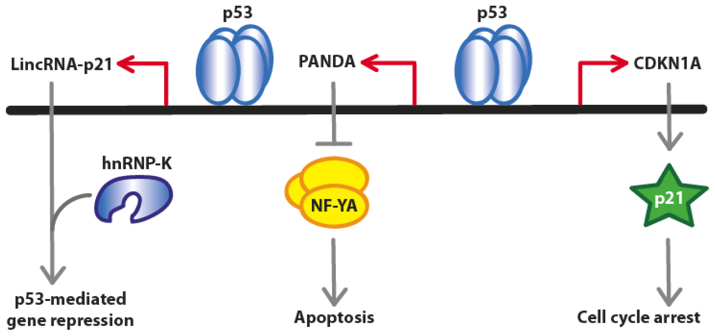
2. Long Non-Coding RNAs at the CDKN1A (p21) Locus
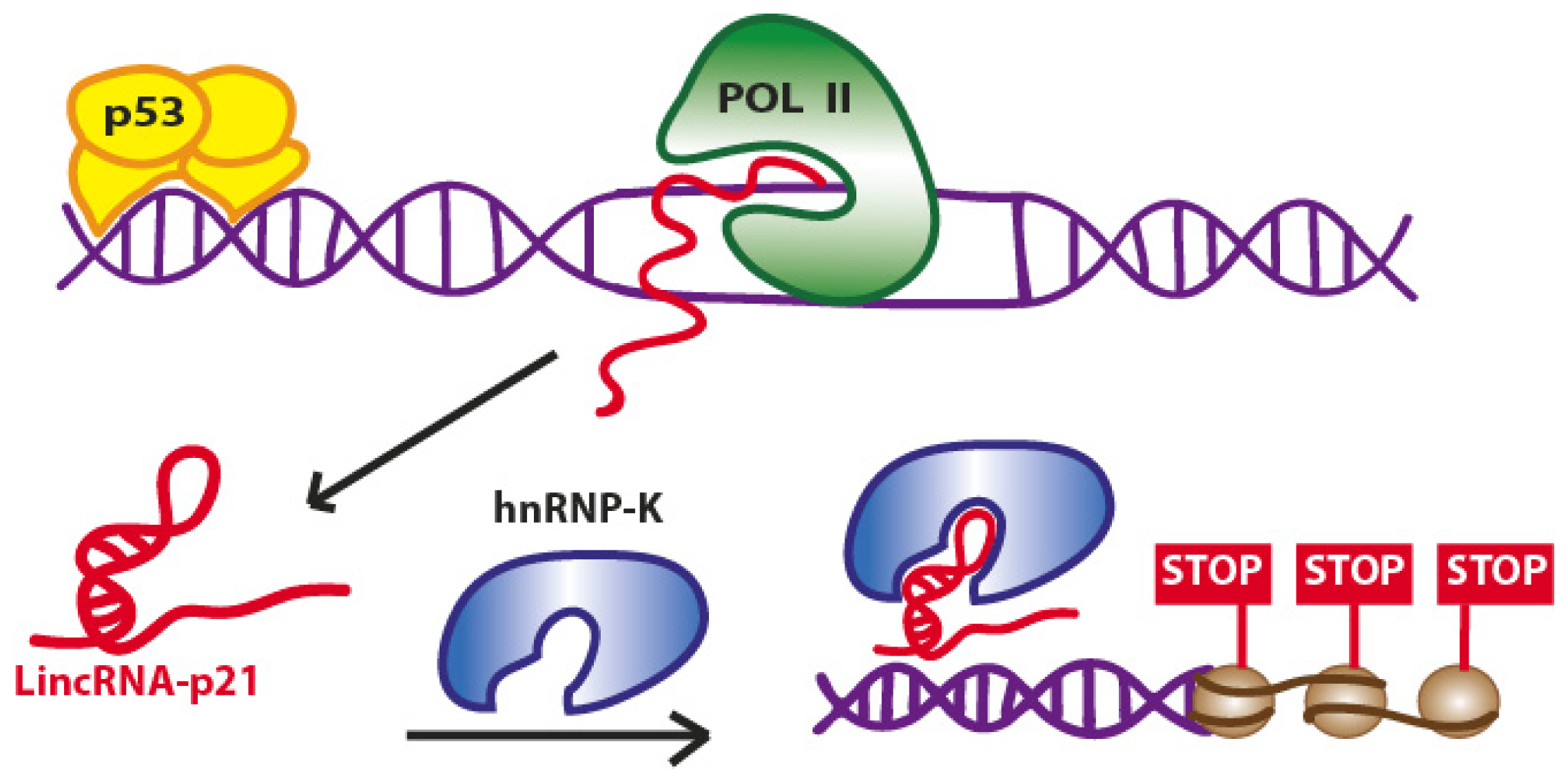
2.1. LincRNA-p21

The main study dealing with LincRNA-p21 and its role in p53 pathway has been reported quite recently [24]. The authors explored the functional roles of this lncRNA intrigued by its properties: genomic location ~15 kb upstream of the gene encoding the critical cell cycle regulator CDKN1A (p21), the presence of a consensus p53 motif for p53-dependent activation and conserved p53-dependent activation of this gene in both human and mouse cell models. Their studies emphasized a role for LincRNA-p21 in a p53-dependent apoptotic response after DNA damage. The authors further observed that siRNA-mediated inhibition of LincRNA-p21 affects the expression of hundreds of target genes, normally repressed by p53, in both the mouse embryonic fibroblasts (MEF) and in lung tumor cell line derived from mice expressing an oncogenic K-Ras mutation (KRAS). Strikingly, upon inhibition of either p53 or LincRNA-p21, the vast majority of these target genes were upregulated suggesting that this lncRNA acts as a downstream repressor in the p53 transcriptional response. Huarte et al. gained also mechanistic clues into how LincRNA-p21 functions to repress such a large subset of genes in the p53 transcriptional response. Biochemical experiments such as RNA-pulldown, native RIP, cross-linked RIP, and deletion-mapping experiments, allowed the authors to identify a specific interaction between the LincRNA-p21 and the heterogeneous nuclear ribonucleoprotein K (hnRNP-K) (Figure 1).
Moreover, they identified a highly conserved 780 nt 5′ region in the sequence of LincRNA-p21 which is required for hnRNP-K binding and subsequent induction of apoptosis. In fact, the two major phenotypic consequences of p53 pathway activation are the growth arrest and the induction of apoptosis [25]. In their work, the authors demonstrated the deregulation of many apoptosis and cell cycle regulator genes by p53 and LincRNA-p21. To mediate gene repression, a physical interaction between LincRNA-p21 and hnRNP-K is required, as the loss of hnRNP-K function induce a reactivation of the same genes repressed by p53 and LincRNA-p21. However, the precise mechanism by which LincRNA-p21 contributes to repression at specific loci has yet to be clarified.

2.2. PANDA (P21 Associated ncRNA DNA Damage Activated)
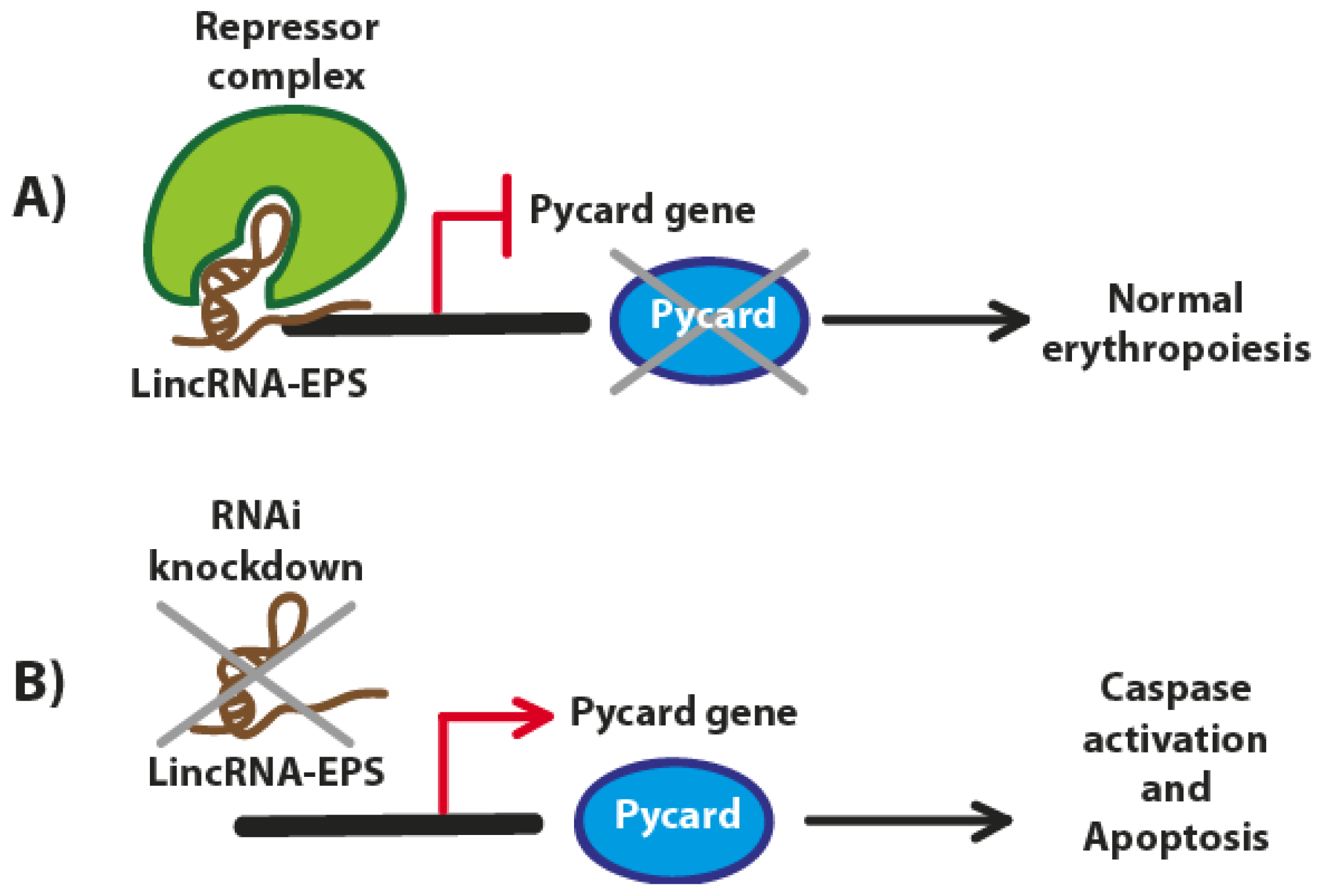
The long non-coding RNA PANDA (a ~1.5-kb transcript) is located ~5 kb upstream of the CDKN1A (p21) transcription start site, is evolutionarily conserved, specifically induced by DNA damage in a p53-dependent manner and mediates anti-apoptotic functions [26]. However, the expression of PANDA is not dependent on p21. It has been observed that PANDA depletion induced several genes encoding canonical activators of apoptosis, such as APAF1, BIK, FAS and LRDD [26].
Promoter regions of p53-dependent cell death genes are characterized by the presence of a binding site for the transcription factor NF-YA, which interacts with PANDA in a highly specific manner [27]. Moreover, the depletion of PANDA substantially increases the occupancy of NF-YA at pro-apoptotic target genes such as CCNB1, FAS, BBC3 (PUMA) and PMAIP1 (NOXA). Therefore, whereas CDKN1A mediates cell cycle arrest, PANDA promotes cell survival by interfering with the apoptotic gene expression program (Figure 2).

3. LincRNA-EPS

During erythropoiesis more than 400 putative lncRNAs are differentially expressed. One of these lncRNAs, named long intergenic non-coding RNA erythroid prosurvival (LincRNA-EPS), is induced during the terminal differentiation of murine erythroid cells and has been recently investigated [28]. LincRNA-EPS is a 2531-nt lncRNA originated from a DNA portion consisting of four exons and three introns, bearing a 5′ end cap structure and a 3′ poly(A) tail. LincRNA-EPS is strongly induced when erythroid precursors begin to synthesize hemoglobin and other lineage-specific proteins. LincRNA-EPS is expressed marginally in other hematopoietic lineages, indicating erythroid specificity. It has been observed that by knocking down LincRNA-EPS, an inhibition of differentiation and the promotion of apoptosis take place. Conversely, ectopic expression of this lncRNA can prevent apoptosis in mouse erythroid cells. Although we still do not know if this LincRNA-EPS is under the direct control of p53, LincRNA-EPS can repress the expression of the proapoptotic gene Pycard, inhibiting programmed cell death [28]. On the other hand, the induction of Pycard (also known as ASC, CARD5, TMS or TMS-1) has been alternatively reported by the same authors to be either dependent on p53 (after exposure to a genotoxic stress in the intrinsic mitochondrial pathway of apoptosis through a p53-Bax network) [29] or p53-independent (under hypoxic conditions in pancreatic cancer cells) (Figure 3) [30]. However, from a preliminary bioinformatics search we found that various p53 regulatory transcription factor binding sites are present in the Pycard gene promoter (data not shown).
To study the functional link between LincRNA-EPS and Pycard and how this lncRNA can inhibit apoptosis, Hu et al. employed microarrays technology to assess the gene expression of lineage-negative (Lin-) fetal liver cells, highly enriched for erythroid progenitors [32] and ectopically expressing LincRNA-EPS. The authors found that many genes involved in apoptosis were downregulated, and the proapoptotic Pycard gene was the most downregulated. These data suggested that Pycard can be one of the targets of LincRNA-EPS and its expression during normal erythropoiesis is inversely correlated with that of the LincRNA-EPS. Moreover, the overexpression of Pycard gene is able to inhibit the proliferation of erythroid cells to promote apoptosis and interfere with their terminal differentiation and enucleation.
The nuclear localization of LincRNA-EPS suggested that the gene expression regulation might occur through nuclear events such as epigenetic modifications, transcription, or mRNA splicing, or simply by its association with chromatin modifiers which repress the transcription of Pycard and the other genes promoting cell apoptosis. Finally, further studies aimed at discovering this lncRNA and other potentially associated factors in humans and novel and more powerful computational methods and prediction algorithms will surely help to identify all the actors involved in this mechanism.

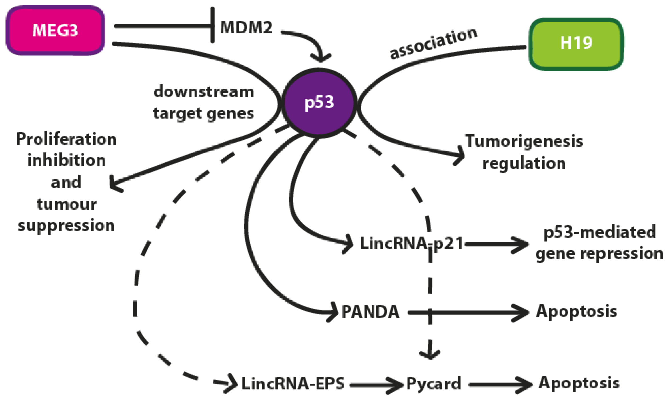
4. p53 Regulation by lncRNAs

4.1. H19

The lncRNA H19 has been recently investigated and found upregulated in many tumors [33,34]. Moreover, it has been found that ectopic expression of H19 increased cell proliferation, whereas H19 silencing (siRNAs) contributed to apoptosis in human gastric cancer cells (AGS cells) [35]. In the latter study, the authors aimed also to investigate if the molecular mechanism by which H19 increases gastric cancer cell growth required p53 activation.
In fact, several groups have studied the link between H19 and p53 [36,37]. Dugimont et al. demonstrated that the H19 promoter is efficiently repressed by p53 [37]. By immunoprecipitation and RNA pulldown experiments, Yang et al. demonstrated the association of H19 and p53, and by transfecting H19 in AGS cells they observed a significant decrease of p53 activity, suppressing also the protein level of the p53 target Bax. Therefore, these data emphasize the fact that the upregulation of the lncRNA H19 contributes to tumorigenesis through p53 activity regulation, at least in gastric cancers.

4.2. MEG3 lncRNA

The transcript of the maternally expressed gene 3 (MEG3) is reported to have several isoforms originating from the human MEG3 gene which contains 10 exons [38]. The length of the different isoforms is variable although their mean value is ~1700 nt. MEG3 lncRNA is expressed in many normal tissues (i.e., human pituitary, including normal gonadotroph cells) but absent in a continuously increasing list of primary human tumors and tumor cell lines [39,40]. MEG3 is expressed during development with higher levels in the paraxial mesoderm, the developing central nervous system, and the epithelia of salivary glands, pancreas and kidney. It is also expressed in adult mouse adrenal and pituitary glands and brain [41]. Gene deletion, promoter hypermethylation and hypermethylation of other intergenic regions are the most probably causes for MEG3 lncRNA loss in these cases. MEG3 lncRNA induces an accumulation of p53 protein, stimulating the transcription from a p53-dependent promoter and selectively regulates the expression of p53 target genes (Figure 4) [42]. In conclusion, MEG3 lncRNA can be considered a novel tumor suppressor lncRNA [43].
Interestingly, Zhang et al. pointed out for the first time that the folding structure of MEG3 lncRNA is more important to its biological function than its primary sequence [38]. In fact, by examining the secondary folding structures of the main MEG3 lncRNA domains, which they identified and called M1, M2 and M3, Zhang et al. assessed that p53-mediated transactivation varies between these different isoforms and that M2 domain has a critical role. As a direct consequence of their results, the most fascinating observation is that also the folding structures of lncRNAs are important factors to consider as they are intimately linked to their functions. We can imagine that, like proteins, also lncRNAs can fold into tertiary structures determining different outcomes and functions. Further crystallographic and RNA-protein studies will surely shed light on these interesting, but still unanswered, questions.

5. Conclusions

Many studies have recently identified and discussed the role of numerous lncRNAs which are involved in p53 regulation or have a role in carcinogenesis or cancer growth [40,4448]. These lncRNAs are emerging as important regulators of gene expression by acting synergistically with other factors in many different and crucial mechanisms. The interplays between lncRNAs and p53 regulation has been discussed in this review and summarized in Figure 4.
The advent of next-generation sequencing techniques has paved the way to important discoveries also in the field of non-coding RNAs, but the exact biological role of this regulatory molecules has still to be clarified. Notwithstanding, many studies are beginning to shed light on lncRNAs and their mechanisms and more information are expected to arise in the next few years.
In this brief review, we emphasized only the role of a selection of these lncRNAs, in particular of those molecules involved in p53 pathway and in its regulation owing to the central importance of this pathway in many conditions. We are aware that many other lncRNAs may play a role in this pathway, although this role is not completely unraveled so far. Moreover, little is known about the expression level (i.e., the threshold) required to a lncRNA to function properly, or the relative abundance in various tissues. To partially answer these questions, a novel integrative approach consisting in a computational approach integrating RNA-seq data with available annotation resources has been recently reported [49]. The result is a reference catalog (Human Body Map lincRNAs) [50] of 8195 human lincRNAs with information about their expression levels in different tissues and linked to the expression of their neighboring genes.
Finally, the bioinformatics tools that will be developed in the near future will surely help researchers to fill the knowledge gaps and to face the present challenges [51]. We believe that the world of lncRNAs, as it was for the “junk” non-coding RNAs such as miRNAs, will open unexpected possibilities, not only for biological and biomedical research, but also for translational research and innovative clinical “theranostics” (therapeutics and diagnostics) applications.

Acknowledgments

The Italian Ministry of Health and the Bambino Gesù Children’s Hospital, IRCCS is gratefully acknowledged for their financial support (grant RC-201202G002799).

References

  1. Mattick, J.S. RNA regulation: A new genetics? Nat. Rev. Genet 2004, 5, 316–323. [Google Scholar]
  2. Mercer, T.R.; Dinger, M.E.; Mattick, J.S. Long non-coding RNAs: Insights into functions. Nat. Rev. Genet 2009, 10, 155–159. [Google Scholar]
  3. Ponting, C.P.; Oliver, P.L.; Reik, W. Evolution and functions of long noncoding RNAs. Cell 2009, 136, 629–641. [Google Scholar]
  4. Wilusz, J.E.; Sunwoo, H.; Spector, D.L. Long noncoding RNAs: Functional surprises from the RNA world. Genes Dev 2009, 23, 1494–1504. [Google Scholar]
  5. Carninci, P. Non-coding RNA transcription: Turning on neighbours. Nat. Cell Biol 2008, 10, 1023–1024. [Google Scholar]
  6. The ENCODE Project Consortium. Identification and analysis of functional elements in 1% of the human genome by the ENCODE pilot project. Nature 2007, 447, 799–816.
  7. The ENCODE Project Consortium. An integrated encyclopedia of DNA elements in the human genome. Nature 2012, 489, 57–74.
  8. Bernstein, E.; Allis, C.D. RNA meets chromatin. Genes Dev 2005, 19, 1635–1655. [Google Scholar]
  9. Bracken, A.P.; Helin, K. Polycomb group proteins: Navigators of lineage pathways led astray in cancer. Nat. Rev. Cancer 2009, 9, 773–784. [Google Scholar]
  10. Faghihi, M.A.; Wahlestedt, C. Regulatory roles of natural antisense transcripts. Nat. Rev. Mol. Cell Biol 2009, 10, 637–643. [Google Scholar]
  11. Whitehead, J.; Pandey, G.K.; Kanduri, C. Regulation of the mammalian epigenome by long noncoding RNAs. Biochim. Biophys. Acta 2009, 1790, 936–947. [Google Scholar]
  12. Kanduri, C.; Whitehead, J.; Mohammad, F. The long and the short of it: RNA-directed chromatin asymmetry in mammalian X-chromosome inactivation. FEBS Lett 2009, 583, 857–864. [Google Scholar]
  13. Lee, J.T. Lessons from X-chromosome inactivation: Long ncRNA as guides and tethers to the epigenome. Genes Dev 2009, 23, 1831–1842. [Google Scholar]
  14. Mohammad, F.; Mondal, T.; Kanduri, C. Epigenetics of imprinted long noncoding RNAs. Epigenetics 2009, 4, 277–286. [Google Scholar]
  15. Clemson, C.M.; Hutchinson, J.N.; Sara, S.A.; Ensminger, A.W.; Fox, A.H.; Chess, A.; Lawrence, J.B. An architectural role for a nuclear noncoding RNA: NEAT1 RNA is essential for the structure of paraspeckles. Mol. Cell 2009, 33, 717–726. [Google Scholar]
  16. Sasaki, Y.T.; Ideue, T.; Sano, M.; Mituyama, T.; Hirose, T. MENepsilon/β noncoding RNAs are essential for structural integrity of nuclear paraspeckles. Proc. Natl. Acad. Sci. USA 2009, 106, 2525–2530. [Google Scholar]
  17. Schoeftner, S.; Blasco, M.A. A “higher order” of telomere regulation: Telomere heterochromatin and telomeric RNAs. EMBO J 2009, 28, 2323–2336. [Google Scholar]
  18. Wong, L.H.; Brettingham-Moore, K.H.; Chan, L.; Quach, J.M.; Anderson, M.A.; Northrop, E.L.; Hannan, R.; Saffery, R.; Shaw, M.L.; Williams, E.; Choo, K.H. Centromere RNA is a key component for the assembly of nucleoproteins at the nucleolus and centromere. Genome Res 2007, 17, 1146–1160. [Google Scholar]
  19. Ferri, F.; Bouzinba-Segard, H.; Velasco, G.; Hube, F.; Francastel, C. Non-coding murine centromeric transcripts associate with and potentiate Aurora B kinase. Nucleic Acids Res 2009, 37, 5071–5080. [Google Scholar]
  20. Willingham, A.T.; Orth, A.P.; Batalov, S.; Peters, E.C.; Wen, B.G.; Aza-Blanc, P.; Hogenesch, J.B.; Schultz, P.G. A strategy for probing the function of noncoding RNAs finds a repressor of NFAT. Science 2005, 309, 1570–1573. [Google Scholar]
  21. Ponjavic, J.; Ponting, C.P.; Lunter, G. Functionality or transcriptional noise? Evidence for selection within long noncoding RNAs. Genome Res 2007, 17, 556–565. [Google Scholar]
  22. Mattick, J.S. The genetic signatures of noncoding RNAs. PLoS Genet 2009, 5, e1000459. [Google Scholar]
  23. Guttman, M.; Amit, I.; Garber, M.; French, C.; Lin, M.F.; Feldser, D.; Huarte, M.; Zuk, O.; Carey, B.W.; Cassady, J.P.; et al. Chromatin signature reveals over a thousand highly conserved large non-coding RNAs in mammals. Nature 2009, 458, 223–227. [Google Scholar]
  24. Huarte, M.; Guttman, M.; Feldser, D.; Garber, M.; Koziol, M.J.; Kenzelmann-Broz, D.; Khalil, A.M.; Zuk, O.; Amit, I.; Rabani, M.; et al. A large intergenic noncoding RNA induced by p53 mediates global gene repression in the p53 response. Cell 2010, 142, 409–419. [Google Scholar]
  25. Levine, A.J.; Hu, W.; Feng, Z. The P53 pathway: What questions remain to be explored? Cell Death Differ 2006, 13, 1027–1036. [Google Scholar]
  26. Hung, T.; Wang, Y.; Lin, M.F.; Koegel, A.K.; Kotake, Y.; Grant, G.D.; Horlings, H.M.; Shah, N.; Umbricht, C.; Wang, P.; et al. Extensive and coordinated transcription of noncoding RNAs within cell-cycle promoters. Nat. Genet 2011, 43, 621–629. [Google Scholar]
  27. Morachis, J.M.; Murawsky, C.M.; Emerson, B.M. Regulation of the p53 transcriptional response by structurally diverse core promoters. Genes Dev 2010, 24, 135–147. [Google Scholar]
  28. Hu, W.; Yuan, B.; Flygare, J.; Lodish, H.F. Long noncoding RNA-mediated anti-apoptotic activity in murine erythroid terminal differentiation. Genes Dev 2011, 25, 2573–2578. [Google Scholar]
  29. Ohtsuka, T.; Ryu, H.; Minamishima, Y.A.; Macip, S.; Sagara, J.; Nakayama, K.I.; Aaronson, S.A.; Lee, S.W. ASC is a Bax adaptor and regulates the p53-Bax mitochondrial apoptosis pathway. Nat. Cell Biol 2004, 6, 121–128. [Google Scholar]
  30. Ohtsuka, T.; Mitsuno, M.; Kitajima, Y.; Ide, T.; Lee, S.W.; Miyazaki, K. Role of ASC in hypoxia-mediated cell death in pancreatic cancer. Mol. Med. Report 2008, 1, 827–831. [Google Scholar]
  31. Paralkar, V.R.; Weiss, M.J. A new “Linc” between noncoding RNAs and blood development. Genes Dev 2011, 25, 2555–2558. [Google Scholar]
  32. Flygare, J.; Rayon Estrada, V.; Shin, C.; Gupta, S.; Lodish, H.F. HIF1alpha synergizes with glucocorticoids to promote BFU-E progenitor self-renewal. Blood 2011, 117, 3435–3444. [Google Scholar]
  33. Matouk, I.J.; DeGroot, N.; Mezan, S.; Ayesh, S.; Abu-lail, R.; Hochberg, A.; Galun, E. The H19 non-coding RNA is essential for human tumor growth. PLoS One 2007, 2, e845. [Google Scholar]
  34. Barsyte-Lovejoy, D.; Lau, S.K.; Boutros, P.C.; Khosravi, F.; Jurisica, I.; Andrulis, I.L.; Tsao, M.S.; Penn, L.Z. The c-Myc oncogene directly induces the H19 noncoding RNA by allele-specific binding to potentiate tumorigenesis. Cancer Res 2006, 66, 5330–5337. [Google Scholar]
  35. Yang, F.; Bi, J.; Xue, X.; Zheng, L.; Zhi, K.; Hua, J.; Fang, G. Up-regulated long non-coding RNA H19 contributes to proliferation of gastric cancer cells. FEBS J 2012, 279, 3159–3165. [Google Scholar]
  36. Park, I.Y.; Sohn, B.H.; Choo, J.H.; Joe, C.O.; Seong, J.K.; Lee, Y.I.; Chung, J.H. Deregulation of DNA methyltransferases and loss of parental methylation at the insulin-like growth factor II (Igf2)/H19 loci in p53 knockout mice prior to tumor development. J. Cell. Biochem 2005, 94, 585–596. [Google Scholar]
  37. Dugimont, T.; Montpellier, C.; Adriaenssens, E.; Lottin, S.; Dumont, L.; Iotsova, V.; Lagrou, C.; Stehelin, D.; Coll, J.; Curgy, J.J. The H19 TATA-less promoter is efficiently repressed by wild-type tumor suppressor gene product p53. Oncogene 1998, 16, 2395–2401. [Google Scholar]
  38. Zhang, X.; Rice, K.; Wang, Y.; Chen, W.; Zhong, Y.; Nakayama, Y.; Zhou, Y.; Klibanski, A. Maternally expressed gene 3 (MEG3) noncoding ribonucleic acid: Isoform structure, expression, and functions. Endocrinology 2010, 151, 939–947. [Google Scholar]
  39. Zhang, X.; Zhou, Y.; Mehta, K.R.; Danila, D.C.; Scolavino, S.; Johnson, S.R.; Klibanski, A. A pituitary-derived MEG3 isoform functions as a growth suppressor in tumor cells. J. Clin. Endocrinol. Metab 2003, 88, 5119–5126. [Google Scholar]
  40. Zhang, X.; Gejman, R.; Mahta, A.; Zhong, Y.; Rice, K.A.; Zhou, Y.; Cheunsuchon, P.; Louis, D.N.; Klibanski, A. Maternally expressed gene 3, an imprinted noncoding RNA gene, is associated with meningioma pathogenesis and progression. Cancer Res 2010, 70, 2350–2358. [Google Scholar]
  41. Schmidt, J.V.; Matteson, P.G.; Jones, B.K.; Guan, X.J.; Tilghman, S.M. The Dlk1 and Gtl2 genes are linked and reciprocally imprinted. Genes Dev 2000, 14, 1997–2002. [Google Scholar]
  42. Zhou, Y.; Zhong, Y.; Wang, Y.; Zhang, X.; Batista, D.L.; Gejman, R.; Ansell, P.J.; Zhao, J.; Weng, C.; Klibanski, A. Activation of p53 by MEG3 non-coding RNA. J. Biol. Chem 2007, 282, 24731–24742. [Google Scholar]
  43. Zhou, Y.; Zhang, X.; Klibanski, A. MEG3 noncoding RNA: A tumor suppressor. J. Mol. Endocrinol 2012, 48, R45–R53. [Google Scholar]
  44. Matouk, I.J.; Mezan, S.; Mizrahi, A.; Ohana, P.; Abu-Lail, R.; Fellig, Y.; Degroot, N.; Galun, E.; Hochberg, A. The oncofetal H19 RNA connection: Hypoxia, p53 and cancer. Biochim. Biophys. Acta 2010, 1803, 443–451. [Google Scholar]
  45. Braconi, C.; Kogure, T.; Valeri, N.; Huang, N.; Nuovo, G.; Costinean, S.; Negrini, M.; Miotto, E.; Croce, C.M.; Patel, T. microRNA-29 can regulate expression of the long non-coding RNA gene MEG3 in hepatocellular cancer. Oncogene 2011, 30, 4750–4756. [Google Scholar]
  46. Kogo, R.; Shimamura, T.; Mimori, K.; Kawahara, K.; Imoto, S.; Sudo, T.; Tanaka, F.; Shibata, K.; Suzuki, A.; Komune, S.; et al. Long noncoding RNA HOTAIR regulates polycombdependent chromatin modification and is associated with poor prognosis in colorectal cancers. Cancer Res 2011, 71, 6320–6326. [Google Scholar]
  47. Lai, M.C.; Yang, Z.; Zhou, L.; Zhu, Q.Q.; Xie, H.Y.; Zhang, F.; Wu, L.M.; Chen, L.M.; Zheng, S.S. Long non-coding RNA MALAT-1 overexpression predicts tumor recurrence of hepatocellular carcinoma after liver transplantation. Med. Oncol 2012, 29, 1810–1816. [Google Scholar]
  48. Benetatos, L.; Vartholomatos, G.; Hatzimichael, E. MEG3 imprinted gene contribution in tumorigenesis. Int. J. Cancer 2011, 129, 773–779. [Google Scholar]
  49. Cabili, M.N.; Trapnell, C.; Goff, L.; Koziol, M.; Tazon-Vega, B.; Regev, A.; Rinn, J.L. Integrative annotation of human large intergenic noncoding RNAs reveals global properties and specific subclasses. Genes Dev 2011, 25, 1915–1927. [Google Scholar]
  50. Cabili, M.N. Human Body Map lincRNAs. Available online: http://www.broadinstitute.org/genome_bio/human_lincrnas/ accessed on 5 December 2012.
  51. Da Sacco, L.; Baldassarre, A.; Masotti, A. Bioinformatics tools and novel challenges in long non-coding RNAs (lncRNAs) functional analysis. Int. J. Mol. Sci 2012, 13, 97–114. [Google Scholar]
Figure 1. Proposed model for the role of LincRNA-p21 in the p53 transcriptional response. The induction of p53 activates the transcription of LincRNA-p21 by binding to its promoter (upper left). LincRNA-p21 binds to hnRNP-K and acts to repress genes that are downregulated as part of the canonical p53 transcriptional response. (Adapted from [24]).
Ijms 13 16708f1
Figure 2. Model of coding and non-coding transcripts at the CDKN1A locus coordinating the DNA damage response. After DNA damage, p53 binding at the CDKN1A locus coordinately activates transcription of CDKN1A as well as non-coding transcripts PANDA and LincRNA-p21. CDKN1A mediates cell cycle arrest, PANDA blocks apoptosis through NF-YA, and LincRNA-p21 mediates gene silencing through recruitment of hnRPK. (Adapted from [26]).
Ijms 13 16708f2
Figure 3. The potential role of LincRNA-EPS in erythropoiesis. (A) LincRNA-EPS represses Pycard gene to inhibit apoptosis of red blood cells progenitors. LincRNA-EPS may bind the Pycard gene and inhibit its transcription by recruiting transcriptional repressor complexes; (B) Knockdown of LincRNA-EPS allows the transcription of Pycard, which in turn activates intercellular caspases and induces apoptosis. (Adapted from [31]).
Ijms 13 16708f3
Figure 4. In the p53 pathway, MEG3 may activate p53 directly by RNA-protein interaction or indirectly by suppressing MDM2, resulting in selective activation of p53 downstream targets such as GDF-15 with both anti-proliferative and tumor suppressive functions. The association of H19 with p53 determines tumorigenesis regulation. The role of the lncRNAs LincRNA-p21, PANDA and LincRNA-EPS have been discussed in the text.
Ijms 13 16708f4

