
Antibacterial Activity and Cytotoxicity of Silver(I) Complexes of Pyridine and (Benz)Imidazole Derivatives. X-ray Crystal Structure of [Ag(2,6-di(CH2OH)py)2]NO3

 and
and
Abstract
:1. Introduction

| Ag(I) Complex Formula (No.) | Ligand Structure | Ref. |
|---|---|---|
| {[Ag(4-pmOpe)]NO3}n (1) |  4-pmOpe | [28] |
| [Ag(2-bimOpe)2]NO3 (2) |  2-bimOpe | [28] |
| [Ag(2,6-di(CH2OH)py)2]NO3 (3) |  2,6-di(CH2OH)py | [29] |
| [Ag(4-CH2OHpy)2]NO3 (4) |  4-CH2OHpy | [29] |
| [Ag(2-CH2OHbim)2]NO3 (5) |  2-CH2OHbim | [29] |
| [Ag(MTZ)2NO3] (6) |  MTZ (metronidazole) | [13] |
2. Results and Discussion
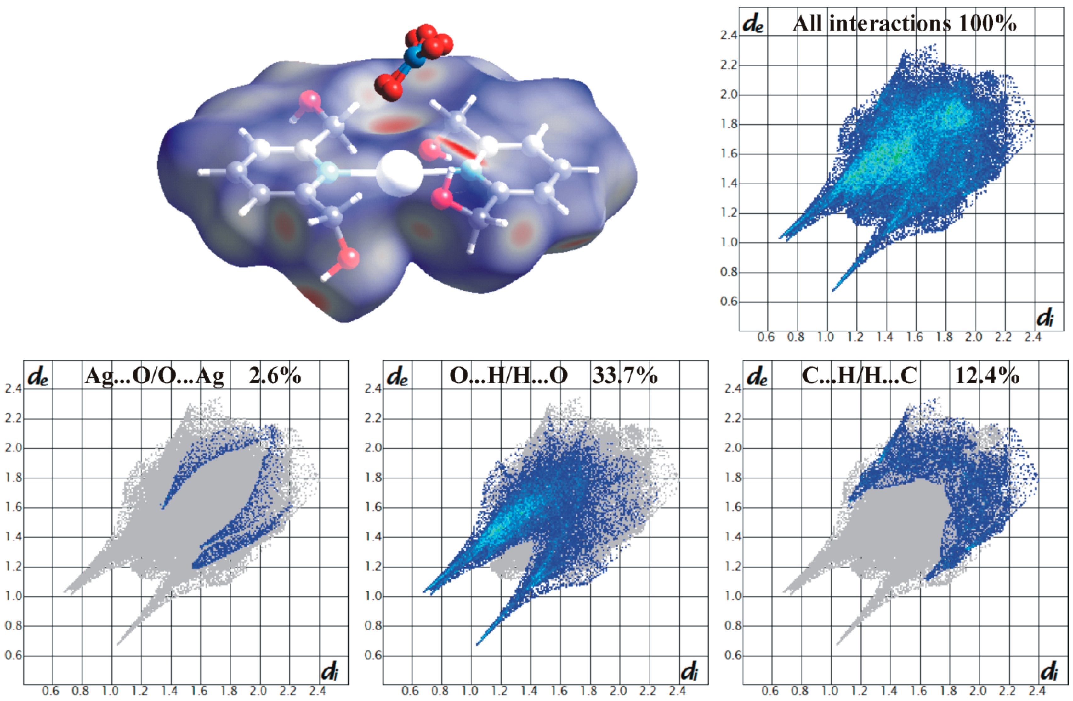
2.1. Crystal and Molecular Structure of 3
| Complex 3 | |
|---|---|
| Empirical formula | C14H18AgN3O7 |
| Formula weight | 448.18 |
| Crystal system | Monoclinic |
| Space group | P21/c |
| a (Å) | 9.7304(1) |
| b (Å) | 14.7348(2) |
| c (Å) | 14.0158(2) |
| α (°) | 90.000 |
| β (°) | 127.803(1) |
| γ (°) | 90.000 |
| V (Å3) | 1587.77(4) |
| Z | 4 |
| T (K) | 100(2) |
| F(000) | 904 |
| Dx (g·cm−3) | 1.875 |
| μ (mm−1) | 1.32 |
| Scan method | ω scan |
| θ range (°) | 2.3–28.0 |
| Measured reflections | 27856 |
| Unique reflections | 3836 |
| Observed reflections [I > 2σ(I)] | 3729 |
| Completeness to θmax (%) | 100 |
| No. of parameters/restraints | 271/18 |
| R [I > 2σ(I)] | 0.0147 |
| wR (all data) | 0.0379 |
| S | 1.09 |
| Largest diff. peak (e·Å−3) | 0.48 |
| Largest diff. hole (e·Å−3) | −0.30 |
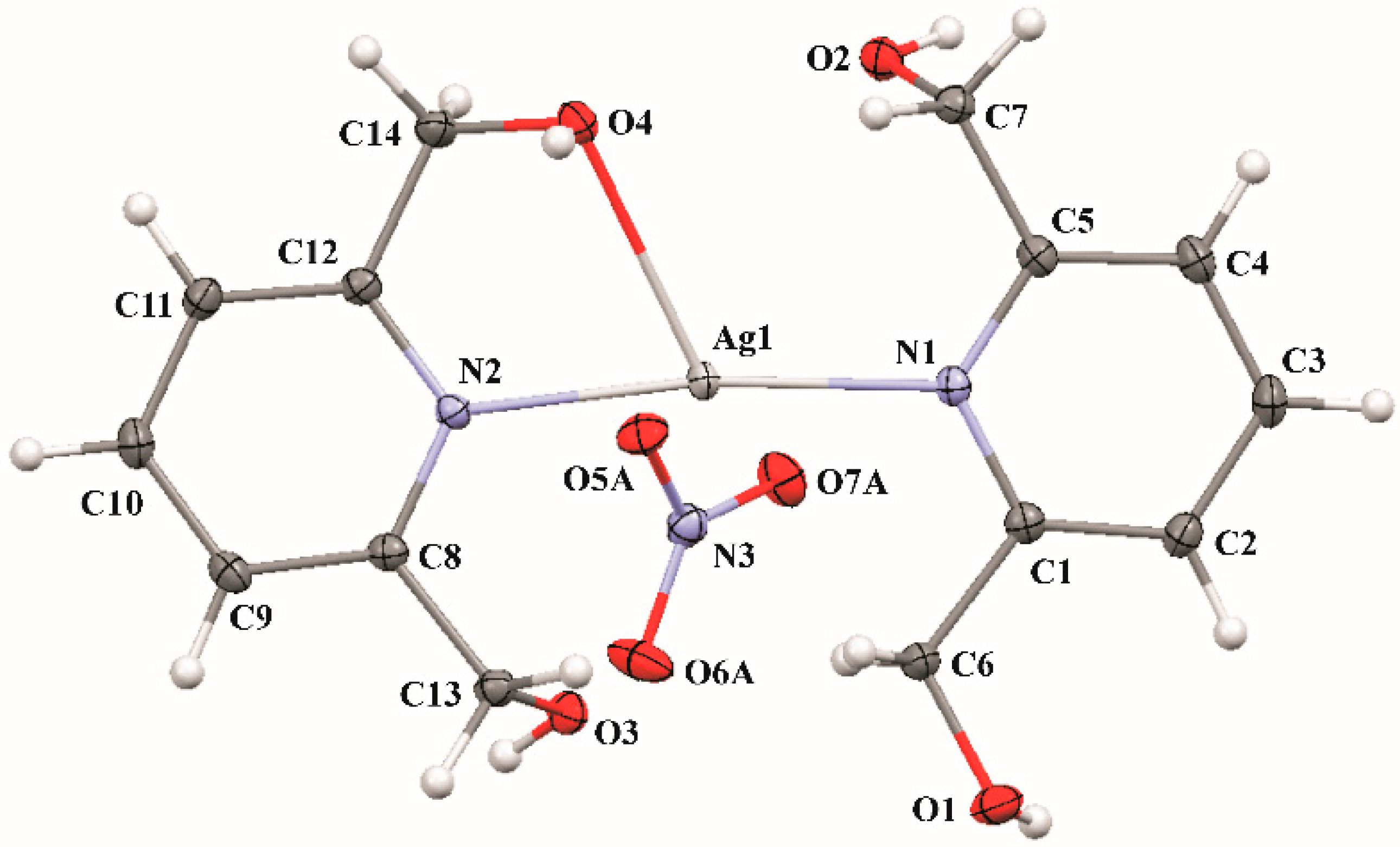
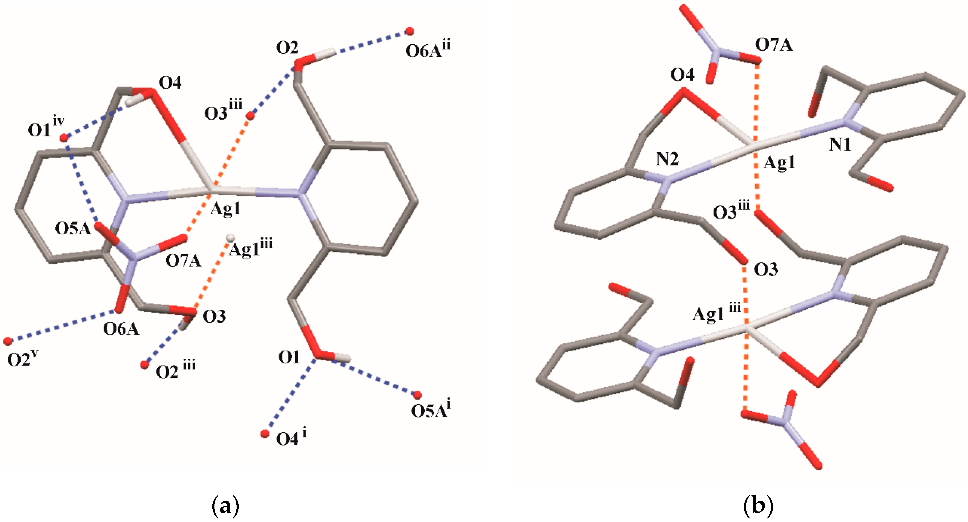
| D-H | H···A | D···A | D-H···A | |
|---|---|---|---|---|
| O1-H1A···O5Ai | 0.81(2) | 1.94(2) | 2.709(5) | 159(2) |
| O2-H2A···O6Aii | 0.78(2) | 1.95(2) | 2.721(5) | 170(2) |
| O3-H3A···O2Aiii | 0.81(2) | 1.89(2) | 2.691(2) | 169(2) |
| O4-H4A···O1Aiv | 0.82(2) | 1.94(2) | 2.726(2) | 163(2) |

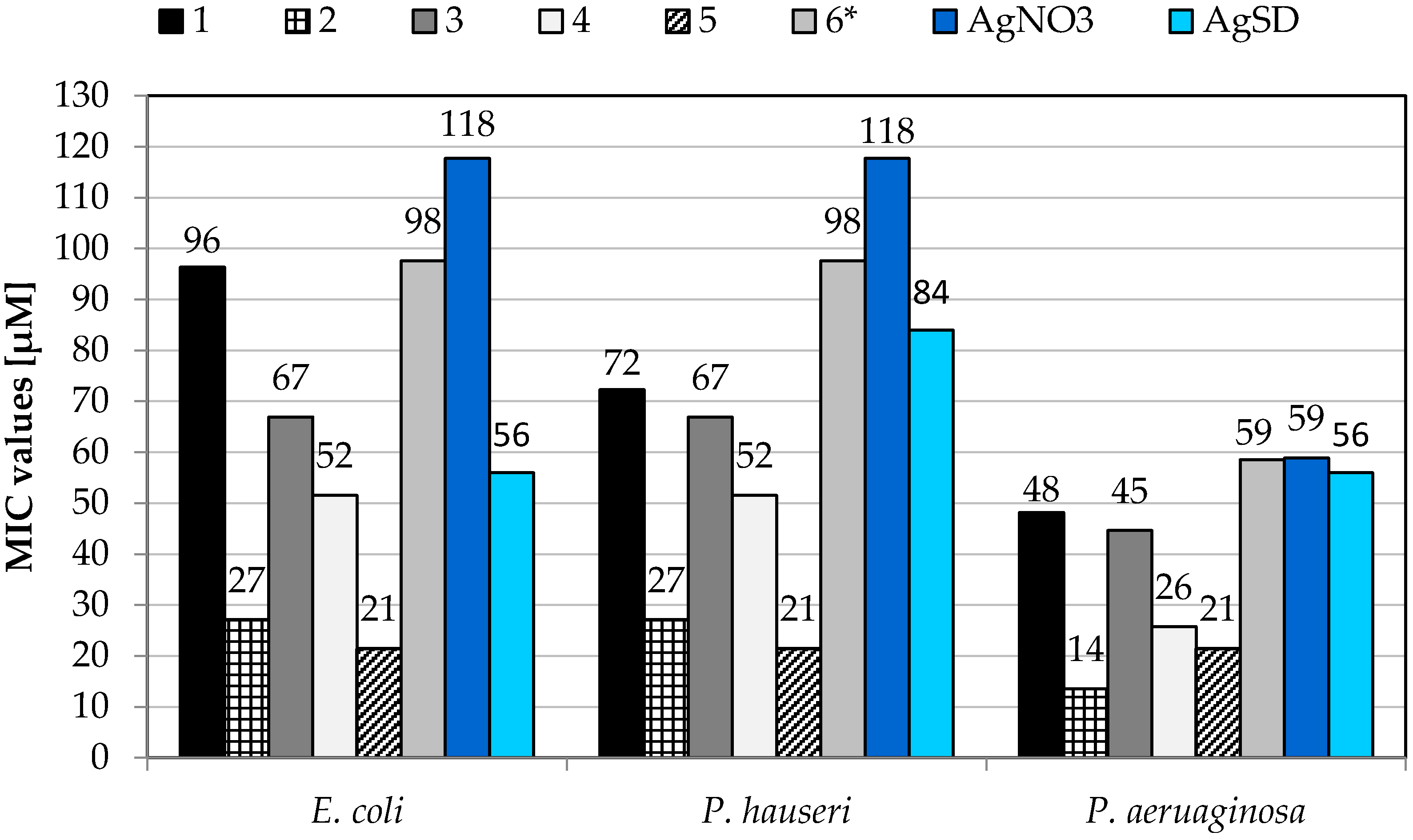
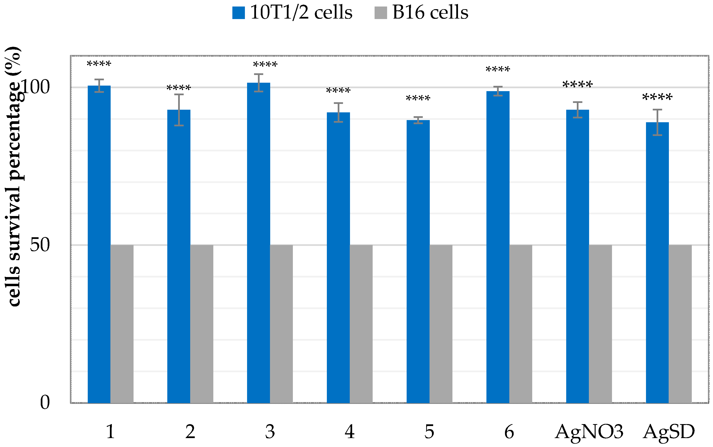
2.2. Antibacterial Activity

| Compound | E. coli ATCC 25992 | P. hauseri ATCC 13315 | P. aeruginosa ATCC 15442 |
|---|---|---|---|
| 1 | 145 | 120 | 72 |
| 2 | 54 | 41 | 27 |
| 3 | 67 | 112 | 67 |
| 4 | 77 | 77 | 52 |
| 5 | 21 | 43 | 21 |
| 6 [13] | 98 | 117 | 117 |
| AgNO3 | 177 | 177 | 59 |
| AgSD | 84 | 140 | 56 |
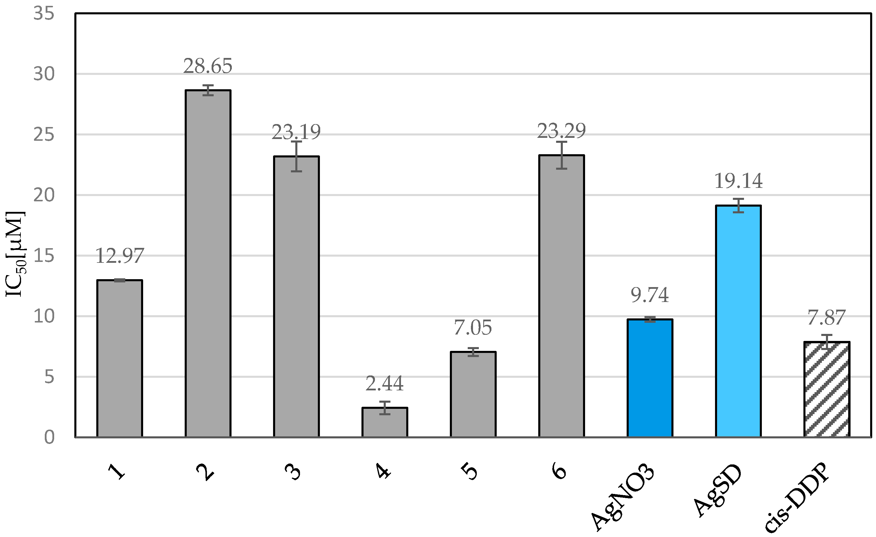
2.3. Cytotoxic Activity

3. Materials and Methods
3.1. Synthesis of the Complexes 1–6
3.2. X-ray Single-Crystal Structure Determination
3.3. Antibacterial Activity Studies
3.4. Cytotoxic Activity. MTT Assays
4. Conclusions
Supplementary Materials
Acknowledgments
Author Contributions
Conflicts of Interest
References
- Bansal, Y.; Silakari, O. The therapeutic journey of benzimidazoles: A review. Bioorg. Med. Chem. 2012, 20, 6208–6236. [Google Scholar] [CrossRef] [PubMed]
- Gholivand, K.; Molaei, F.; Oroujzadeh, N.; Mobasseri, R.; Naderi-Manesh, H. Two novel Ag(I) complexes of N-nicotinyl phosphoric triamide derivatives: Synthesis, X-ray crystal structure and in vitro antibacterial and cytotoxicity studies. Inorg. Chim. Acta 2014, 423, 107–116. [Google Scholar] [CrossRef]
- Kadayat, T.M.; Song, C.; Shin, S.; Magar, T.B.; Bist, G.; Shrestha, A.; Thapa, P.; Na, Y.; Kwon, Y.; Lee, E.S. Synthesis, topoisomerase I and II inhibitory activity, cytotoxicity, and structure-activity relationship study of 2-phenyl- or hydroxylated 2-phenyl-4-aryl-5H-indeno[1,2-b]pyridines. Bioorg. Med. Chem. 2015, 23, 3499–3512. [Google Scholar] [CrossRef] [PubMed]
- Adhami, F.; Safavi, M.; Ehsani, M.; Ardestani, S.K.; Emmerling, F.; Simyari, F. Synthesis, crystal structure, and cytotoxic activity of novel cyclic systems in [1,2,4]thiadiazolo[2,3-a]pyridine benzamide derivatives and their copper(II) complexes. Dalton Trans. 2014, 43, 7945–7957. [Google Scholar] [CrossRef] [PubMed]
- Sun, G.-X.; Yang, M.-Y.; Shi, Y.-X.; Sun, Z.-H.; Liu, X.-H.; Wu, H.-K.; Li, B.-J.; Zhang, Y.-G. Microwave assistant synthesis, antifungal activity and dft theoretical study of some novel 1,2,4-triazole derivatives containing pyridine moiety. Int. J. Mol. Sci. 2014, 15, 8075. [Google Scholar] [CrossRef] [PubMed]
- Sontakke, V.A.; Kate, A.N.; Ghosh, S.; More, P.; Gonnade, R.; Kumbhar, N.M.; Kumbhar, A.A.; Chopade, B.A.; Shinde, V.S. Synthesis, DNA interaction and anticancer activity of 2-anthryl substituted benzimidazole derivatives. New J. Chem. 2015, 39, 4882–4890. [Google Scholar] [CrossRef]
- Silver, S.; Phung le, T.; Silver, G. Silver as biocides in burn and wound dressings and bacterial resistance to silver compounds. J. Ind. Microbiol. Biotechnol. 2006, 33, 627–634. [Google Scholar] [CrossRef] [PubMed]
- Lansdown, A.B.G. Silver in healthcare: Its antimicrobial efficacy and safety in use. RSC Publishing: Cambridge, UK, 2010; pp. 24–42. [Google Scholar]
- De Gracia, C.G. An open study comparing topical silver sulfadiazine and topical silver sulfadiazine-cerium nitrate in the treatment of moderate and severe burns. Burns 2001, 27, 67–74. [Google Scholar] [CrossRef]
- Kyros, L.; Banti, C.N.; Kourkoumelis, N.; Kubicki, M.; Sainis, I.; Hadjikakou, S.K. Synthesis, characterization, and binding properties towards CT-DNA and lipoxygenase of mixed-ligand silver(I) complexes with 2-mercaptothiazole and its derivatives and triphenylphosphine. J. Biol. Inorg. Chem. 2014, 19, 449–464. [Google Scholar] [CrossRef] [PubMed]
- Haque, R.A.; Choo, S.Y.; Budagumpi, S.; Iqbal, M.A.; Al-Ashraf Abdullah, A. Silver(I) complexes of mono- and bidentate N-heterocyclic carbene ligands: Synthesis, crystal structures, and in vitro antibacterial and anticancer studies. Eur. J. Medi. Chem. 2015, 90, 82–92. [Google Scholar] [CrossRef] [PubMed]
- Alisir, S.H.; Demir, S.; Sariboga, B.; Buyukgungor, O. A disparate 3-D silver(I) coordination polymer of pyridine-3,5-dicarboxylate and pyrimidine with strong intermetallic interactions: X-ray crystallography, photoluminescence and antimicrobial activity. J. Coord. Chem. 2015, 68, 155–168. [Google Scholar] [CrossRef]
- Kalinowska-Lis, U.; Felczak, A.; Chęcińska, L.; Zawadzka, K.; Patyna, E.; Lisowska, K.; Ochocki, J. Synthesis, characterization and antimicrobial activity of water-soluble silver(I) complexes of metronidazole drug and selected counter-ions. Dalton Trans. 2015, 44, 8178–8189. [Google Scholar] [CrossRef] [PubMed]
- McCann, M.; Curran, R.; Ben-Shoshan, M.; McKee, V.; Devereux, M.; Kavanagh, K.; Kellett, A. Synthesis, structure and biological activity of silver(I) complexes of substituted imidazoles. Polyhedron 2013, 56, 180–188. [Google Scholar] [CrossRef]
- Haque, R.A.; Asekunowo, P.O.; Razali, M.R. Dinuclear silver(I)-N-heterocyclic carbene complexes of n-allyl substituted (benz)imidazol-2-ylidenes with pyridine spacers: Synthesis, crystal structures, nuclease and antibacterial studies. Trans. Met. Chem. 2014, 39, 281–290. [Google Scholar] [CrossRef]
- Klasen, H.J. A historical review of the use of silver in the treatment of burns. II. Renewed interest for silver. Burns 2000, 26, 131–138. [Google Scholar] [CrossRef]
- Mohan, M.; Gupta, S.K.; Kalra, V.K.; Vajpayee, R.B.; Sachdev, M.S. Topical silver sulphadiazine—A new drug for ocular keratomycosis. Br. J. Ophthalmol. 1988, 72, 192–195. [Google Scholar] [CrossRef] [PubMed]
- Hamilton-Miller, J.M.T.; Shah, S.; Smith, C. Silver sulphadiazine: A comprehensive in vitro reassessment. Chemotherapy 1993, 39, 405–409. [Google Scholar] [CrossRef] [PubMed]
- Fuller, F.W. The side effects of silver sulfadiazine. J. Burn Care Res. 2009, 30, 464–470. [Google Scholar] [CrossRef] [PubMed]
- Sandri, G.; Bonferoni, M.C.; Ferrari, F.; Rossi, S.; Aguzzi, C.; Mori, M.; Grisoli, P.; Cerezo, P.; Tenci, M.; Viseras, C.; et al. Montmorillonite–chitosan–silver sulfadiazine nanocomposites for topical treatment of chronic skin lesions: In vitro biocompatibility, antibacterial efficacy and gap closure cell motility properties. Carbohydr. Polym. 2014, 102, 970–977. [Google Scholar] [CrossRef] [PubMed]
- Cooper, M.L.; Boyce, S.T.; Hansbrough, J.F.; Foreman, T.J.; Frank, D.H. Cytotoxicity to cultured human keratinocytes of topical antimicrobial agents. J. Surg. Res. 1990, 48, 190–195. [Google Scholar] [CrossRef]
- Cooper, M.L.; Laxer, J.A.; Hansbrough, J.F. The cytotoxic effects of commonly used topical antimicrobial agents on human fibroblasts and keratinocytes. J. Trauma 1991, 31, 775–782; discussion 782–784. [Google Scholar] [CrossRef] [PubMed]
- Banti, C.N.; Hadjikakou, S.K. Anti-proliferative and anti-tumor activity of silver(I) compounds. Metallomics 2013, 5, 569–596. [Google Scholar] [CrossRef] [PubMed]
- Zhu, H.-L.; Zhang, X.-M.; Liu, X.-Y.; Wang, X.-J.; Liu, G.-F.; Usman, A.; Fun, H.-K. Clear Ag–Ag bonds in three silver(I) carboxylate complexes with high cytotoxicity properties. Inorg. Chem. Commun. 2003, 6, 1113–1116. [Google Scholar] [CrossRef]
- Liu, X.Y.; Zhu, H.L. Strong silver-silver interactions in three silver(I) carboxylate complexes with high cytotoxicity properties. Synth. React. Inorg. Met. Org. Nano Met. Chem. 2005, 35, 325–332. [Google Scholar] [CrossRef]
- Santini, C.; Pellei, M.; Papini, G.; Morresi, B.; Galassi, R.; Ricci, S.; Tisato, F.; Porchia, M.; Rigobello, M.P.; Gandin, V.; et al. In vitro antitumour activity of water soluble Cu(I), Ag(I) and Au(I) complexes supported by hydrophilic alkyl phosphine ligands. J. Inorg. Biochem. 2011, 105, 232–240. [Google Scholar] [CrossRef] [PubMed]
- McKeage, M.J.; Papathanasiou, P.; Salem, G.; Sjaarda, A.; Swiegers, G.F.; Waring, P.; Wild, S.B. Antitumor activity of gold(I), silver(I) and copper(I) complexes containing chiral tertiary phosphines. Met. Based Drugs 1998, 5, 217–223. [Google Scholar] [CrossRef] [PubMed]
- Kalinowska-Lis, U.; Szewczyk, E.M.; Chęcińska, L.; Wojciechowski, J.M.; Wolf, W.M.; Ochocki, J. Synthesis, characterization, and antimicrobial activity of silver(I) and copper(II) complexes of phosphate derivatives of pyridine and benzimidazole. Chemmedchem 2014, 9, 169–176. [Google Scholar] [CrossRef] [PubMed]
- Kalinowska-Lis, U.; Felczak, A.; Chęcińska, L.; Lisowska, K.; Ochocki, J. Synthesis, characterization and antimicrobial activity of silver(I) complexes of hydroxymethyl derivatives of pyridine and benzimidazole. J. Organomet. Chem. 2014, 749, 394–399. [Google Scholar] [CrossRef]
- Bodor, E.T.; Offermanns, S. Nicotinic acid: An old drug with a promising future. Br. J. Pharmacol. 2008, 153 (Suppl. 1), S68–S75. [Google Scholar] [CrossRef] [PubMed]
- Brown, B.G.; Zhao, X.Q. Nicotinic acid, alone and in combinations, for reduction of cardiovascular risk. Am. J. Cardiol. 2008, 101, 58b–62b. [Google Scholar] [CrossRef] [PubMed]
- Freeman, C.D.; Klutman, N.E.; Lamp, K.C. Metronidazole. A therapeutic review and update. Drugs 1997, 54, 679–708. [Google Scholar] [CrossRef] [PubMed]
- Kaper, J.B.; Nataro, J.P.; Mobley, H.L. Pathogenic escherichia coli. Nat. Rev. Microbiol. 2004, 2, 123–140. [Google Scholar] [CrossRef] [PubMed]
- Driscoll, J.A.; Brody, S.L.; Kollef, M.H. The epidemiology, pathogenesis and treatment of pseudomonas aeruginosa infections. Drugs 2007, 67, 351–368. [Google Scholar] [CrossRef] [PubMed]
- Bereket, W.; Hemalatha, K.; Getenet, B.; Wondwossen, T.; Solomon, A.; Zeynudin, A.; Kannan, S. Update on bacterial nosocomial infections. Eur. Rev. Med. Pharmacol. Sci. 2012, 16, 1039–1044. [Google Scholar] [PubMed]
- Macrae, C.F.; Bruno, I.J.; Chisholm, J.A.; Edgington, P.R.; McCabe, P.; Pidcock, E.; Rodriguez-Monge, L.; Taylor, R.; van de Streek, J.; Wood, P.A. Mercury CSD 2.0—New features for the visualization and investigation of crystal structures. J. Appl. Crystallogr. 2008, 41, 466–470. [Google Scholar] [CrossRef]
- Bell, W.; Coupar, P.I.; Ferguson, G.; Glidewell, C. Multiple hydrogen bonds in pyridine-2,6-dimethanol and benzene-1,3,5-trimethanol. Acta Crystallogr. Sect. C 1996, 52, 2520–2524. [Google Scholar] [CrossRef]
- Groom, C.R.; Allen, F.H. The cambridge structural database in retrospect and prospect. Angew. Chem. 2014, 53, 662–671. [Google Scholar] [CrossRef] [PubMed]
- Winter, S.; Seichter, W.; Weber, E. Syntheses and crystal structures of cobalt and nickel complexes of 2,6-bis(hydroxymethyl)pyridine. J. Coord. Chem. 2004, 57, 997–1014. [Google Scholar] [CrossRef]
- Vlahopoulou, G.C.; Alexandropoulos, D.I.; Raptopoulou, C.P.; Perlepes, S.P.; Escuer, A.; Stamatatos, T.C. A tetranuclear complex from the employment of pyridine-2,6-dimethanol in copper(II) nitrate chemistry: Synthetic, structural and magnetic studies. Polyhedron 2009, 28, 3235–3242. [Google Scholar] [CrossRef]
- Winter, S.; Seichter, W.; Weber, E. Complexes of 2, 6-bis(hydroxymethyl)pyridine with different copper(II) salts involving the anions chloride, perchlorate, nitrate and acetate. Synthesis and crystal structures of the complexes. Z. Anorg. Allg. Chem. 2004, 630, 434–442. [Google Scholar] [CrossRef]
- Hamamci, S.; Yilmaz, V.T.; Harrison, W.T.A. Silver(I)-saccharinato complexes with 2-(aminomethyl)pyridine and 2-(2-aminoethyl)pyridine ligands: Ag(sac)(ampy) and Ag2(sac)2(μ-aepy)2. Z. Naturforschung Sect. B 2005, 60, 978–983. [Google Scholar]
- Abbas, H.; Streb, C.; Pickering, A.L.; Neil, A.R.; Long, D.-L.; Cronin, L. Molecular growth of polyoxometalate architectures based on [−Ag{Mo8}Ag−] synthons: Toward designed cluster assemblies. Cryst. Growth Des. 2008, 8, 635–642. [Google Scholar] [CrossRef]
- Spackman, M.A.; Jayatilaka, D. Hirshfeld surface analysis. CrystEngComm 2009, 11, 19–32. [Google Scholar] [CrossRef]
- Wolff, S.K.; Grimwood, D.J.; McKinnon, J.J.; Turner, M.J.; Jayatilaka, D.; Spackman, M.A. CrystalExplorer 3.1, University of Western Australia: Perth, Australia, 2012.
- Spackman, M.A.; Byrom, P.G. A novel definition of a molecule in a crystal. Chem. Phys. Lett. 1997, 267, 215–220. [Google Scholar] [CrossRef]
- McKinnon, J.J.; Spackman, M.A.; Mitchell, A.S. Novel tools for visualizing and exploring intermolecular interactions in molecular crystals. Acta Crystallogr. Sect. B 2004, 60, 627–668. [Google Scholar] [CrossRef] [PubMed]
- McKinnon, J.J.; Jayatilaka, D.; Spackman, M.A. Towards quantitative analysis of intermolecular interactions with Hirshfeld surfaces. Chem. Commun. 2007, 3814–3816. [Google Scholar] [CrossRef]
- Spackman, M.A.; McKinnon, J.J. Fingerprinting intermolecular interactions in molecular crystals. CrystEngComm 2002, 4, 378–392. [Google Scholar] [CrossRef]
- Chęcińska, L.; Grabowsky, S.; Małecka, M.; Rybarczyk-Pirek, A.J.; Jóżwiak, A.; Paulmann, C.; Luger, P. Experimental and theoretical electron-density study of three isoindole derivatives: Topological and hirshfeld surface analysis of weak intermolecular interactions. Acta Crystallogr. Sect. B 2011, 67, 569–581. [Google Scholar] [CrossRef] [PubMed]
- Haque, R.A.; Asekunowo, P.O.; Budagumpi, S. Binuclear silver(I) complexes of p-xylyl/2,6-lutidinyl linked bis-N-heterocyclic carbene ligands: Synthesis, crystal structures and biological evaluation. Inorg. Chem. Commun. 2014, 47, 56–59. [Google Scholar] [CrossRef]
- Gok, Y.; Sar, Y.; Akko, S.; Ozdemir, I.; Gunal, S. Antimicrobial studies of N-heterocyclic carbene silver complexes containing benzimidazol-2-ylidene ligand. Int. J. Inorg. Chem. 2014, 2014. [Google Scholar] [CrossRef]
- Wright, B.D.; Shah, P.N.; McDonald, L.J.; Shaeffer, M.L.; Wagers, P.O.; Panzner, M.J.; Smolen, J.; Tagaev, J.; Tessier, C.A.; Cannon, C.L.; et al. Synthesis, characterization, and antimicrobial activity of silver carbene complexes derived from 4,5,6,7-tetrachlorobenzimidazole against antibiotic resistant bacteria. Dalton Trans. 2012, 41, 6500–6506. [Google Scholar] [CrossRef] [PubMed]
- Puszynska-Tuszkanow, M.; Grabowski, T.; Daszkiewicz, M.; Wietrzyk, J.; Filip, B.; Maciejewska, G.; Cieslak-Golonka, M. Silver(I) complexes with hydantoins and allantoin synthesis, crystal and molecular structure, cytotoxicity and pharmacokinetics. J. Inorg. Biochem. 2011, 105, 17–22. [Google Scholar] [CrossRef] [PubMed]
- Tan, X.-J.; Liu, H.-Z.; Ye, C.-Z.; Lou, J.-F.; Liu, Y.; Xing, D.-X.; Li, S.-P.; Liu, S.-L.; Song, L.-Z. Synthesis, characterization and in vitro cytotoxic properties of new silver(I) complexes of two novel schiff bases derived from thiazole and pyrazine. Polyhedron 2014, 71, 119–132. [Google Scholar] [CrossRef]
- Papathanasiou, P.; Salem, G.; Waring, P.; C. Willis, A. Synthesis of gold(I), silver(I) and copper(I) complexes containing substituted (2-aminophenyl)phosphines. Molecular structures of [AuI(2-H2NC6H4PPh2)], [AuI{(±)-2-H2NC6H4PMePh}] and (±)-[Cu(2-H2NC6H4PPh2)2];PF6. J. Chem. Soc. Dalton Trans. 1997, 3435–3443. [Google Scholar] [CrossRef]
- Kalinowska, U.; Chęcińska, L.; Małecka, M.; Erxleben, A.; Lippert, B.; Ochocki, J. Synthesis and spectroscopy of diethyl (pyridinylmethyl) phosphates and their palladium(II) complexes. X-ray crystal structures of Pd(II) complexes. Inorg. Chim. Acta 2005, 358, 2464–2472. [Google Scholar] [CrossRef]
- Kufelnicki, A.; Woźniczka, M.; Kalinowska-Lis, U.; Jezierska, J.; Ochocki, J. Synthesis, acid–base and complexing properties with Cu(II), Co(II) and Zn(II) in aqueous solution of a novel 1H-benzimidazol-2-ylmethyl diethyl phosphate ligand: Comparison with other 2-substituted benzimidazole ligands. Polyhedron 2013, 53, 20–25. [Google Scholar] [CrossRef]
- CrysAlisPRO Ver. 1.171.36.32; Agilent Technologies: Yarnton, Oxfordshire, UK, 2013.
- Sheldrick, G.M. A short history of shelx. Acta Crystallogr. Sect. A 2008, 64, 112–122. [Google Scholar] [CrossRef] [PubMed]
- Miłoszewska, J.; Szaniawska, B.; Trembacz, H.; Gos, M.; Przybyszewska, M.; Swoboda, P.; Małecki, M.; Janik, P. Effect of transfection with a gene coding for the fibronectin FNIII/10 fragment upon contact inhibition of C3H10T1/2 fibroblasts. Cell Prolif. 2006, 39, 195–203. [Google Scholar] [CrossRef] [PubMed]
- Sample Availability: Samples of the compounds are available from the authors.
© 2016 by the authors. Licensee MDPI, Basel, Switzerland. This article is an open access article distributed under the terms and conditions of the Creative Commons by Attribution (CC-BY) license ( http://creativecommons.org/licenses/by/4.0/).
Share and Cite
Kalinowska-Lis, U.; Felczak, A.; Chęcińska, L.; Szabłowska-Gadomska, I.; Patyna, E.; Małecki, M.; Lisowska, K.; Ochocki, J. Antibacterial Activity and Cytotoxicity of Silver(I) Complexes of Pyridine and (Benz)Imidazole Derivatives. X-ray Crystal Structure of [Ag(2,6-di(CH2OH)py)2]NO3. Molecules 2016, 21, 87. https://doi.org/10.3390/molecules21020087
Kalinowska-Lis U, Felczak A, Chęcińska L, Szabłowska-Gadomska I, Patyna E, Małecki M, Lisowska K, Ochocki J. Antibacterial Activity and Cytotoxicity of Silver(I) Complexes of Pyridine and (Benz)Imidazole Derivatives. X-ray Crystal Structure of [Ag(2,6-di(CH2OH)py)2]NO3. Molecules. 2016; 21(2):87. https://doi.org/10.3390/molecules21020087
Chicago/Turabian StyleKalinowska-Lis, Urszula, Aleksandra Felczak, Lilianna Chęcińska, Ilona Szabłowska-Gadomska, Emila Patyna, Maciej Małecki, Katarzyna Lisowska, and Justyn Ochocki. 2016. "Antibacterial Activity and Cytotoxicity of Silver(I) Complexes of Pyridine and (Benz)Imidazole Derivatives. X-ray Crystal Structure of [Ag(2,6-di(CH2OH)py)2]NO3" Molecules 21, no. 2: 87. https://doi.org/10.3390/molecules21020087