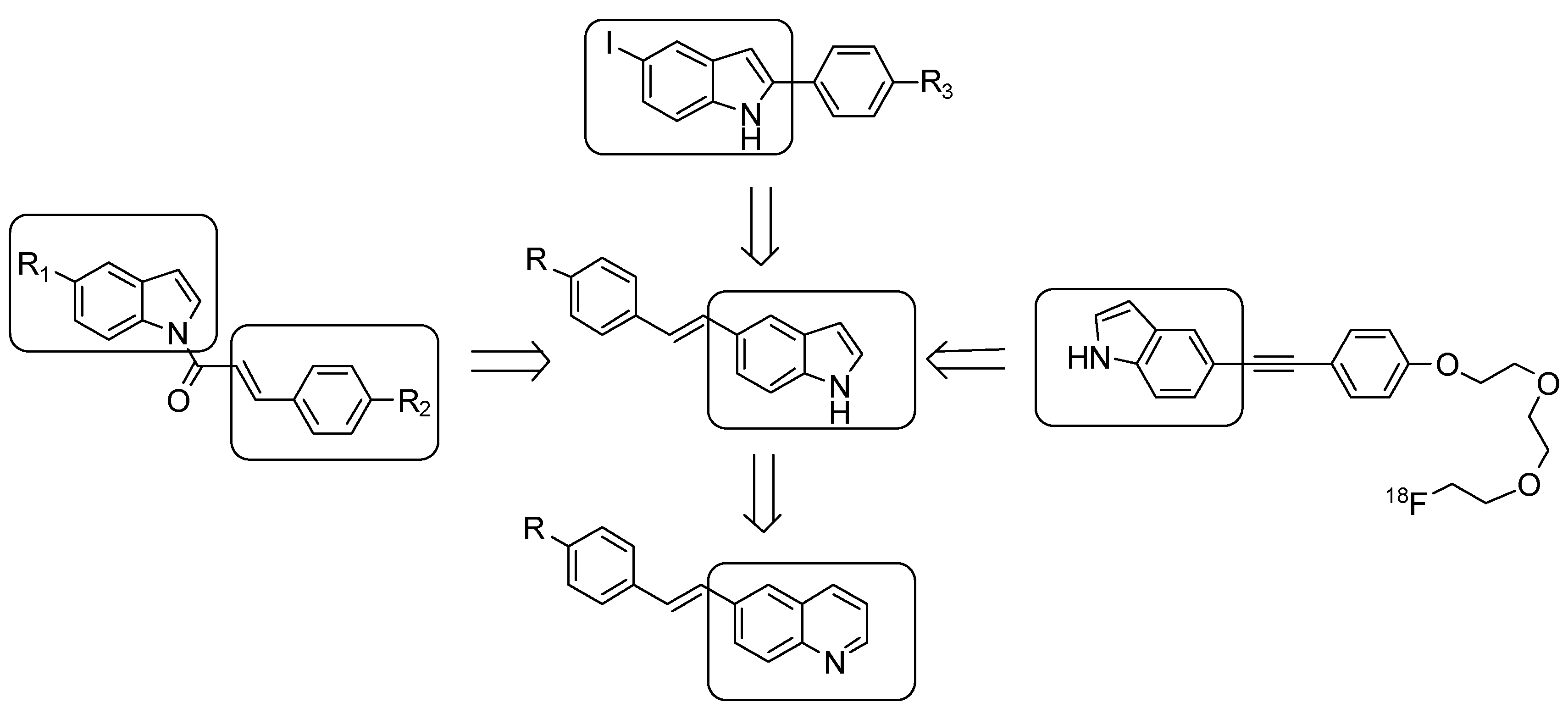
(E)-5-Styryl-1H-indole and (E)-6-Styrylquinoline Derivatives Serve as Probes for β-Amyloid Plaques

Abstract
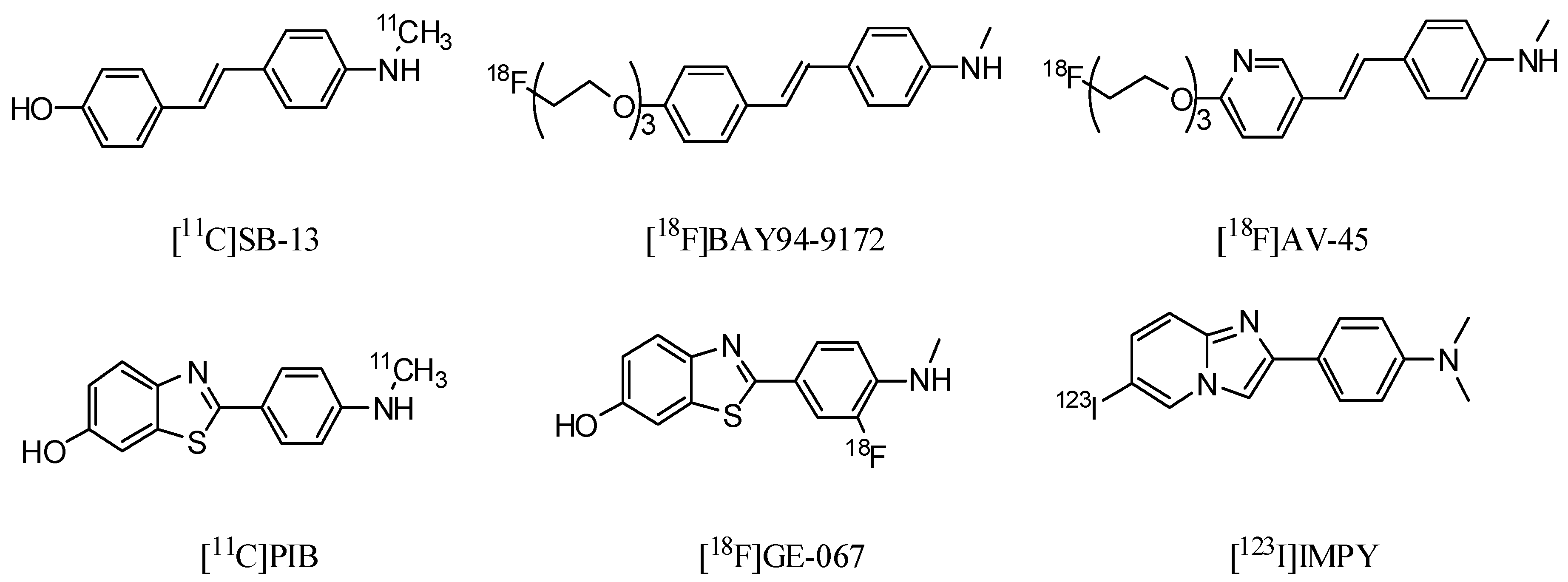
:1. Introduction

2. Results and Discussion
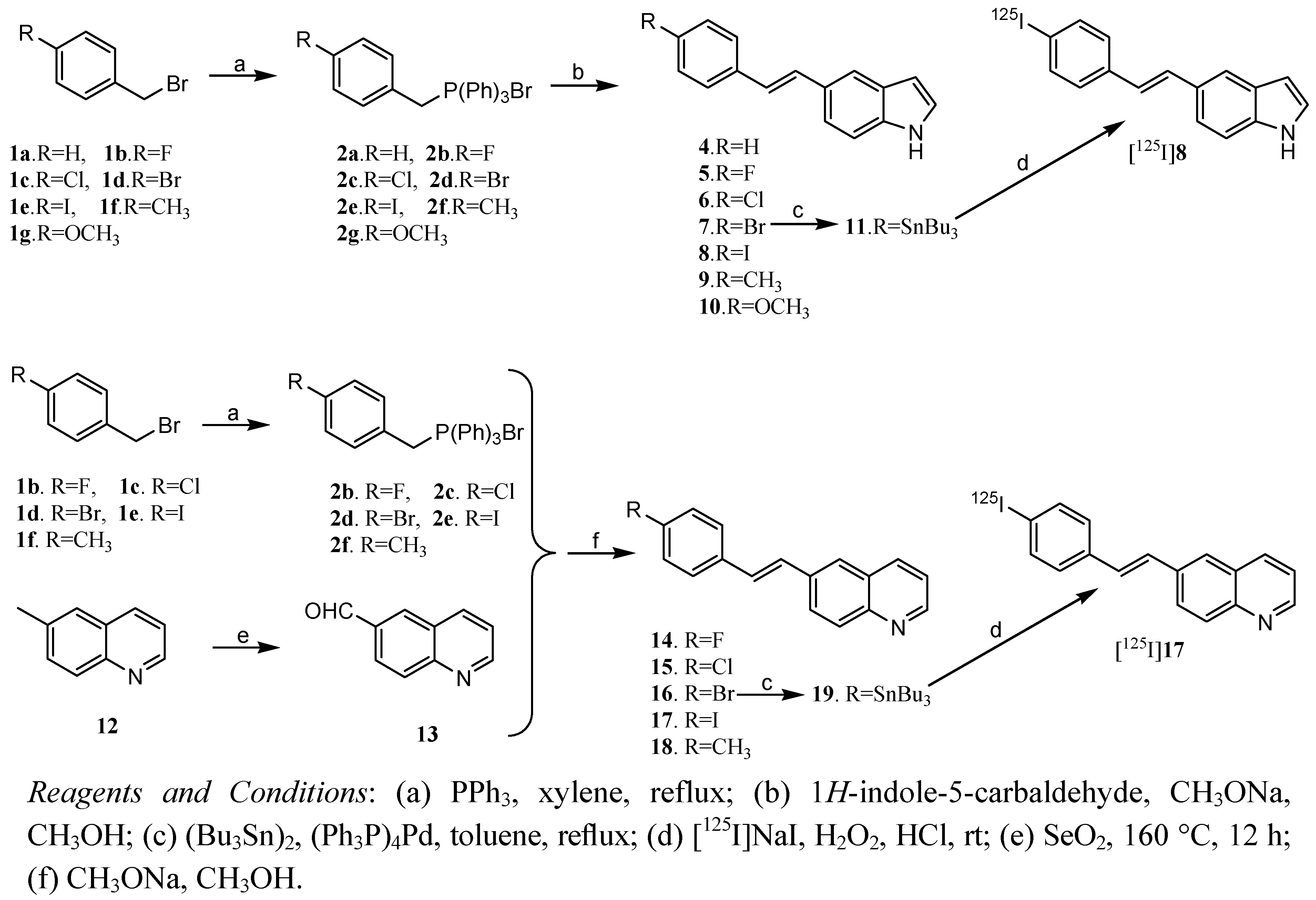
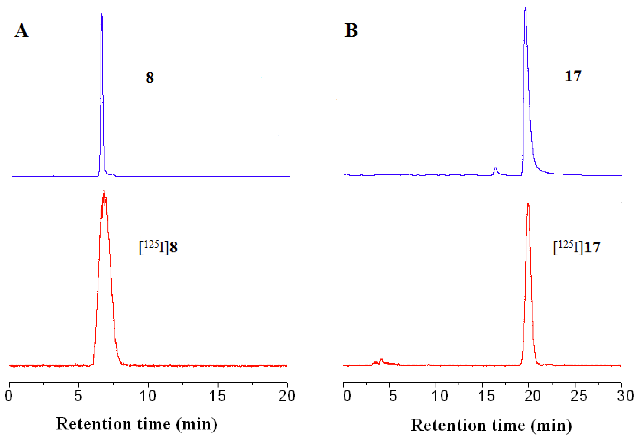
2.1. Chemistry and Radiochemistry
2.2. In Vitro Binding Studies Using the Aggregated Aβ1–40

| Compound | Ki (nM) a | Compound | Ki (nM) a |
|---|---|---|---|
| 4 | 25.1 ± 2.1 | 10 | 32.4 ± 1.9 |
| 5 | 89.3 ± 2.6 | 14 | 270.4 ± 1.5 |
| 6 | 51.5 ± 1.0 | 15 | 45.0 ± 1.3 |
| 7 | 16.3 ± 1.7 | 16 | 23.5 ± 1.3 |
| 8 | 4.1 ± 0.2 | 17 | 8.6 ± 1.2 |
| 9 | 15.8 ± 1.5 | 18 | 288.4 ± 1.3 |
| TZDM | 4.2 ± 0.4 | TZDM b | 0.9 ± 0.2 |
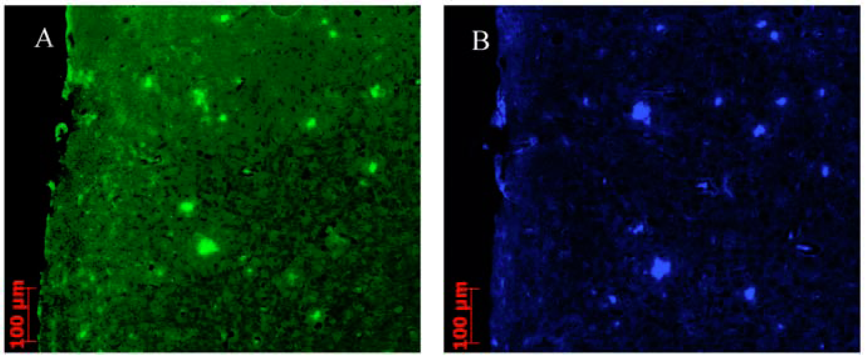
2.3. In Vitro Fluorescent Staining of Amyloid Plaques in Brain Sections from Transgenic Mouse

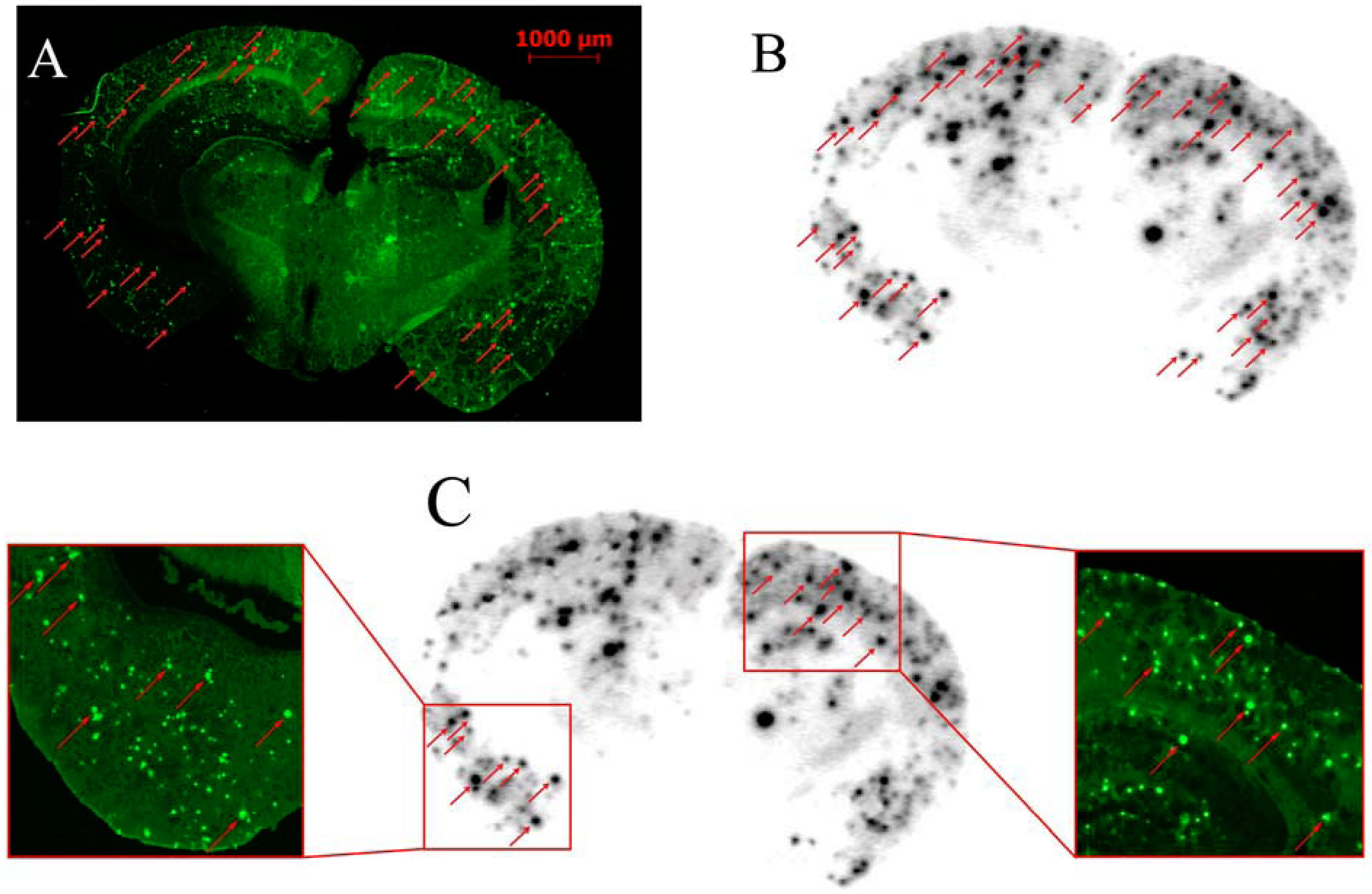
2.4. In Vitro Labeling of Brain Sections from Transgenic Mouse by Autoradiography
2.5. In Vivo Biodistribution Studies
| Organ | 2 min | 15 min | 30 min | 60 min | 120 min | 240 min |
|---|---|---|---|---|---|---|
| [125I]8 (log D = 2.52 ± 0.04) | ||||||
| Blood | 11.91 ± 0.62 | 12.29 ± 1.30 | 7.64 ± 0.62 | 4.49 ± 0.28 | 2.93 ± 0.30 | 1.50 ± 0.13 |
| Brain | 4.27 ± 0.49 | 1.37 ± 0.16 | 0.64 ± 0.11 | 0.28 ± 0.06 | 0.20 ± 0.08 | 0.10 ± 0.02 |
| Heart | 5.76 ± 0.38 | 3.70 ± 0.40 | 2.55 ± 0.52 | 1.59 ± 0.28 | 1.47 ± 0.48 | 0.65 ± 0.16 |
| Liver | 14.73 ± 0.66 | 10.66 ± 0.31 | 7.14 ± 1.13 | 4.45 ± 0.23 | 4.19 ± 0.61 | 3.06 ± 0.36 |
| Spleen | 4.38 ± 0.33 | 4.21 ± 0.26 | 3.44 ± 0.21 | 2.44 ± 0.12 | 1.87 ± 0.28 | 1.37 ± 0.09 |
| Lung | 10.76 ± 0.63 | 8.22 ± 0.88 | 5.39 ± 0.83 | 3.28 ± 0.10 | 2.30 ± 0.20 | 1.43 ± 0.52 |
| Kidney | 11.66 ± 1.52 | 14.89 ± 4.23 | 9.46 ± 1.95 | 4.65 ± 0.98 | 1.93 ± 0.76 | 1.34 ± 0.36 |
| Stomach a | 1.23 ± 0.56 | 4.82 ± 0.46 | 3.50 ± 0.19 | 1.72 ± 0.21 | 3.48 ± 0.79 | 2.31 ± 1.44 |
| Muscle | 2.64 ± 0.40 | 2.01 ± 0.24 | 1.25 ± 0.04 | 0.81 ± 0.12 | 0.61 ±0.24 | 0.35 ± 0.09 |
| [125I]17 (log D = 2.73 ± 0.03) | ||||||
| Blood | 11.39 ± 1.56 | 6.31 ± 0.51 | 5.80 ± 0.37 | 3.86 ± 0.74 | 1.97 ± 0.35 | 1.38 ± 0.24 |
| Brain | 2.05 ± 0.25 | 1.18 ± 0.17 | 0.93 ± 0.13 | 0.55 ± 0.11 | 0.26 ± 0.03 | 0.14 ± 0.02 |
| Heart | 7.70 ± 0.86 | 3.80 ± 0.12 | 3.21 ± 0.11 | 2.55 ± 0.21 | 1.33 ± 0.06 | 0.80 ± 0.19 |
| Liver | 22.45 ± 1.79 | 9.95 ± 0.18 | 9.12 ± 0.53 | 6.97 ± 0.28 | 3.93 ± 0.46 | 2.96 ± 0.28 |
| Spleen | 5.88 ± 0.30 | 5.91 ± 0.58 | 4.66 ± 0.52 | 4.71 ± 0.97 | 2.25 ± 0.36 | 1.59 ± 0.17 |
| Lung | 13.56 ± 1.71 | 6.42 ± 0.47 | 5.64 ± 0.41 | 4.55 ± 0.49 | 2.01 ± 0.18 | 1.43 ± 0.28 |
| Kidney | 15.01 ± 1.56 | 7.47 ± 1.01 | 6.57 ± 0.50 | 4.72 ± 0.69 | 2.20 ± 0.34 | 1.64 ± 0.23 |
| Stomach a | 4.05 ± 0.09 | 15.84 ± 0.78 | 8.11 ± 1.21 | 6.75 ± 0.43 | 11.12 ± 2.48 | 7.03 ± 1.51 |
| Muscle | 2.78 ± 0.42 | 1.68 ± 0.20 | 2.30 ± 0.32 | 1.40 ± 0.34 | 0.72 ± 0.21 | 0.61 ± 0.08 |
3. Experimental
3.1. General
3.1.1. General Procedure for Preparing Substituted Triphenyl Phosphonium Ylide 2 (2a–g)
3.1.2. General Procedure for Preparing 4–11, 13
3.1.3. General procedure for preparing 14–19
3.1.4. Preparation of Radioiodinated Ligands
3.2. Partition Coefficient Determination
3.3. In Vitro Binding Studies Using the Aggregated Aβ1–40
3.4. In Vitro Fluorescent Staining of Amyloid Plaques in Brain Sections from Transgenic Mouse
3.5. In Vitro Labeling of Brain Sections from Transgenic Mouse by Autoradiography
3.6. In Vivo Biodistribution in Normal Mice
4. Conclusions
Supplementary Materials
Acknowledgments
References and Notes
- Selkoe, D.J. The origins of Alzheimer disease: Aβ is for amyloid. J. Am. Med. Assoc. 2000, 283, 1615–1617. [Google Scholar]
- Hardy, J.; Selkoe, D.J. The amyloid hypothesis of Alzheimer’s disease: Progress and problems on the road to therapeutics. Science 2002, 297, 353–356. [Google Scholar]
- Hardy, J.A.; Higgins, G.A. Alzheimer’s disease: The amyloid cascade hypothesis. Science 1992, 256, 184–185. [Google Scholar]
- Nordberg, A. PET imaging of amyloid in Alzheimer’s disease. Lancet Neurol. 2004, 3, 519–527. [Google Scholar]
- Cai, L.S.; Innis, R.B.; Pike, V.W. Radioligand development for PET Imaging of β-amyloid (Aβ)-current status. Curr. Med. Chem. 2007, 14, 19–52. [Google Scholar]
- Mathis, C.A.; Wang, Y.; Klunk, W.E. Imaging β-amyloid plaques and neurofibrillary tangles in the aging human brain. Curr. Pharm. Des. 2004, 10, 1469–1492. [Google Scholar]
- Ono, M.; Wilson, A.; Nobrega, J.; Westaway, D.; Verhoeff, P.; Zhuang, Z.P.; Kung, M.P.; Kung, H.F. 11C-Labeled stilbene derivatives as Aβ-aggregate-specific PET imaging agents for Alzheimer’s disease. Nucl. Med. Biol. 2003, 30, 565–571. [Google Scholar] [CrossRef]
- Verhoeff, N.P.; Wilson, A.A.; Takeshita, S.; Trop, L.; Hussey, D.; Singh, K.; Kung, H.F.; Kung, M.P.; Houle, S. In vivo imaging of Alzheimer disease β-amyloid with [11C]SB-13 PET. Am. J. Geriatr. Psychiatry 2004, 12, 584–595. [Google Scholar]
- Rowe, C.C.; Ackerman, U.; Browne, W.; Mulligan, R.; Pike, K.L.; O’Keefe, G.; Tochon-Danguy, H.; Chan, G.; Berlangieri, S.U.; Jones, G.; et al. Imaging of amyloid β inAlzheimer’s diseasewith 18F-BAY94-9172, a novel PET tracer: Proof of mechanism. Lancet Neurol. 2008, 7, 129–135. [Google Scholar] [CrossRef]
- Choi, S.R.; Golding, G.; Zhuang, Z.P.; Zhang, W.; Lim, N.; Hefti, F.; Benedum, T.E.; Kilbourn, M.R.; Skovronsky, D.; Kung, H.F. Preclinical properties of 18F-AV-45: A PET agent for Aβ plaques in the brain. J. Nucl. Med. 2009, 50, 1887–1894. [Google Scholar]
- Kung, H.F.; Choi, S.R.; Qu, W.C.; Zhang, W.; Skovronsky, D. 18F Stilbenes and styrylpyridines for PET imaging of Aβ plaques in Alzheimer’s disease: A miniperspective. J. Med. Chem. 2010, 53, 933–941. [Google Scholar]
- Mathis, C.A.; Wang, Y.M.; Holt, D.P.; Huang, G.F.; Debnath, M.L.; Klunk, W.E. Synthesis and evaluation of 11C-labeled 6-substituted 2-arylbenzothiazoles as amyloid imaging agents. J. Med. Chem. 2003, 46, 2740–2754. [Google Scholar]
- Klunk, W.E.; Engler, H.; Nordberg, A.; Wang, Y.M.; Blomqvist, G.; Holt, D.P.; Bergström, M.; Savitcheva, I.; Huang, G.F.; Estrada, S.; et al. Imaging brain amyloid in Alzheimer’s disease with Pittsburgh Compound-B. Ann. Neurol. 2004, 55, 306–319. [Google Scholar] [CrossRef]
- Koole, M.; Lewis, D.M.; Buckley, C.; Nelissen, N.; Vandenbulcke, M.; Brooks, D.J.; Vandenberghe, R.; Laere, K.V. Whole-body biodistribution and radiation dosimetry of 18F-GE067: A radioligand for in vivo brain amyloid imaging. J. Nucl. Med. 2009, 50, 818–822. [Google Scholar]
- Kung, M.P.; Hou, C.; Zhuang, Z.P.; Zhang, B.; Skovronsky, D.; Trojanowski, J.Q.; Lee, V.M.; Kung, H.F. IMPY: An improved thioflavin-T derivative for in vivo labeling of β-amyloid plaques. Brain Res. 2002, 956, 202–210. [Google Scholar]
- Zhuang, Z.P.; Kung, M.P.; Wilson, A.; Lee, C.W.; Plössl, K.; Hou, C.; Holtzman, D.M.; Kung, H.F. Structure-activity relationship of imidazo[1,2-a]pyridines as ligands for detecting β-amyloid plaques in the brain. J. Med. Chem. 2003, 46, 237–243. [Google Scholar]
- Newberg, A.B.; Wintering, N.A.; Plössl, K.; Hochold, J.; Stabin, M.G.; Watson, M.; Skovronsky, D.; Clark, C.M.; Kung, M.P.; Kung, H.F. Safety, biodistribution and dosimetry of 123I-IMPY: A novel amyloid plague-imaging agent for the diagnosis of Alzheimer’s disease. J. Nucl. Med. 2006, 47, 748–754. [Google Scholar]
- Yang, Y.; Duan, X.H.; Deng, J.Y.; Bing, J.; Jia, H.M.; Liu, B.L. Novel imaging agents for β-amyloid plaque based on the N-benzoylindole core. Bioorg. Med. Chem. Lett. 2011, 21, 5594–5597. [Google Scholar]
- Qu, W.C.; Choi, S.R.; Hou, C.; Zhuang, Z.P.; Oya, S.; Zhang, W.; Kung, M.P.; Manchandra, R.; Skovronsky, D.M.; Kung, H.F. Synthesis and evaluation of indolinyl- and indolylphenylacetylenes as PET imaging agents for β-amyloid plaques. Bioorg. Med. Chem. Lett. 2008, 18, 4823–4827. [Google Scholar]
- Watanabe, H.; Ono, M.; Haratake, M.; Kobashi, N.; Saji, H.; Nakayama, M. Synthesis and characterization of novel phenylindoles as potential probes for imaging of β-amyloid plaques in the brain. Bioorg. Med. Chem. 2010, 18, 4740–4746. [Google Scholar]
- Zhuang, Z.P.; Kung, M.P.; Hou, C.; Skovronsky, D.M.; Gur, T.L.; Plössl, K.; Trojanowski, J.Q.; Lee, V.M.; Kung, H.F. Radioiodinated styrylbenzenes and thioflavins as probes for amyloid aggregates. J. Med. Chem. 2001, 44, 1905–1914. [Google Scholar] [CrossRef]
- Mikhail, K.; Dmitry, P.; Denis, L.; Dmitry, K. Synthesis and practical use of 1H-1,2,3-benzotriazole-5-carboxaldehyde for reductive amination. Synth. Commun. 2005, 35, 2587–2595. [Google Scholar]
- Wu, C.Y.; Wei, J.J.; Gao, K.Q.; Wang, Y.M. Dibenzothiazoles as novel amyloid-imaging agents. Bioorg. Med. Chem. 2007, 15, 2789–2796. [Google Scholar]
- Klunk, W.E.; Wang, Y.M.; Huang, G.F.; Debnath, M.L.; Holt, D.P.; Shao, L.; Hamilton, R.L.; Ikonomovic, M.D.; DeKosky, S.T.; Mathis, C.A. The binding of 2-(4'-methylaminophenyl)benzothiazole to postmortem brain homogenates is dominated by the amyloid component. J. Neurosci. 2003, 23, 2086–2092. [Google Scholar]
- Cheng, Y.C.; William, H.P. Relationship between the inhibition constant (Ki) and the concentration of inhibitor which causes 50 per cent inhibition (IC50) of an enzymatic reaction. Biochem. Pharmacol. 1973, 22, 3099–3108. [Google Scholar]
- Sample Availability: Samples of the compounds are available from the authors.
© 2012 by the authors; licensee MDPI, Basel, Switzerland. This article is an open-access article distributed under the terms and conditions of the Creative Commons Attribution license (http://creativecommons.org/licenses/by/3.0/).
Share and Cite
Yang, Y.; Jia, H.-M.; Liu, B.-L. (E)-5-Styryl-1H-indole and (E)-6-Styrylquinoline Derivatives Serve as Probes for β-Amyloid Plaques. Molecules 2012, 17, 4252-4265. https://doi.org/10.3390/molecules17044252
Yang Y, Jia H-M, Liu B-L. (E)-5-Styryl-1H-indole and (E)-6-Styrylquinoline Derivatives Serve as Probes for β-Amyloid Plaques. Molecules. 2012; 17(4):4252-4265. https://doi.org/10.3390/molecules17044252
Chicago/Turabian StyleYang, Yang, Hong-Mei Jia, and Bo-Li Liu. 2012. "(E)-5-Styryl-1H-indole and (E)-6-Styrylquinoline Derivatives Serve as Probes for β-Amyloid Plaques" Molecules 17, no. 4: 4252-4265. https://doi.org/10.3390/molecules17044252