1. Introduction
For ensure the pharmacological activity of an active pharmaceutical ingredient (API), the solubility of the API in physiological liquids is required, so that the API can be available at the place of absorption. Solubility in various solvents is a characteristic property of a particular compound. The solubility of a compound in water correlates to a great extent with the solubility in physiological liquids and is the first limiting factor for good absorption and biodistribution. Contrary to these facts, over the last ten years, the number of poorly soluble drugs has steadily increased. Estimates suggest that 40% of the drugs in the pipelines have solubility problems. Literature states that about 60% of all drugs coming directly from synthesis nowadays are poorly soluble [
1,
2,
3].
One of the progressive ways how to increase the solubility of an APIs is the preparation of drug nanoparticles. The technique of nanoparticle drug delivery allows many pharmacological agents to reach the desired site of action. APIs are either adjusted alone till nano size and administered in nanoparticle oral dosage forms or attached/incorporated into nanoparticles prepared from inert materials which serve as a universal drug delivery system. The advantages of nanotechnology are as follows: (i) increased bioavailability (quick dissolution; improved penetration through membranes); (ii) lower doses; (iii) lower toxicity; (iv) targeted biodistribution; (v) reduction of influence of food on variability; (vi) quicker development of formulations [
2,
4,
5,
6,
7]. Nanoparticles of less than 200 nm are of practical importance [
8,
9,
10,
11,
12,
13]. A great problem is the insufficiently investigated possible toxicity of nanoparticles. The toxicity is dependent on the shape and surface properties of nanoparticles, because both can influence nanoparticle-cell interactions as well as the rate of penetration to cells. Among the various nanoparticle forms nanotubes were found to be one of the most toxic nanoparticle shapes [
14,
15,
16,
17].
A wide range of techniques have been developed for the preparation of nanomaterials. These methods are typically grouped into two categories: top-down (generally dispergation processes) [
11,
12,
13,
18,
19,
20,
21] and bottom-up (generally precipitation processes) [
11,
12,
13,
18,
22,
23,
24], whereas the latter is by far the most popular in the preparation of nanoparticles. In bottom-up methods, nanoparticles can be produced by crystallization/precipitation and solvent evaporation. Spray drying, evaporative precipitation into aqueous solution, microemulsions or supercritical fluid technology belong to the solvent evaporation methods. The liquid antisolvent (LAS) precipitation process is a noteworthy method that has been extensively studied. An excellent review dealing with this technique was published by Thorat
et al. [
25]. The current paper is aimed at verification of conditions of an effective and an affordable technique for the preparation of nanoparticles by solvent evaporation as was discussed recently [
26].
A polar and nonpolar solvent were used in our research, therefore the exact principle of the applied solvent evaporation method is dependent on the water-based system, including or not an aqueous miscible organic solvent. The polar acetone (AC) and nonpolar dichloromethane (DCM) were chosen as the most suitable solvents for easy dissolution of the APIs, so two different possible mechanisms can be supposed for the nanoparticle synthesis. When API is dissolved in AC and then mixed with water containing a stabilizer, nanoparticles are formed spontaneously and immediately upon mixing. This method can be called antisolvent precipitation/solvent evaporation, and the procedure is in principle similar to the evaporative precipitation into aqueous solution [
27,
28] or the liquid antisolvent precipitation [
25]. When the API is dissolved in DCM and then mixed with water containing stabilizers, an emulsion (o/w type) is formed; API is clustered by the excipient, which results in the encapsulation of the API into nano-vesicula. This combination of emulsification and solvent evaporation nanoparticle synthesis can be called emulsion/solvent evaporation [
19,
29].
The model APIs candesartan cilexetil (
I) and atorvastatin calcium (
II) were chosen as representatives of poorly aqueous soluble compounds belonging to class II drugs of the biopharmaceutical classification system (BCS). Drugs of the mentioned class are characterized by low aqueous solubility and high permeability [
30]. Candesartan (2-ethoxy-1-({4-[2-(2H-1,2,3,4-tetrazol-5-yl)phenyl]phenyl}methyl)-1
H-1,3-benzodiazole-6-carboxylic acid) is an angiotensin II receptor antagonist used mainly for the treatment of hypertension. The prodrug candesartan cilexetil, see
Figure 1, is marketed by AstraZeneca and Takeda Pharmaceuticals, commonly under the trade names Blopress
®, Atacand
®, Amias
®, and Ratacand
®. The use of a prodrug form increases the bioavailability of candesartan. Despite this, its absolute oral bioavailability is relatively poor (approx. 15%) [
31,
32]. Atorvastatin [(3
R,5
R)-7-[2-(4-fluorophenyl)-3-phenyl-4-(phenylcarbamoyl)-5-propan-2-ylpyrrol-1-yl]-3,5-dihydroxyheptanoic acid] inhibits HMG-CoA reductase and thus causes a decrease of cholesterol in the body. Atorvastatin is used as a calcium salt, see
Figure 1, and is marketed by Pfizer under the trade names Lipitor
® or Sortis
®. The low plasma concentration (approx. 12%) of atorvastatin is especially caused by an extensive first-pass metabolism in the liver, nevertheless the overall solubility of atorvastatin is strictly pH-dependent (many atorvastatin solid dosage forms are buffered, e.g., by carbonates), and administration with food produces a 25% reduction of atorvastatin absorption [
33,
34].
As mentioned, both APIs are BCS class II drugs, hence their oral bioavailability is solubility rate limited [
30,
31,
32,
35,
36,
37]. For enhancement of solubility of candesartan cilexetil various approaches can be used, such as pectin complexes [
38], self-emulsifying drug delivery systems [
39] or development of nanoparticle formulations [
7,
40]. The solubility of atorvastatin calcium can be enhanced, for example, using an amorphous API [
41], by application of the liquisolid technique [
42], formulation of self-microemulsifying drug delivery systems [
43], utilization of drug-polymer interactions found due to physical mixing [
44] or preparation of amorphous nanoparticles [
45].
Various types of surface-active excipients were used as nanoparticle stabilizers and relationships between a substance, a solvent and a used excipient are discussed. Used excipients represent various classes of pharmaceutical adjutants (emulsifiers/viscosity modifiers/thickeners, nonionic or anionic surfactants) that can be utilized as solubility modifying compounds/nanoparticle stabilizers, such as Tween 80 (TW), sodium dodecyl sulfate (SDS), macrogol 6000 (PEG), sodium carboxymethyl cellulose (SCMC) and sodium salt of carboxymethyl dextran (SCMD). The main criteria for excipient selection were its pharmaceutical safety (all excipients are GRAS, Generally Recognized as Safe, substances) and their affordability. Based on a previous study 5% and 10% concentrations of each excipient were chosen [
26]. The optimal concentration of surfactant is important for optimal particles wetting. If the concentration is too low, particles float on the surface. If the concentration is too high bubbles appear [
46].
Figure 1.
Structures of candesartan cilexetil as prodrug and atorvastatin calcium salt.
Figure 1.
Structures of candesartan cilexetil as prodrug and atorvastatin calcium salt.
2. Results and Discussion
Both model APIs
I and
II dissolved in dichloromethane and acetone (2% concentration) were added to aqueous solutions (5%, 10% concentration) of excipients,
i.e., eight samples were prepared with each excipient. The final API:excipient ratios were 1:2.5 (2%:5%), 1:5 (2%:10%). The systems were stirred for 10 min at 35 °C; afterwards the mixtures were transferred to an ultrasonic bath, where they were mixed again for 40 min, and simultaneously the organic solvent was evaporated (to final 10 mL sample volume) by self-warming of the ultrasonic bath. Then all the samples were characterized by dynamic light scattering [
46]. All the results are presented in
Table 1,
Table 2,
Table 3,
Table 4,
Table 5 and
Figure 2,
Figure 3,
Figure 4,
Figure 5,
Figure 6,
Figure 7,
Figure 8.
Table 1.
Particle size (x10, x90 [nm]) of APIs I, II and concentration [%] of Tween 80 in dichloromethane (DCM) or acetone (AC). All the presented results are reported as the medium value of four independent measurements. The results of nano-size samples are expressed as the mean ± SD (n = 4 measurements). The SDs of micro-size samples are not indicated due to the measurability range of Nanophox. Samples that contained nanoparticles <200 nm are bolded; nanoparticles <10 nm are indicated by asterisk. (S.No. = sample number).
| API/Solvent | Tween 80 | |
|---|
| S.No. | 5% | S.No. | 10% | |
|---|
| x10 | x90 | x10 | x90 | Particle size [nm] |
|---|
| I/DCM | 1 | 160 ± 4.8 | 219 ± 5.6 | 2 | 14 ± 0.4 | 16 ± 0.5 * |
| I/AC | 3 | 3183 | 6531 | 4 | 2 ± 0.1 | 3 ± 0.1 * |
| II/DCM | 5 | 97 ± 2.9 | 142 ± 4.3 | 6 | 145 ± 4.4 | 213 ± 6.4 |
| II/AC | 7 | 101 ± 3.0 | 111 ± 3.3 | 8 | 3 ± 0.1 | 4 ± 0.1 * |
Table 2.
Particle size (x10, x90 [nm]) of APIs I, II and concentration [%] of sodium dodecyl sulfate in dichloromethane (DCM) or acetone (AC). All the presented results are reported as the medium value of four independent measurements. The results of nano-size samples are expressed as the mean ± SD (n = 4 measurements). The SDs of micro-size samples are not indicated due to the measurability range of Nanophox. Samples that contained nanoparticles <200 nm are bolded; nanoparticles <10 nm are indicated by asterisk. (S.No. = sample number).
| API/Solvent | Sodium dodecyl sulfate | |
|---|
| S.No. | 5% | S.No. | 10% | |
|---|
| x10 | x90 | x10 | x90 | Particle size [nm] |
|---|
| I/DCM | 9 | 90 ± 2.7 | 99 ± 3.0 | 10 | 2 ± 0.1 | 3 ± 0.1 * |
| I/AC | 11 | 4 ± 0.1 | 5 ± 0.2 * | 12 | 1 ± 0.03 | 2 ± 0.1 * |
| II/DCM | 13 | 1 ± 0.03 | 2 ± 0.1 * | 14 | 90 ± 2.7 | 99 ± 3.0 |
| II/AC | 15 | 2 ± 0.1 | 2 ±0.1 * | 16 | 2 ± 0.1 | 4 ± 0.1 * |
Table 3.
Particle size (x10, x90 [nm]) of APIs I, II and concentration [%] of macrogol 6000 in dichloromethane (DCM) or acetone (AC). All the presented results are reported as the medium value of four independent measurements. The results of nano-size samples are expressed as the mean ± SD (n = 4 measurements). The SDs of micro-size samples are not indicated due to the measurability range of Nanophox. Samples that contained nanoparticles <200 nm are bolded; nanoparticles <10 nm are indicated by asterisk. (S.No. = sample number).
| API/Solvent | Macrogol 6000 | |
|---|
| S.No. | 5% | S.No. | 10% | |
|---|
| x10 | x90 | x10 | x90 | Particle size [nm] |
|---|
| I/DCM | 17 | 2 ± 0.1 | 3 ± 0.1 * | 18 | 2 ± 0.1 | 3 ± 0.1 * |
| I/AC | 19 | 2 ± 0.1 | 3 ± 0.1 * | 20 | 156 ± 4.7 | 206 ± 6.2 |
| II/DCM | 21 | 1639 | 1804 | 22 | 5231 | 5755 |
| II/AC | 23 | 6 ± 0.2 | 8 ± 0.2 * | 24 | 4 ± 0.1 | 6 ± 0.2 * |
Table 4.
Particle size (x10, x90 [nm]) of APIs I, II and concentration [%] of sodium carboxymethyl cellulose in dichloromethane (DCM) or acetone (AC). All the presented results are reported as the medium value of four independent measurements. The results of nano-size samples are expressed as the mean ± SD (n = 4 measurements). The SDs of micro-size samples are not indicated due to the measurability range of Nanophox. Samples that contained nanoparticles <200 nm are bolded; nanoparticles <10 nm are indicated by asterisk. (S.No. = sample number).
| API/Solvent | Sodium carboxymethyl cellulose | |
| S.No. | 5% | S.No. | 10% | |
| x10 | x90 | x10 | x90 | Particle size [nm] |
| I/DCM | 25 | 11 ± 0.3 | 13 ± 0.4 | 26 | 2 ± 0.1 | 3 ± 0.1 * |
| I/AC | 27 | 1 ± 0.03 | 2 ± 0.1 * | 28 | 32 ± 1.0 | 35 ± 1.1 |
| II/DCM | 29 | 401 ± 12 | 574 ± 17 | 30 | 1 ± 0.03 | 2 ± 0.1 * |
| II/AC | 31 | 6 ± 0.2 | 7 ± 0.2 * | 32 | 27 ± 0.8 | 30 ± 0.9 |
Table 5.
Particle size (x10, x90 [nm]) of APIs I, II and concentration [%] of sodium carboxymethyl dextran in dichloromethane (DCM) or acetone (AC). All the presented results are reported as the medium value of four independent measurements. The results of nano-size samples are expressed as the mean ± SD (n = 4 measurements). The SDs of micro-size samples are not indicated due to the measurability range of Nanophox. Samples that contained nanoparticles <200 nm are bolded; nanoparticles <10 nm are indicated by asterisk. (S.No. = sample number).
| API/Solvent | Sodium carboxymethyl dextran | |
|---|
| S.No. | 5% | S.No. | 10% | |
|---|
| x10 | x90 | x10 | x90 | Particle size [nm] |
|---|
| I/DCM | 33 | 2 ± 0.1 | 2 ± 0.1 * | 34 | 1 ± 0.03 | 1 ± 0.03 * |
| I/AC | 35 | 3 ± 0.1 | 4 ± 0.1 * | 36 | 39 ± 1.2 | 43 ± 1.3 |
| II/DCM | 37 | 2 ± 0.1 | 2 ± 0.1 * | 38 | 9345 | 10281 |
| II/AC | 39 | 2 ± 0.1 | 3 ± 0.1 * | 40 | 70 ± 2.1 | 77 ± 2.3 |
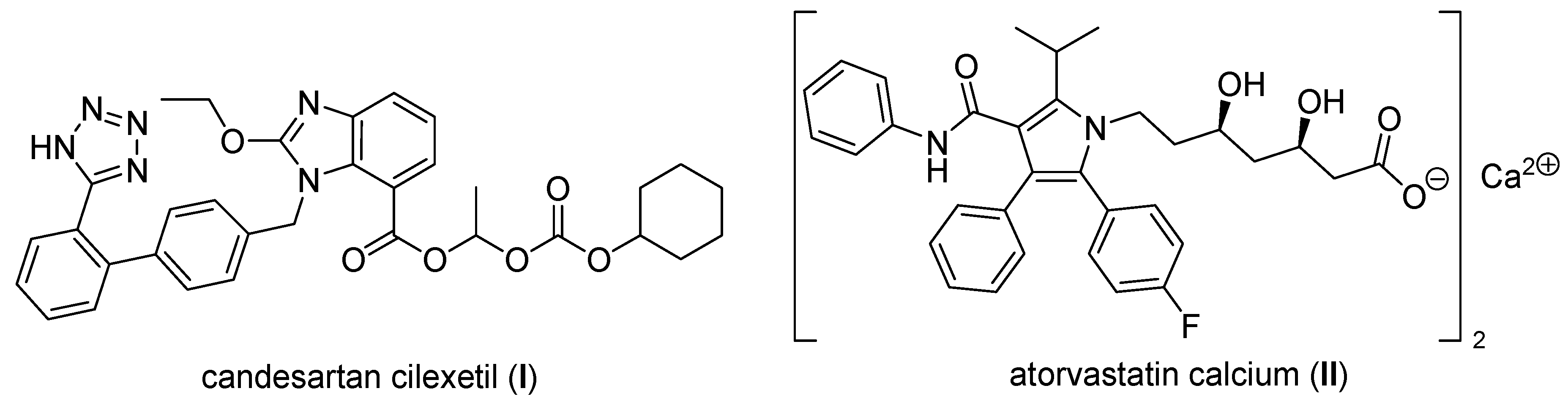
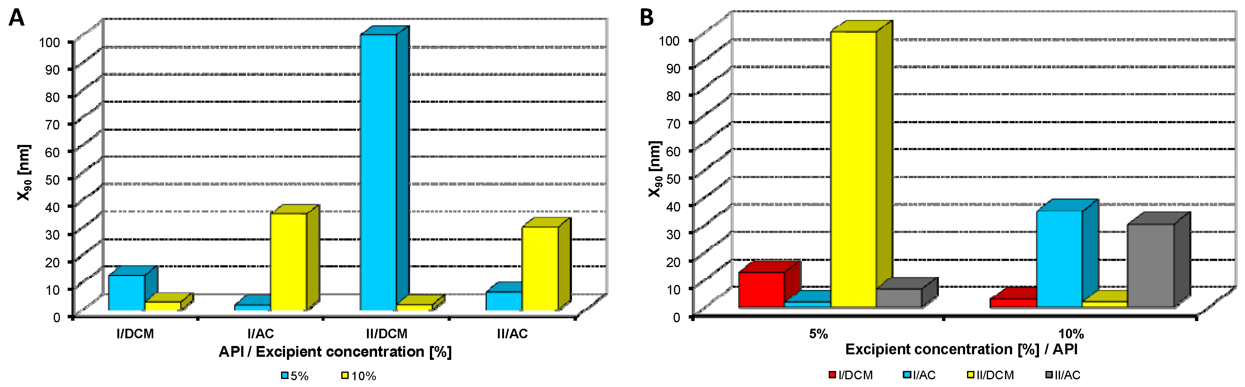
Figure 2.
Dependence of particle size (x90 [nm]) of model APIs I, II on concentration [%] of Tween 80 in dichloromethane (DCM) or acetone (AC). (A) Samples are grouped according to APIs; (B) samples are grouped according to excipient percentage. For clarity sake, the values on y-axis are only to 300 nm.
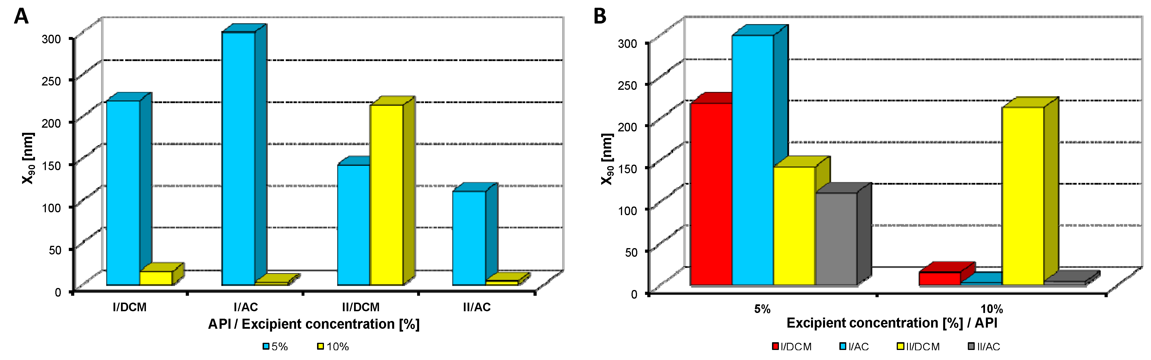
Figure 3.
Dependence of particle size (x90 [nm]) of model APIs I, II on concentration [%] of sodium dodecyl sulfate in dichloromethane (DCM) or acetone (AC). (A) Samples are grouped according to APIs; (B) samples are grouped according to excipient percentage. For clarity sake, the values on y-axis are only to 100 nm.
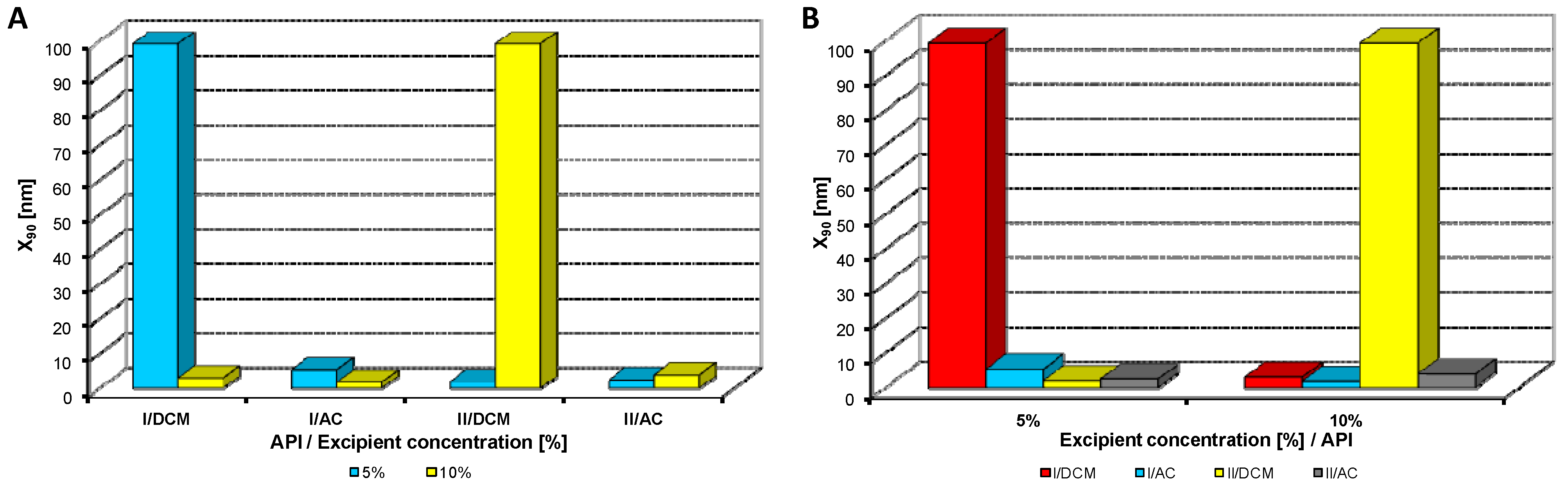
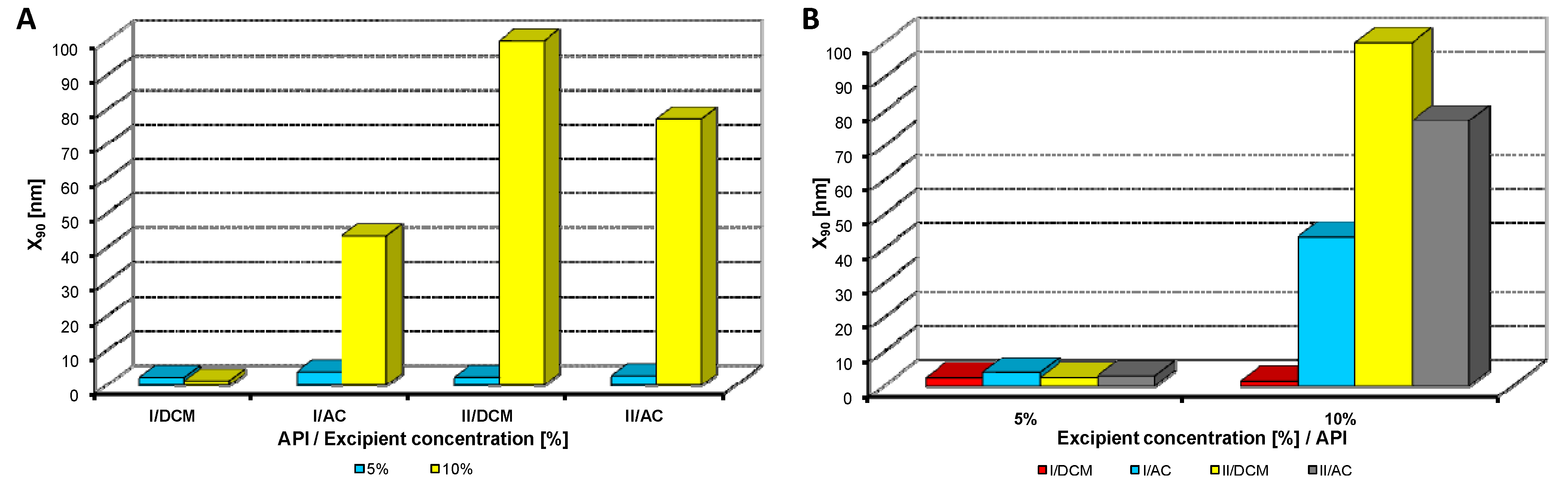
Figure 4.
Dependence of particle size (x90 [nm]) of model APIs I, II on concentration [%] of macrogol 6000 in dichloromethane (DCM) or acetone (AC). (A) Samples are grouped according to APIs; (B) samples are grouped according to excipient percentage. For clarity sake, the values on y-axis are only to 300 nm.
Figure 5.
Dependence of particle size (x90 [nm]) of model APIs I, II on concentration [%] of sodium carboxymethyl cellulose in dichloromethane (DCM) or acetone (AC). (A) Samples are grouped according to APIs; (B) samples are grouped according to excipient percentage. For clarity sake, the values on y-axis are only to 100 nm.
Figure 6.
Dependence of particle size (x90 [nm]) of model APIs I, II on concentration [%] of sodium carboxymethyl dextran in dichloromethane (DCM) or acetone (AC). (A) Samples are grouped according to APIs; (B) samples are grouped according to excipient percentage. For clarity sake, the values on y-axis are only to 100 nm.
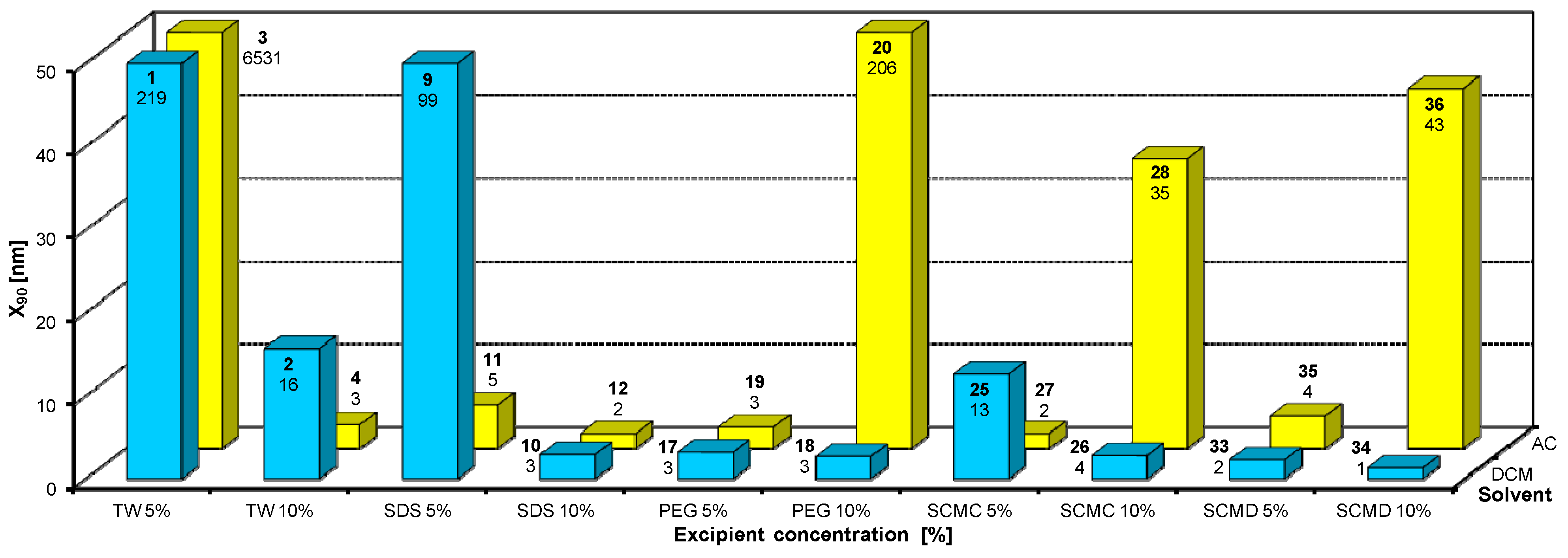
Figure 7.
Dependence of particle size (x90 [nm]) of candesartan cilexetil (I) on concentration [%] of Tween 80 (TW), sodium dodecyl sulfate (SDS), macrogol 6000 (PEG), sodium carboxymethyl cellulose (SCMC), sodium carboxymethyl dextran (SCMD) in dichloromethane (DCM) or acetone (AC). For clarity sake, the values on y-axis are only to 50 nm.
Figure 8.
Dependence of particle size (x90 [nm]) of atorvastatin calcium (II) on concentration [%] of Tween 80 (TW), sodium dodecyl sulfate (SDS), macrogol 6000 (PEG), sodium carboxymethyl cellulose (SCMC), sodium carboxymethyl dextran (SCMD) in dichloromethane (DCM) or acetone (AC). For clarity sake, the values on y-axis are only to 50 nm.
Figure 2,
Figure 3,
Figure 4,
Figure 5,
Figure 6 illustrate the dependence of particle size expressed as the cumulative distribution x
90 [nm] of the APIs
I,
II on the concentration [%] of an individual excipient, whereas in Figures A samples are grouped according to individual APIs
I,
II, while in Figures B individual APIs are always separated according to the percentage of the excipient. The particle size x
90 was used for evaluation of the method success, since this value represents 90% of the cumulative particle size distribution in the measured sample. The dispersity is a measure/degree of the homogeneity/heterogeneity of sizes of particles in a mixture/system. It is possible to see this feature on the width of the particle-size distribution, which is described as differences between cumulative distribution x
10 and x
90, see
Table 1,
Table 2,
Table 3,
Table 4,
Table 5. According to the results, when micro-size samples (
3,
21,
22,
38) were eliminated, the average relation of the cumulative distribution x
10/x
90 ranged from 0.6 to 0.9. It is possible to suppose that nanoparticles are spheres, because the size in dynamic light scattering represents the hydrodynamic diameter of the particle. All samples were dispersed by ultrasonics directly before the measurement to avoid possible re-agglomeration. Stabilization of the dispersed samples was achieved by surfactants and by the constant temperature. The measuring cell was equilibrated at 25 °C, so the Brown motion of nanoparticles is influenced just by their size.
From
Figure 2A,
Figure 3A,
Figure 4A,
Figure 5A,
Figure 6A it can be stated that generally particle size is not dependent on the type of model API, but it is partially influenced by the type and concentration of the excipient utilized. Nevertheless, it can be supposed that in the case of candesartan cilexetil (
I) smaller particles were found, especially when atorvastatin calcium (
II) and SDS, PEG and SCMC in dichloromethane were used, as it is illustrated in
Figure 7 and
Figure 8, where the dependences of the particle size of individual APIs
I and
II on the concentrations of individual excipients are shown. This fact is probably caused by the solvent used, because dichloromethane has less advantageous properties in comparison with acetone, as discussed below.
Table 6 summarizes results of all the samples of nanoparticles under 900 nm size depending on solvents and the type and amount of excipients. As the aim of this contribution is specification of suitable conditions for nanoparticles preparation, in
Table 6 generated nanoparticles are not divided according to used APIs.
Table 6.
View of formed samples of nanoparticles (≤900 nm) depending on solvents and type and amount of excipients. (conc. = concentration; excp. = excipient; dichloromethane = DCM; acetone = AC; Tween 80 = TW; sodium dodecyl sulfate = SDS; macrogol 6000 = PEG; sodium carboxymethyl cellulose = SCMC; sodium carboxymethyl dextran = SCMD).
| Excp. conc./type | DCM | Sum total | Overall averagex90 [nm] | AC | Sum total | Overall averagex90 [nm] |
|---|
| 5% | 10% | 5% | 10% |
|---|
| number of nanop. samples | number of nanop. samples |
|---|
| TW | 2 | 2 | 4 | 147 | 1 | 2 | 3 | 39 |
| SDS | 2 | 2 | 4 | 51 | 2 | 2 | 4 | 3 |
| PEG | 1 | 1 | 2 | 3 | 2 | 2 | 4 | 56 |
| SCMC | 2 | 2 | 4 | 148 | 2 | 2 | 4 | 18 |
| SCMD | 2 | 1 | 3 | 2 | 2 | 2 | 4 | 32 |
| Sum total | 9 | 8 | 17 | 351 | 9 | 10 | 19 | 148 |
| Overall averagex90 [nm] | 117 | 42 | 160 | ![Molecules 17 13221 i001]() | 16 | 41 | 57 | ![Molecules 17 13221 i002]() |
After summation of all the results it can be concluded that from 40 prepared mixtures 36 samples contained nanoparticles (see
Table 1,
Table 2,
Table 3,
Table 4,
Table 5), from which 32 samples contained nanoparticles smaller than 200 nm (see
Table 1,
Table 2,
Table 3,
Table 4,
Table 5, bolded values). Nanoparticles under 10 nm were determined in 22 samples from 32, see
Table 1,
Table 2,
Table 3,
Table 4,
Table 5 (asterisked bolded values).
Based on the results listed in
Table 6 and
Figure 7 and
Figure 8 it can be generally stated that the solvent used plays the crucial role in generation of nanoparticles. This fact was not so evident in the previous study, where only steroid-like compounds were investigated [
26]. This effect of solvent was significant in the case of atorvastatin calcium (
II), which is a salt and thus by its chemical nature absolutely different from other investigated model compounds. It depends on the used solvent, if the system is single-phase (acetone/water) or biphasic (dichloromethane/water, o/w type), thus whether nanoparticles will be formed spontaneously and immediately upon mixing or if emulsions will be generated and nanoparticles will not be formed spontaneously but after energy input, e.g., ultrasonic. As the way of preparation was the same (mixing and ultrasounding), it is evident from the results that the polar solvent acetone is preferable to nonpolar dichloromethane,
i.e., that antisolvent precipitation/solvent evaporation method is a more convenient/versatile way for preparation of nanoparticles than the emulsion/solvent evaporation technique. Results with APIs dissolved in acetone provided more nanoparticle samples comparable with dichloromethane (19/17), and the particle size of APIs dissolved in acetone was significantly smaller than that of APIs dissolved in dichloromethane (148/351).
From all the results (see
Figure 7 and
Figure 8) it is evident that the usage of Tween 80, especially at 5% concentration (ratio 1:2.5), and sodium carboxymethyl dextran, especially at 10% concentration (ratio 1:5), was the least advantageous as discussed previously [
26]. In other cases both 5% and 10% concentrations of excipients provided similar results. Surprisingly, macrogol 6000 did not afford as good results as expected [
26]. Sodium dodecyl sulfate and sodium carboxymethyl cellulose can be universally used as nanoparticle stabilizers both in dichloromethane and acetone.


