Review

Is Endothelial Nitric Oxide Synthase a Moonlighting Protein Whose Day Job is Cholesterol Sulfate Synthesis? Implications for Cholesterol Transport, Diabetes and Cardiovascular Disease †

1
Computer Science and Artificial Intelligence Laboratory, Massachusetts Institute of Technology, Cambridge, MA 01890, USA
2
Independent Researcher, Houston, TX 77084, USA
3
Internal Medicine Group Practice, PhyNet, Inc. Longview, TX 75605, USA
4
Biochemistry Laboratory Director, Mount Holyoke College, South Hadley, MA 01075, USA
*
Author to whom correspondence should be addressed.
Note added by the Publisher: The editors of the journal have been alerted to concerns over potential bias in opinions and bias in the choice of citation sources used in this article. We note that the authors stand by the content as published. Since the nature of the claims against the paper concern speculation and opinion, and not fraud or academic misconduct, the editors would like to issue an Expression of Concern to make readers aware that the approach to collating literature citations for this article was likely not systematic and may not reflect the spectrum of opinions on the issues covered by the article. Please refer to our policy regarding <a href=\\\'https://www.mdpi.com/about/controversial-articles\\\'>possibly controversial articles.
Entropy 2012, 14(12), 2492-2530; https://doi.org/10.3390/e14122492
Submission received: 8 October 2012 / Revised: 28 November 2012 / Accepted: 4 December 2012 / Published: 7 December 2012
(This article belongs to the Special Issue Biosemiotic Entropy: Disorder, Disease, and Mortality)

Abstract

:
Theoretical inferences, based on biophysical, biochemical, and biosemiotic considerations, are related here to the pathogenesis of cardiovascular disease, diabetes, and other degenerative conditions. We suggest that the “daytime” job of endothelial nitric oxide synthase (eNOS), when sunlight is available, is to catalyze sulfate production. There is a striking alignment between cell types that produce either cholesterol sulfate or sulfated polysaccharides and those that contain eNOS. The signaling gas, nitric oxide, a well-known product of eNOS, produces pathological effects not shared by hydrogen sulfide, a sulfur-based signaling gas. We propose that sulfate plays an essential role in HDL-A1 cholesterol trafficking and in sulfation of heparan sulfate proteoglycans (HSPGs), both critical to lysosomal recycling (or disposal) of cellular debris. HSPGs are also crucial in glucose metabolism, protecting against diabetes, and in maintaining blood colloidal suspension and capillary flow, through systems dependent on water-structuring properties of sulfate, an anionic kosmotrope. When sunlight exposure is insufficient, lipids accumulate in the atheroma in order to supply cholesterol and sulfate to the heart, using a process that depends upon inflammation. The inevitable conclusion is that dietary sulfur and adequate sunlight can help prevent heart disease, diabetes, and other disease conditions.
PACS Codes:
87.18.Nq; 87.15.bk; 87.19.xw; 87.18.Vf; 87.18.Ed; 47.63.Cb; 87.19.ug; 87.19.uj

1. Introduction

The nitric oxide synthases (NOSs) are a family of three distinct highly regulated isoforms of an enzyme class that produces nitric oxide (NO) [1,2]. The class consists of two constitutive forms, endothelial NOS (eNOS) and neuronal NOS (nNOS), which are activated by calcium-bound calmodulin, and an inducible form, iNOS, which is insensitive to calcium. eNOS, even when activated, produces only a small fraction of the amount of NO typically produced by iNOS. eNOS and nNOS are also capable of producing superoxide (O2) in the absence of their substrate L-arginine, and this is generally viewed as a pathological response, although we will argue otherwise in this paper.
Although eNOS is an extremely well-studied protein, much remains mysterious about its function. It is expressed not only in the endothelial cells lining blood vessel walls, but also in fibroblasts [3], cardiomyocytes [4], platelets [5], and red blood cells (RBCs) [6], among others. It maintains a “resting” state bound to the protein caveolin in the small invaginations in the membrane surface known as caveolae associated with lipid rafts on the plasma membrane [4,7]. Lipid rafts are organized subdomains of the plasma membrane that are enriched in cholesterol and that play an important role in both signal transduction and nutrient transport across the membrane [8,9].
Caveolin-binding of eNOS disables NO synthesis. Activation of eNOS for NO production requires a complex sequence of events, beginning with calcium entry into the cell, followed by calcium binding to calmodulin, calmodulin binding to eNOS (triggering its disengagement from caveolin and the membrane), and phosphorylation of eNOS. Activation also requires the presence of tetrahydrobiopterin (BH4) as a cofactor, which increases the binding of the substrate, L-arginine [10]. When BH4 levels are low, or when L-arginine is unavailable, eNOS produces superoxide (O2) instead of NO [11], due to an uncoupling mechanism.
NO was the first signaling gas to be identified (others are CO and H2S). NO has diverse biological functions, some beneficial but others clearly pathological. NO induces relaxation of the artery wall via a guanylyl cyclase-dependent mechanism, and it promotes endothelial cell migration and neovascularization via activation of the phosphatidyl inositol 3 kinase (PI3K)/Akt pathway [12]. However, NO can react with superoxide to form the highly reactive agent, peroxynitrite (ONOO). ONOO is destructive to the iron-sulfur clusters associated with mitochondrial electron transport and DNA repair systems [13].
A less well-known property of NO is its potent effect on glyceraldehyde 3-phosphate dehydrogenase (GAPDH), the rate-limiting enzyme in glycolysis. NO inactivates this enzyme through nitrosylation [14], causing it to migrate to the nucleus and activate synthesis of phase II detoxifying genes. The expression of these genes is associated with increased tumor resistance to several anticancer drugs [15]. Excess NO leads to the synthesis of S-nitrosoglutathione (GSNO) from glutathione (GSH) [16], which is metabolized to release ammonia, leading to increased permeability of the blood-brain barrier and toxic effects in the brain [17]. Perhaps the most disturbing aspect of NO is its negative effect on autophagy [18], which can lead to accumulation of toxic cellular debris, ultimately resulting in loss of cell viability. Impaired autophagy is a key factor in heart failure [19].
Here, we show that eNOS is a dual-purpose enzyme, i.e., a moonlighting enzyme [20], with complex signaling systems in place to control its switch between two distinct (and complementary) biological roles. One role is the synthesis of nitric oxide, which has already been well characterized. The other role, which we infer and document here, is the synthesis of sulfate from sulfane or sulfide sulfur and superoxide. In Section 3.2, we show how eNOS bound to caveolin at the plasma membrane can facilitate sulfate formation, provided sunlight is available. Sulfate enables cholesterol transport, and, furthermore, maintenance of sulfation in the extracellular matrix proteins is crucial for the health of the endothelial wall and of red blood cells and platelets. It has been established that insulin resistance and impaired autophagy can result from inadequate supply of cholesterol and sulfate to the plasma membrane and to lysosomes.

2. The Central Inference

The novel inference, well supported by research cited in the following sections, is that cholesterol sulfate (Ch-S) deficiencies throughout the blood stream and the tissues can lead to diabetes and cardiovascular disease. Due to its amphiphilic character, Ch-S is able to supply both cholesterol and sulfate to tissues. The central theoretical inference is this: The "daylight job" of eNOS, mediated by sunlight exposure, is to synthesize Ch-S in the skin, while the “moonlighting job” switches to nitrate synthesis (via NO production) under adverse conditions (inadequate sunlight and/or insufficient membrane cholesterol).
Heparan sulfate (HS) proteoglycans (HSPGs) play a central role. From the biosemiotic perspective, they control various signaling cascades [21], and respond differentially, depending upon the availability of cholesterol and sulfate. When sulfation levels are adequate, HSPGs temporarily buffer glucose, while protecting proteins from glycation damage. Thus, insufficient sulfate supply leads to insulin resistance. HSPGs in the glycocalyx stabilize the blood. Sulfate deficiencies therefore lead to increased risk of thrombosis and/or hemorrhage [22,23]. We suggest for the first time here, based on reasonable theoretical inferences from empirical research findings, that the atheroma in cardiovascular disease enables a repair system to synthesize both Ch-S and HS, alleviating deficiencies and protecting from thrombosis. Lysosomes, evolving from the budding-off of membrane caveolae, as is known, depend upon adequate supplies of both cholesterol and sulfate to function [24,25,26,27,28,29].
Supporting the central inference about the switching of eNOS from its daylight to its moonlighting job; the Hofmeister series shows sulfate and nitrate as polar opposites with vastly different effects on local water structure. Nitric oxide interferes with autophagy [18]. It follows that a system based predominantly on nitrate derived from NO is not sustainable owing to cumulative impairment of lysosomal-based autophagy. Briefly put; cells would eventually be overwhelmed with their own debris.
In Section 3 below, the many important functions of Ch-S are first presented. With those functions in mind, plausible processes by which eNOS can synthesize the sulfate associated with Ch-S are inferred and described. In Section 4, deficiency in the supply of Ch-S is shown to be a reasonable (though novel) explanation for the etiology of cardiovascular disease. The jumping off point for the reasoning leading to the conclusion about cardiovascular disease is the controversy surrounding the role of eNOS in red blood cells (RBCs). The surprising finding of research suggests that cardiovascular plaque can (serendipitously) synthesize Ch-S through an alternative pathway involving reactive oxygen species (ROS) in the artery wall. Our central thesis explains and expands on that unexpected finding. Section 5 discusses the Hofmeister series and some additional results on the effects of dissolved sulfate on water structure. Those results show why a cell must switch from sulfate synthesis to nitrate synthesis when membrane cholesterol is depleted. Section 6 discusses sulfated glycosaminoglycans (GAGs), and some of their known essential functions. Several cell types known to synthesize eNOS also have high demand for sulfate to produce sulfated polysaccharides. It follows that sulfate plays an essential role in the lysosomal breakdown of cellular debris. Section 7 discusses the three gasotransmitters, NO, CO, and H2S, emphasizing the fact that H2S does not react with superoxide to produce a highly reactive molecule, by contrast with the problematic formation of peroxynitrite from NO and superoxide. Section 8 discusses the role that NO plays in suppressing glycolysis and autophagy, resulting in impaired lysosomal processing. The final section offers some conclusions.

3. Cholesterol Sulfate and eNOS

Cholesterol sulfate (Ch-S), a mysterious and poorly understood molecule, is constantly present in the blood plasma in small amounts with rapid turnover [30]. It is the most abundant of all the sterol sulfates in the blood, and the research we bring to bear shows that it provides colloidal stability to suspended particles and membrane stability to many cell types. Ch-S, in comparison to mere cholesterol, is amphiphilic, due in part to its negative charge, which imparts mobility through water-based media such as the blood plasma and the cytoplasm. Ch-S is an overlooked supplier of both cholesterol and sulfate to lipoproteins and cells suspended in the blood and to all the tissues. When its supply is impaired, the tissues develop deficiencies in both cholesterol and sulfate. The remainder of this section first documents key functions of Ch-S, and then shows how eNOS attached to caveolae may produce Ch-S. A schema for an alternative mode of Ch-S production in the atheroma, one that invariably also induces inflammation in the artery wall, is also presented.

3.1. Biological Roles for Cholesterol Sulfate

Due to its ionic charge and amphiphilic property, Ch-S has a rate of inter-membrane exchange that is approximately ten times faster than that for cholesterol [31], allowing it to more readily enter plasma membranes. Cholesterol in the cell membrane plays a crucial role in protecting from membrane leaks [32]. A cell normally expends a significant portion of its ATP supply operating the Na/K-ATPase pump, which maintains the ion gradients for sodium and potassium [33]. These two ions, being in the first column of the periodic table, are very small, and therefore can readily migrate passively through the membrane, especially when cholesterol is depleted [34]. The chronic excess ion pumping in order to maintain the ionic gradient (high potassium concentration inside the cell, and high sodium concentration outside the cell) can exhaust the ATP supply chain, and impairment in the ATPase pump has been proposed to be a key early factor in many neurological diseases [35]. Most critically, free cholesterol is an essential component of lysosomal membranes, where it maintains low fluidity and reduces permeability to both potassium ions and protons [36,25], as well as maintaining osmotic stability [36]. Lysosomes are the highly acidic cellular organelles responsible for breakdown of endocytosed nutrients as well as cellular debris. Protection from proton leaks is essential for maintaining the low pH associated with lysosomes, required for the breakdown of metabolites. Loss of lysosomal membrane impermeability has been proposed as an early event in amyloid beta pathogenesis in Alzheimer’s disease [37] and in various related conditions (some of which we discuss in later sections).
Sulfate also plays an essential role in lysosomes, initially bound to the polysaccharide chains of HSPGs, but ultimately detached from the sugars via a large number of different sulfatases that are localized to the lysosome [26]. Various lysosomal storage disorders related to genetically defective sulfatases are associated with severe physical and mental disabilities, attesting to the importance of these sulfatases, and therefore of the sulfates, for cellular viability [28,29]. Chondrocytes from animals with a dermatan sulfate storage disease related to impaired sulfatase function release nitric oxide and inflammatory cytokines, leading to degenerative joint disease [38]. These defective chondrocytes become filled with vacuoles containing undegraded chondroitin sulfate GAGs.
Ch-S is produced in large amounts in the epidermis, which, according to [30], is likely the major supplier to the blood stream. In X-linked ichthyosis, Ch-S accumulates in the skin, due to the defective steroid sulfatase associated with this disease. Both RBCs and platelets also produce Ch-S, which helps these cells maintain the negative charge that keeps them dispersed and protects from agglutination and thrombosis. Its presence in RBCs induces a change in shape from discoid to echinocytic, caused by the tendency of Ch-S to migrate to the outer membrane layer [39]. An impairment in such deformability due to glycated hemoglobin is associated with diabetes [40].
Ch-S plays a role in the maintenance of the skin barrier, by stimulating the production of profilaggrin [41]. Profillagrin is the precursor to filaggrin, the highly cross-linked protein that forms a tight mesh to maintain impermeability of the skin to water and microbes. Ichythosis results in atopic dermatitis due to impaired filaggrin function [42].
Leukotrienes are important mediators of the inflammatory response associated with asthma, atherosclerosis, and several types of cancer. It has been demonstrated that Ch-S suppresses the release of leukotrienes in neutrophils [43].
Ch-S plays an essential role in fertilization, as it is critically involved in the sperm capacitation stage that allows the sperm to fertilize the egg [44]. It also accumulates in high concentrations in the placental villi during the third trimester of pregnancy, likely supplying both cholesterol and sulfate to the fetus [45,46]. Both cholesterol and sulfate deficiencies are associated with autism [47,48].

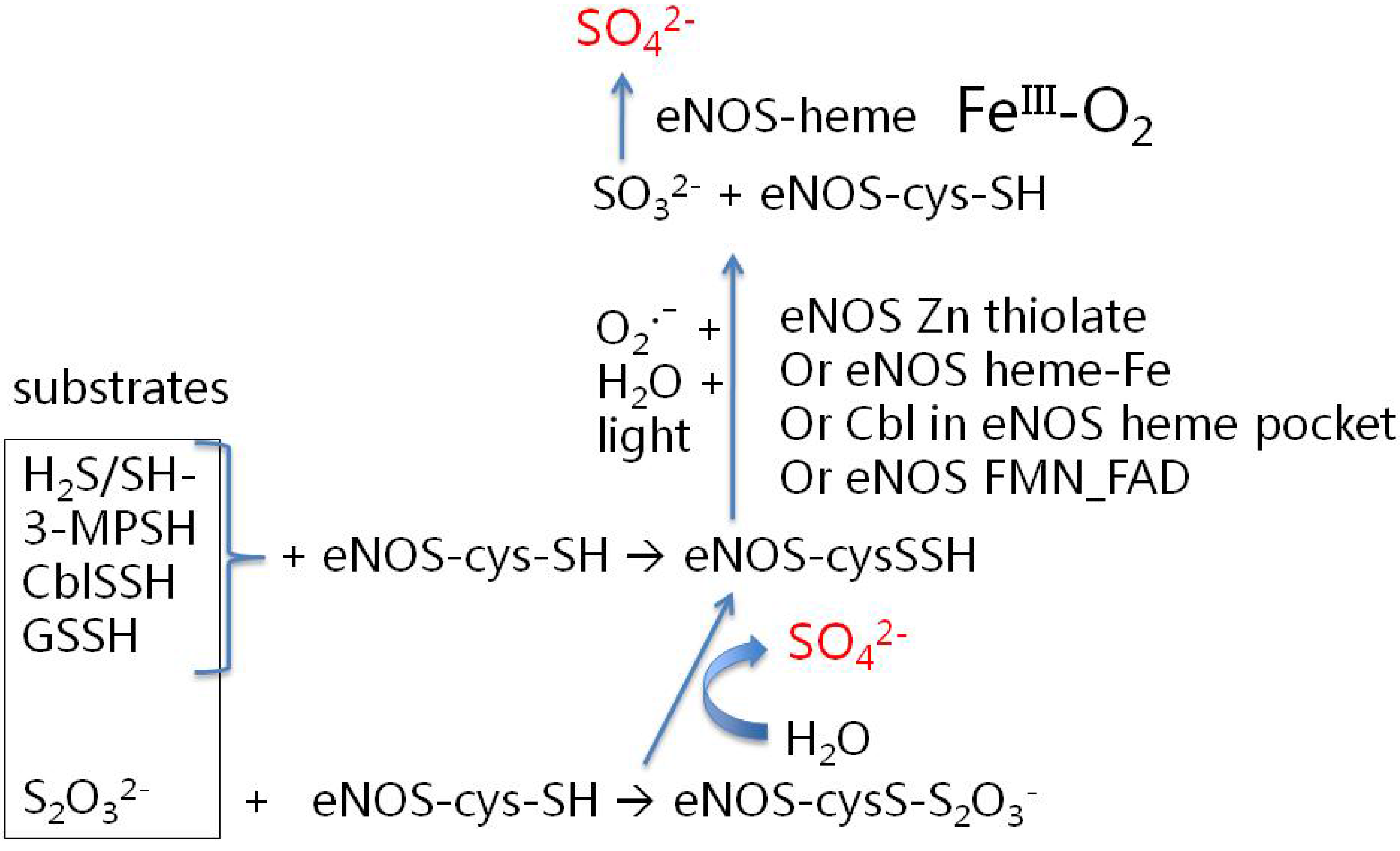
3.2. How Might eNOS Synthesize Sulfate? Some Possibilities

eNOS is an exceedingly complex and highly regulated enzyme [49]. eNOS consists of a homodimer, where each monomer contains two major regions, an N-terminal heme oxygenase domain and a C-terminal FMN/FAD binding reductase domain. The reductase domain is homologous to NADPH:cytochrome P450 reductase and similar flavoproteins. An interdomain linker provides a hinge which is modified from a closed position to an open position upon calmodulin binding. It has been proposed that the closed structure would support simultaneous two-electron transfer, whereas the open position favors one-electron transfer. It has been puzzling to researchers as to why eNOS requires such complexity and so much regulation [50]. If it is a dual-purpose enzyme, then the complexity can be explained as a constant need to determine whether it should synthesize sulfate or nitric oxide (which quickly reacts with oxygen to form nitrate [51] or with superoxide to form peroxynitrite [13]).
One of the simplest life forms utilizes sulfur as a source of energy for metabolism, the sulfur metabolizing bacteria [52,53,54], famously known by their colorful hues in sulfur hot springs [55]. In fact, it has been hypothesized that the porphyrin ring in heme, an ancient biological molecular structure, was originally developed before the existence of an aerobic atmosphere, to support sulfur-based electron transport [56]. Most photosynthetic bacteria can use a variety of inorganic sulfur compounds such as sulfide, elemental sulfur, polysulfides, thiosulfate, or sulfite as electron donors to grow photoautotrophically [57]. Several species of purple sulfur bacteria can also oxidize sulfur compounds to obtain energy as well as electrons for carbon dioxide reduction.
In contrast with the vast number of microbial metabolic pathways reported for sulfate synthesis, the established routes by which humans and other mammals achieve this same end under normal, non-inflammatory conditions seem to be limited to catabolism of cysteine, via cysteine-sulfinate, to pyruvate and sulfate [58] and mitochondrial conversion of H2S to thiosulfate, followed by oxidation to sulfite and sulfate [59,60]. There seems to be an additional, essential route for sulfate formation in humans: eNOS, which is predominantly expressed in cells that also make cholesterol sulfate, can catalyze the conversion of reduced sulfur to sulfate.
In the remainder of this section, we justify certain additional inferences and testable hypotheses regarding possible substrates and mechanistic pathways for eNOS-catalyzed sulfate production. The ideas discussed have been partly inspired by studies on eNOS structure and function [1,11] and evidence on mitochondrial and microbial pathways for inorganic sulfur oxidation [59,60,61,62]. Testing these hypotheses will clearly require well-designed experiments featuring the following:
(a)
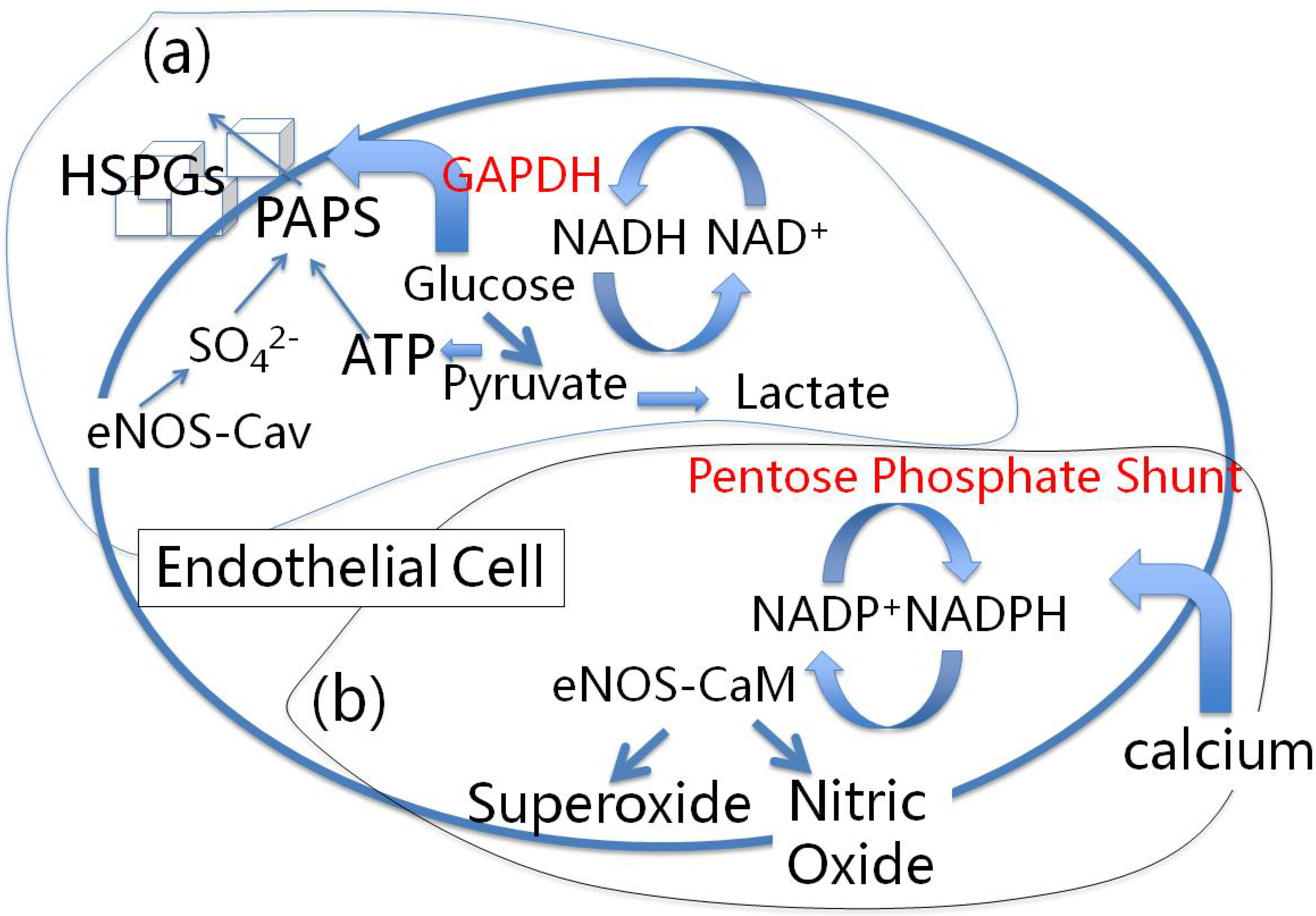
use of caveolin-bound, calmodulin-free eNOS;
(b)
use of various candidate reduced sulfur substrate compounds (see discussion below), preferably at physiologically-realistic concentrations;
(c)
activity assays aimed at detecting sulfate (not just NO); and
(d)
appropriate controls to suppress the facile non-enzymatic oxidation of reduced sulfur compounds in the presence of oxygen and trace metals [63].
Candidates for entry-level substrates include thiols such as HS and 3-mercaptopyruvate (3-MP), plus several sulfane sulfur species, including glutathione persulfide (GSSH), cobalamin persulfide (CblSSH), and thiosulfate (S2O32−). Most of these compounds figure prominently as starting points or intermediates in mitochondrial and microbial sulfur oxidation mechanisms [59,60,61,62]. However, inclusion of CblSSH in this list merits additional comment.
Cobalamin, the heart of the vitamin B12 molecule, contains a cobalt cation embedded in a corrin ring, which is related to the heme group of hemoglobin. Our suggestion of CblSSH as a candidate substrate for eNOS-catalyzed sulfate synthesis is based on evidence that a reasonably stable cobalamin persulfide derivative can be prepared in aqueous solution at pH 7 [64]. Moreover, structural studies indicate that cobalamin, an inhibitor of NO synthesis, can fit well into the heme pocket of NOS isoforms [65]. In addition, a cobalt-phthalocyanine complex (a nitrogen-containing ring system comparable to a heme group) can facilitate photocatalyic oxidation of sulfur dioxide, in an aqueous environment, to produce sulfate. A two-electron transfer from sulfite leads to a Co(III)-sulfito-superoxide complex, via a photo-assisted reaction on a titanium dioxide surface [66]. This aligns well with the two-electron transfer supported by eNOS in the closed hinge structure, when calmodulin is not present, as discussed above.
We propose that one or more of these candidate substrates can transfer sulfur to a reactive cysteine (cys) residue in eNOS to form a cys-persulfide or, if thiosulfate is the substrate, a cys-thiosulfate intermediate, as schematized in Figure 1. Hydrolysis of the cys-thiosulfate moiety yields sulfate and cys-persulfide. eNOS contains a number of highly preserved reactive cys residues [11]. Likely candidates for participation in sulfate synthesis include cys-184, the axial ligand to the heme iron in the oxygenase domain, the cys-94 and cys-99 residues from each chain that form the Zn tetrathiolate group, or cys-689 and cys-908, located near the FAD- and FMN-binding regions. Cys-689 and cys-908 have recently been shown to undergo S-glutathionylation by oxidized glutathione (GSSG), resulting in eNOS uncoupling and superoxide generation in the reductase domain [67,68].
Figure 1. Potential substrates and intermediates for eNOS-catalyzed sulfate synthesis. See discussion in text.
Entropy 14 02492 g001
The next proposed step in our mechanistic scheme involves photocatalyzed reaction of the eNOS cys-persulfide intermediate with superoxide and water to generate sulfite and restore the cys residue to the thiol form (Figure 1). Sunlight may act directly to activate the water needed for this reaction, as discussed in Section 5 below. Additional assistance with electron transfer and superoxide formation may be provided by the Fe-heme group in the oxidase domain, the zinc from the Zn tetrathiolate group, the cobalamin moiety (in the case of a Cbl-SSH substrate), and/or the FMN-FAD electron transfer apparatus in the reductase domain (in the case of a glutathione persulfide substrate reacting with cys-689 or cys-908). Sulfite can be further oxidized to sulfate by spontaneous reaction with oxygen or superoxide, or with assistance from heme-Fe(III)-O2 in the oxidase domain.
As will be discussed further in Section 5 below, there are several ways in which water, with its special properties, may contribute to the “eNOS-catalyzed” synthesis of sulfate. Heterogeneous catalysis by the “on-water” effect that occurs at the interface of water and hydrophobic surfaces may stabilize the transition state(s) for the oxidation to sulfate [69]. An acidic pH afforded by the gel-like water exclusion zones (EZs) and Grotthuss-style “proton hopping” may further catalyze the oxidation [70]. Relative immobilization of substrates by long-range dynamic structuring of interfacial hydration water may also be operative [71,72]. The sulfate dianion has a long-range effect on water structure, and this effect is likely to be an important factor in its Hofmeister behavior [73].
With regard to the union of sulfate with cholesterol to produce cholesterol sulfate, eNOS’s binding to caveolin is significant, because caveolin is also intimately involved in cholesterol transport both from HDL particles into cellular stores and from the endoplasmic reticulum (ER) to the plasma membrane [74]. Caveolin is therefore well positioned to bring cholesterol (whether retrieved from serum HDL or internally synthesized) into close proximity to sulfate synthesized by eNOS. The attachment of sulfate to cholesterol via the sulfotransferase SULT2B1b [75] requires an activation step which consumes ATP to produce 3’-phosphoadenosine-5’-phosphosulfate (PAPS). As discussed later, ATP can be provided by glycolysis. It is reasonable to infer also that sulfation is an essential step in the transport of cholesterol from the Golgi apparatus to the plasma membrane.

4. Red Blood Cells and Cardiovascular Disease

According to the Framingham heart study [76,77], low cholesterol in HDL is the single strongest lipid predictor of cardiovascular disease. Furthermore, both cardiovascular disease and diabetes are associated with impaired zinc utilization and increased oxidative stress [78]. In a state of zinc deficiency, it follows that eNOS, which depends upon zinc, may provide insufficient sulfate, an essential mediator of cholesterol transport to HDL. As a consequence, superoxide, released into the vessel lumen, supports the synthesis of sulfate from homocysteine as a back-up supplier. This, then, can explain why homocysteine is a risk factor for cardiovascular disease [79].
Diabetes, also a known risk factor for cardiovascular disease, is associated with excess cholesterol in RBCs, leading to morphological changes and reduced deformability [40], thus impeding passage of RBCs through capillaries. Furthermore, a significant inverse correlation was found between RBC cholesterol content and serum ApoA1-HDL levels. This implies that the transporting of cholesterol from epidermal cells and RBCs to HDL-A1, as mediated by cholesterol sulfate, is impaired, leading to excess membrane cholesterol in these cells. Ordinarily, cells in the epidermis are major suppliers of cholesterol to HDL-A1. Due to the limited solubility of cholesterol esters in aqueous environments, cholesterol trafficking would be expected to require vesicular transport, yet no such vesicles have been identified [74]. Sulfation would solve the problem of cholesterol’s hydrophobicity, allowing it to traverse the water-based cytoplasm from the ER and/or Golgi bodies to the plasma membrane, and to travel from the plasma membrane through water-based blood to HDL-A1.
The scavenger receptor, SR-BI, integrated into membrane caveolae, plays many roles in mediating cholesterol transport, as detailed in [80]. It is responsible for the uptake of oxidized or acetylated LDL by macrophages in cardiovascular plaque. However, it also binds HDL, and HDL outcompetes LDL when both are present in the medium. In the case of LDL, its binding leads to the endocytosis of the entire particle and subsequent processing in lysosomes. However, in the case of HDL, both free cholesterol from the lipid monolayer and cholesterol esters from the internal stores are transported into the cell, in a process termed “selective lipid uptake”. In [80], p. 794, it is stated, “The detailed molecular mechanism underlying SR-BI–mediated selective lipid uptake has not yet been elucidated.” The problem is lipid transport through water-based cytoplasm, one potentially solved through sulfation.
Moreover, selective lipid uptake involving cholesterol sulfation is likely to be reversible, since macrophages deliver cholesterol to HDL particles via SR-BI, whereas steroidogenesis cells in the adrenal glands take up cholesterol from HDL particles via SR-BI for processing into steroids. Erythrocytes from mice with knock-out SR-BI function exhibit an irregular shape and macrocytosis, implying that RBCs make use of an SR-BI mediated mechanism as well. The presence of large autophagosomes in the defective erythrocytes implies impaired autophagy.
Thus, it can be inferred that both cholesterol import as well as export depend upon sulfation. Caveolin binds to cholesterol esters and transports them from caveolae to intracellular membranes [74]. eNOS, also bound to caveolin, could provide the sulfate that imparts amphiphilic character to cholesterol, thereby enabling cholesterol transport through the aqueous medium and facilitating systemic cholesterol bioavailability. One of the key features of cardiovascular disease is the accumulation of lipid-laden macrophages, which eventually succumb to excess cholesterol loading, leading to apoptosis. Macrophages in atherosclerotic plaque undergo apoptosis following excess accumulation of free cholesterol in the ER, which induces the unfolded protein response [81]. This is due in part to the inability to export cholesterol to HDL-A1 [82]. The key problem may be insufficient sulfate supply to alleviate their cholesterol burden. Blood sulfate levels may be biophysically-determined by the Jones-Ray effect [83] and the long-range dynamic water structuring property of sulfate [73].

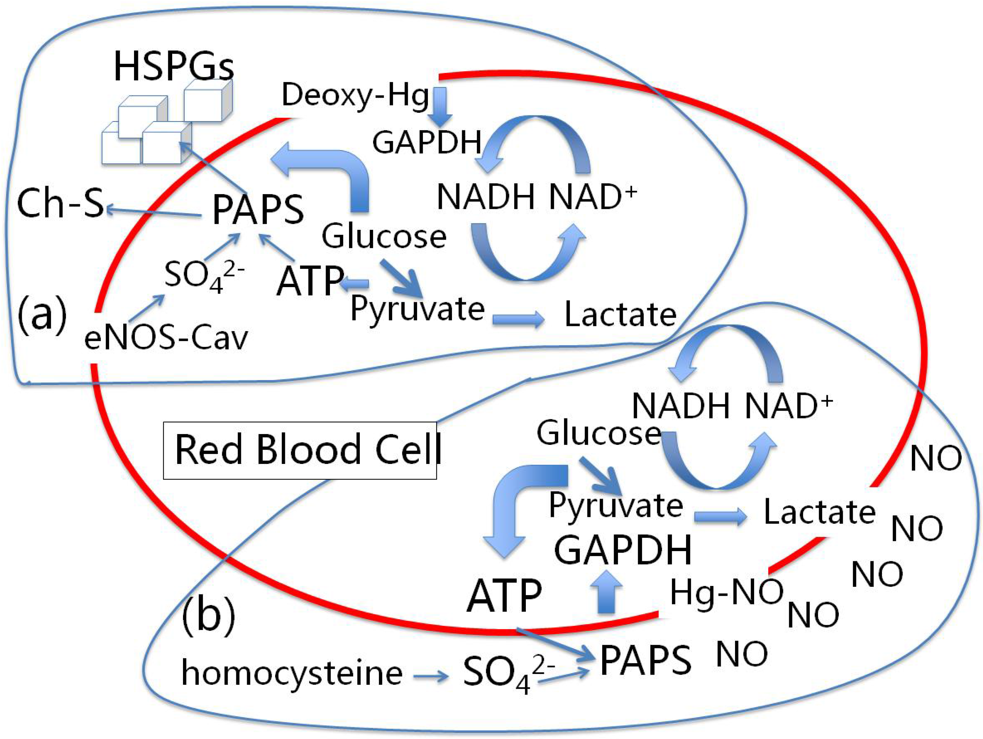
4.1. Red Blood Cells, eNOS and Cholesterol Sulfate

Ever since it was discovered that RBCs contain eNOS, researchers have struggled to understand what function it could possibly serve in these cells [6,84,85]. It is chemically implausible that eNOS in RBCs could produce nitric oxide that would escape binding to the massive amounts of hemoglobin contained in RBCs. Binding to the iron in hemoglobin would render the hemoglobin inactive in oxygen transport, an effect that resembles the toxic effect of carbon monoxide. Thus, both the NO and the hemoglobin would be impaired in their normal function. Indeed, RBCs exhibit a very low rate of transport of the substrate L-arginine into the cell, and they contain a significant amount of the enzyme L-arginase, which degrades any L-arginine that manages to gain entry. It has been confirmed that erythrocytic NOS is inactive – that it is disabled in producing NO [85]. Furthermore, the eNOS in RBCs co-localizes with the plasma membrane [6], and it has been well established through studies on endothelial cells that membrane-bound eNOS is inactive with respect to NO synthesis.
RBCs also contain a membrane-bound protein, band3, which binds, thereby inactivating, several enzymes involved with the glycolytic pathway, such as phosphofructokinase (PFK), aldolase, glyceraldehyde-3-phosphate dehydrogenase (GAPDH) and lactate dehydrogenase (LDH) [86,87]. Deoxyhemoglobin (deoxyHb) also binds to band3 and competes with the glycolytic enzymes, reactivating them by dislodging them from the membrane [88]. It has been argued in [86] that this technique of gathering the enzymes in an inactive state near the membrane allows them to become more concentrated once they are dislodged from the membrane by deoxyHb, thus increasing their reactivity.
However, glycolytic enzymes are needed near the membrane to produce the ATP that can enable sulfate transfer to cholesterol. The sulfate dianion is assembled at the membrane by eNOS in the RBC, as previously discussed. Such a process can fixate oxygen and sulfur in the sulfate anion, and provides a second method besides oxyhemoglobin to transport oxygen. RBCs are among the short list of cell types that produce Ch-S [30], and we argue here that Ch-S synthesis is the main purpose of eNOS in RBCs, which explains its presence there.
Ch-S also stabilizes the RBC membrane and protects it from hemolysis [89]. The concentration of Ch-S in RBC plasma membranes is approximately 2-fold higher than the concentration in blood plasma. We propose that the sulfate is first produced from thiosulfate or another form of sulfane sulfur by eNOS, with superoxide as a reactive intermediate (see Figure 1), and then reacts with ATP provided through glycolysis to form PAPS, which is subsequently used to produce Ch-S from cholesterol. PAPS is formed from ATP and sulfate, where one molecule of ATP is converted to pyrophosphate to activate the reaction, and a second ATP molecule converts APS to PAPS by phosphorylation at the 3’ position. Glycolysis localized to the cytoplasmic area near the membrane can provide the ATP.
In addition to its role in binding hemoglobin and glycolytic enzymes, band3 also plays an important role in the transport of anions across the membrane, and it has been shown that a chloride-sulfate exchange mediated by band3 is associated with the import of hydrogen ions, thus exporting the sulfate ions [90]. Since RBCs also export ATP [91], it becomes feasible to synthesize PAPS directly in the endothelial wall from ATP and sulfate. The PAPS, synthesized by endothelial cells, then becomes substrate for Ch-S synthesis by platelets adhering to the endothelial artery wall, as will be further discussed in section 4.3 on the atheroma.

4.2. Nitrite-Induced Vasodilation by RBCs

It has long been recognized that RBCs can enhance the vasodilation effects of serum nitric oxide, but most of the ideas developed to account for the mechanism are derailed by the problem of how to get the NO out of the RBC, as it will be quickly taken up by hemoglobin (Hb). The most plausible idea along these lines involves the reduction of nitrite back to NO under deoxygenated conditions. Nitrite reacts with deoxyHb to form various nitrosylated products [92], and it has been conjectured that these reaction products might bind to the RBC membrane and subsequently give up their bound NO to the bloodstream [93,94,95,96,97].
A more appealing solution has been proposed in [98], which elegantly bypasses the problem of the release of NO by RBCs by invoking a signaling mechanism involving ATP instead. These authors demonstrated that metHb, an oxidation product of oxyHb with nitrite, has a much higher affinity to the membrane than oxyHb. Furthermore, nitrite reaction products with deoxyHb have much higher affinity for the membrane than deoxyHb itself, which in turn has significantly greater affinity than oxyHb. Hence, under both oxygenated and deoxygenated conditions, Hb reaction with nitrite results in an increased tendency for Hb to bind to the membrane, displacing glycolytic enzymes and inducing glycolysis, thus generating ATP. The authors also showed that nitrite injection into plasma yielded a large increase in serum ATP, demonstrating that RBCs release the additional ATP into the medium. Finally, ATP reacts with purinergic receptors in the endothelium to directly stimulate NO synthesis by eNOS. Hence, this indirect mechanism leads to the observed increase in NO caused by RBCs.

4.3. A Proposed Role for the Atheroma

In this section, atherosclerosis, the inflammatory buildup of abnormal fatty deposits (atheromas) in the arteries, is considered as providing a serendipitous alternate route to Ch-S synthesis under conditions of sulfate deficiency. Specifically, homocysteine can provide a source of sulfate that becomes operative when the eNOS in RBCs, endothelial cells and platelets fails to produce adequate supplies of sulfate, likely due to deficiencies in zinc or cobalamin and/or to insufficient sunlight exposure.
Like RBCs, platelets also inexplicably contain a functional eNOS [5], and they also produce Ch-S [99]. Platelets produce an abundance of Ch-S from cholesterol in the presence of PAPS. A quote from [99] captures the level of surprise that the researchers experienced in their experiments on platelets: “The remarkable increase in platelet cholesterol sulfate content (300-fold) after the addition of exogenous PAPS to the incubation medium was totally unexpected.” [99]
Chronically inflamed tissues support high levels of lymphocyte extravasation from the blood, and the initial adhesion of these immune cells to the vessel wall is mediated by selectins [100], whose ligands are sulfated sialomucins. In [101], it was demonstrated that endothelial cells in inflamed tissues possess active expression of the gene for ATP sulfurylase, which catalyzes the synthesis of PAPS from sulfate. This is viewed as a limiting step in the sulfation of selectin ligands, and so lymphocyte adhesion depends critically on the supply of PAPS by ATP sulfurylase. Thus, we conjecture that endothelial cells in the inflamed artery wall of atherosclerosis may be supplying PAPS to adhering platelets, utilizing as substrates sulfate and the ATP released by RBCs.
Thus, both ATP and sulfate are needed for endothelial cells to produce PAPS, which can then be combined with cholesterol by platelets, using a sulfotransferase, to produce abundant Ch-S. This then provides another reason for RBCs to release ATP to the serum in the presence of nitrite–to provide substrate for PAPS synthesis by endothelial cells and subsequent Ch-S synthesis by platelets. Note that the presence of nitrite implies excess NO synthesis, which, in turn, reflects, according to our hypothesis, impaired sulfate synthesis by eNOS: eNOS has switched from sulfate to NO synthesis, and therefore the atheroma is tasked with supplying Ch-S.
We therefore propose that a need for Ch-S synthesis by platelets may in fact be a controlling factor in atherogenesis, and that homocysteine, as a source of sulfate, can mediate this goal, but inflammation is necessary for the homocysteine-to-sulfate conversion to occur. Indeed, Ch-S has been shown to be present in atherosclerotic lesions [102]. Homocysteine, a strong risk factor for atherosclerosis [79], is associated with increased presence of ROS in the artery wall [103]. It has been demonstrated that excess homocysteine in the blood is converted into homocysteine thiolactone, which can become a source for sulfate in the presence of superoxide, with vitamins A and C serving as catalysts [104].
Since macrophages in the plaque readily take up cholesterol from oxidized small dense LDL particles (ox-LDL) and later release it into HDL-A1 [105,106], cholesterol would be available in the plaque to form Ch-S from PAPS, a service that would be performed by platelets trapped in the plaque. In [99], it was demonstrated that platelets will only accept cholesterol from HDL-A1 as a source for Ch-S synthesis–they reject not only all forms of LDL, but also other apolipoproteins bound to HDL such as apoE and apoA-II.
Several lines of evidence point to the idea that homocysteine actively promotes the survival of O2 in the artery wall [107]. It does this in part by inhibiting the expression and secretion of antioxidant enzymes such as glutathione peroxidase (GPx) and superoxide dismutase (SOD) [108]. Homocysteine also inhibits the binding of SOD to the HSPGs at the surface of endothelial cells [109]. Finally, homocysteine activates cytokines such as nuclear factor κB (NF-κB) and protein kinase C (PKC) [110], which in turn upregulate NAD(P)H oxidase and NOS, further enhancing O2 production [111].
In parallel, homocysteine upregulates expression of HMG-CoA reductase, increasing the biosynthesis and uptake of cholesterol [112], and thus enhancing the bioavailability of cholesterol to react with the PAPS that will be derived from homocysteine in the presence of O2 via the reaction presented in [104]. While these activities of homocysteine have in the past been viewed as pathological, a reinterpretation of the observed behavior suggests that, instead, such activities are essential for the establishment of a critically needed supply of cholesterol sulfate.
It has been proposed [113] that elevated homocysteine is a marker for elevated asymmetrical dimethylarginine (ADMA), a known marker of renal disease, and that ADMA, as an endogenous inhibitor of NO, is the true factor that negatively impacts heart health, since serum levels of homocysteine and ADMA are strongly correlated. In [114], it was shown that impaired supply of heparan-sulfate rich anionic sites in glomerular basement membranes is associated with congenital nephrotic syndrome, which is characterized by kidney failure and protein loss through urine. It is likely that severe sulfate deficiency in the glomeruli characterizes other forms of renal disease as well. Hence, suppressed synthesis of NO by eNOS due to excess ADMA would result in the synthesis of superoxide instead, due to uncoupling [115]. The superoxide would supply substrate for the reaction that leads to sulfate synthesis from homocysteine [104], which could then supply the glomeruli with sulfate and improve kidney function. Redirection of homocysteine towards methionine and methylation pathways through supplementation with vitamins B6, B12, and folic acid would reduce the bioavailability of homocysteine as a source of sulfate, thus impairing cholesterol sulfate synthesis in the artery wall and promoting further kidney failure, as well as further deficiencies in the supply of these critical nutrients to the heart. This could explain why trials attempting to lower serum homocysteine pharmacologically have met with little success in reducing heart disease risk [113].
Prostacyclin (PGI2) is a potent vasodilator and inhibitor of platelet aggregation. It also promotes an increased hydrolysis of cholesterol esters in smooth muscle cells, allowing the release of cholesterol to HDL-A1. Experiments have shown [116] that HDL-A1 prolongs the half-life of PGI2 from 5 minutes to 22 minutes. These authors showed that apo-A1 bound to HDL forms a complex with PGI2, thus delaying its degradation, and therefore promoting vasodilation and platelet stability. Apolipoproteins A-II, C-I, C-II, C-III, D and E, as well as LDL and Apo-A1 in isolation (not bound to HDL), did not possess this property.
It has repeatedly been argued that HDL-A1’s benefit in cardiovascular disease is that it takes up cholesterol from macrophages in the plaque and returns it home to the liver. However, we propose instead that the benefit lies in its delivery of cholesterol to the platelets to promote the synthesis of Ch-S, in the presence of PAPS provided by homocysteine. This then explains the need for ROS in the artery wall, because homocysteine requires superoxide for sulfate production [104].
HDL's well-established protective role in cardiovascular disease is proposed to be partly due to the fact that it co-transports with paraoxonase 1 (PON-1), a potent antioxidant enzyme [117], which, however, is inactivated by oxidized LDL [118]. One of the key targets of PON-1 is homocysteine thiolactone [119]. PON1 possesses the ability to hydrolyze and therefore inactivate lactones, thus converting homocysteine thiolactone back to homocysteine [120]. PON-1 activity is inhibited through nitrosylation of ApoA-1 [121], so excess synthesis of nitric oxide would disrupt its function. Suppression of PON-1’s antioxidant effects would lead to enhanced superoxide production, and the increased bioavailability of homocysteine thiolactone will promote sulfate synthesis from the superoxide. Unfortunately, this will also expose the artery wall to oxidation damage, but ultimately enables the synthesis of Ch-S by platelets.

5. The Hofmeister Series, the Glycocalyx and the Effect of Sulfate on Water Structure

There has recently been a resurging interest in the study of the unique properties of water, catalyzed in part by the efforts of Gerald Pollack to popularize this important concept [122]. In our research, we have come to realize that the unique ability of water to gel in the presence of certain solutes is essential to life.
A careful examination of a living blood vessel, which is feasible by adding dyes and observing where they penetrate, reveals that there is a thick layer of impenetrable water surrounding the interior of the capillary wall [123]. Furthermore, this water is in a gel-like state, as it does not participate in the flow. Red blood cells remain in the center of the capillary, well separated from the physical wall of endothelial cells. This gel-like layer consists of water surrounding the glycocalyx, a layer of sulfated polymeric glycoproteins on the outer surface of most cells. The gel layer plays an essential role in maintaining blood flow and in protecting the endothelial wall [124]. The gel prevents the GAGs that are embedded within it from direct physical contact with the particles suspended in the blood. It also keeps out solutes, reducing the leakage of ions across the membranes of all the cells that are decorated with the GAGs, both those lining the capillary wall and those suspended in the blood.
A key component of the glycocalyx is heparan sulfate, and it is the presence of sulfates in the heparan sulfate that maintains the gel, due to the physical properties of sulfate as a kosmotropic ion in the Hofmeister series [73,125,126]. By direct contrast, nitrate is a chaotrope–on the opposite end of the Hofmeister scale–and this distinction between nitrate and sulfate is likely a key reason why it is sulfates rather than nitrates that attach to the sugars in the glycocalyx. The Hofmeister series defines a continuum between the extremes of salting in and salting out, where chaotropes like nitrate promote disorder in the surrounding water and cause proteins to dissolve, whereas kosmotropes like sulfate promote a regular liquid-crystalline gel-like structure in the water and cause proteins to precipitate out. More importantly, anionic kosmotropes create an exclusion zone (EZ) in the water in their neighborhood, a region that is largely impenetrable both to dissolved ions and to suspended particles [127]. This exclusion zone is the gel that encases and protects the cell.
Voeikov and Del Giudice [128] have pointed out the remarkable properties of such EZ water, in creating a negative electrical potential with regard to the bulk water adjacent to it (down to −150 mV), as well as a concentration of protons at the interface between EZ-water and bulk water. EZ water also absorbs light at 270 nm, and emits fluorescence when excited at this frequency. The thickness of the EZ water layer increases dramatically upon exposure to IR radiation [127].
In this context, it is interesting that eNOS attaches to the membrane at caveolae, and that HSPGs also attach at the lipid raft regions associated with the caveolae. According to [128], a key property of EZ water is the generation of electrons in a much higher state of excitation than normal, which can then presumably catalyze the reactions needed to oxidize thiosulfate to sulfate. They wrote: “As radiation, especially light in the IR part of the spectrum, increases the thickness of the layer of EZ-water, thereby increasing its electron-donating capacity, EZ-water becomes a practically inexhaustible source of electrons.” [128].
As a consequence of EZ’s surrounding sulfate molecules, the concentration of suspended particles and ions in the remaining bulk water is increased, and this is what leads to the property of salting out proteins–causing them to precipitate out of solution. We conjecture and will see later that the long-range dynamic water-structuring property of sulfates and relatively weak magnetic field exposures [73,129] play an important role in concentrating enzymes in the lysosome to support the breakdown of proteins for recycling via heterogeneous “on-water” catalysis [69]. Both in the blood system and in the cytoplasm of individual cells, it is critical to maintain the proper balance of chaotropes and kosmotropes to support the proper folding and functioning of active proteins.
Every cell suspended in the blood–the platelets, RBCs and white blood cells–is surrounded by a matrix of glycoproteins that maintains a negative charge and a kosmotropic water environment in order to protect the cell from aggregating with other cells in suspension and to minimize the rate of leakage of ions across the cell membrane. Through this strategy, the amount of energy needed to pump ions across the membrane in order to maintain, for example, a high internal concentration of potassium, is minimized. The sulfate ions in the GAGs are crucial for achieving this effect.
Dianionic sulfate has long-range dynamic water-structuring properties [73], that protect from thrombophilia, a condition that is prevalent in the Western world, which manifests as an increased risk to thrombosis [130]. Sulfate deficiency is likely to lessen this water-structuring effect, throughout the entire vasculature. When sulfate is depleted, platelets will more readily activate, bind to fibrinogen, aggregate, and form blood clots, and cells will experience a depletion of their ATP levels as they are required to constantly maintain the Na/K-ATPase pump.
Further support for our arguments concerning sulfate deficiency in the glycocalyx comes from the observation that hyaluronan overexpression is associated with both hypertrophic cardiomyopathy [131] and cardiovascular disease [132]. Hyaluronan is the only unsulfated glycosaminoglycan—it has an abundance of carboxylate groups instead, replacing the sulfates as kosmotropic anions. Hyaluronan is overexpressed specifically in the artery wall in the early stages of cardiovascular disease [132].

6. Heparan Sulfate Proteoglycans

Having presented above our hypotheses as to how an inadequate supply of cholesterol sulfate can give rise to heart disease, we will now consider how deficiency in another important set of biological sulfates–the heparan sulfate proteoglycans (HSPGs)–can lead to equally serious health problems. In this section, we will first discuss the extensive role that HSPGs play in regulatory systems in the cell. We then develop the novel hypothesis that HSPGs function as a temporary store for glucose, maintained just outside the cell, analogous to glycogen which stores glucose internally. We suggest that diabetes arises as a consequence of an impaired ability to produce HSPGs. Next, we mention briefly several distinct cell types that both contain eNOS and produce sulfated proteoglycans. Finally, we consider the essential role that HSPGs play in the lysosome, an organelle involved in the crucial process of breaking down accumulated cellular debris. This requirement for HSPGs in the lysosome implies that HSPG synthesis impairment leads to lysosomal impairment.

6.1. Heparan Sulfate Proteoglycans and Glucose Metabolism

Researchers have long been aware that all cells are decorated around their exterior wall by a collection of glycosaminoglycans (GAGs) generously modified by sulfate attachment, and often bound to membrane-associated proteins such as syndecans and glypicans [133]. However, it has only been in the last decade or so that an awareness has developed of the critical role these extracellular matrix glycoproteins play in regulatory systems. Particularly noteworthy are the HSPGs, which consist of heparan sulfate polymers made up of glucose and galactose, modified by nitrogen (glucosamine, galactosamine) and by additional acetylation (N-acetylglucosamine) and/or O- or N-linked sulfation. Recently, the term heparanome has been used to characterize the repertoire of heparan sulfate sequence patterns associated with a particular cell type [134,135]. Researchers are only recently developing the necessary tools to be able to sequence the individual HSPG chains and experiment with genetic mutations in mice to try to determine more precisely the many roles they play in cell signaling systems [136].
The syndecans are cell surface proteins that regulate intracellular signaling as well as complex cellular activities such as adhesion, motility and invasion [137]. Syndecan shedding–the release of syndecan from the cell membrane into the interstitial spaces–is well-established as a pathological response associated with inflammation, microbial infection, and cancer [133,138,139,140]. High levels of shed syndecans in the blood plasma correlate with poor outcome in cancer patients [141,142]. Insulin induces a signaling cascade that results in the release of proteases that cleave the core protein of syndecan and induce shedding. Activation of protein tyrosine kinases leads to phosphorylation of the cytoplasmic domain, and subsequent shedding [143,144]. ROS also attack the syndecans, inducing shedding. Ch-S, on the other hand, is a potent inhibitor of proteases, including trypsin, chymotrypsin and elastase [145].
Recent research has revealed an important role for heparan sulfate in stabilizing syndecans on the cell surface [146]. In experiments involving several cell types, both normal and cancerous, depletion of heparan sulfate chains from the syndecan resulted in a 3- to 10-fold increase in the rate of syndecan shedding, depending on the cell type. Heparan sulfate depletion also stimulates the cell to increase the rate of syndecan synthesis, presumably to quickly replace the shed syndecans. Hence, heparan sulfate chains attached to syndecan play a crucial role in protecting the syndecan from destruction. It was hypothesized that the heparan sulfate chains form a physical barrier that protects the cleavage sites on syndecans, a hypothesis that aligns well with our proposal that sulfate forms solute-free exclusion zones in the surrounding water.
Maintenance of a healthy intestinal epithelial barrier is essential for protection from influx of bacteria into the blood stream and subsequent sepsis. The heparan sulfate chains in syndecan-1 have been shown to be essential in maintaining a healthy barrier in both mice and humans [147]. Profound abnormalities in the GAGs, in both the connective and vascular tissues, have been found in association with colitis and Crohn’s disease [148], and this disruption likely is a significant factor in leaky gut syndrome. GAGs have also been shown to contribute to the pathology of rheumatoid arthritis and autoimmune disease [149]. In lupus nephritis, GAGs, and, specifically, heparan sulfate, are excreted in the urine [150]. Pancreatic beta cells produce an extraordinary amount of heparan sulfate, which protects them from damage due to ROS [151]. Heparan sulfate degradation by infiltrating immune cells is a mechanism for induction of type 1 diabetes. Furthermore, in type-1 diabetes, pancreatic beta cells are killed by activated macrophages, in part by the release of excess NO [152].
One hypothesis regarding cardiovascular disease is that the damage to the vasculature accrues mainly as a consequence of cytotoxins resulting from an immune response to infective agents known to be present in the plaque [153]. The most common such agent is Chlamydia pneumoniae [154,155]. Chlamydia pneumoniae can only grow and survive inside a host cell, where it accumulates in vacuolae and produces a glycosaminoglycan which bears an extremely strong resemblance to heparan sulfate [156]. We hypothesize that C. pneumoniae may be playing a protective role in providing heparan sulfate to the vasculature. Indeed, attempts to treat cardiovascular disease with antibiotics have met with failure, the treated group suffered increased cardiovascular mortality compared to the control [157]. An explanation could be that the microbes are assisting in the task of maintaining heparan sulfate supplies.
Aberrant insulin receptor signaling has been proposed as a major contributor to the oxidative stress associated with aging [158], and we suggest that this impairment may be attributed to impaired heparan sulfate synthesis. People with diabetes are at high risk of developing nephropathy, leading to proteinurea and, ultimately, renal failure. Heparan sulfate constitutes over 80% of the proteoglycans in the glomeruli basement membrane of the kidneys [159]. Diabetes is associated with a reduction in heparan sulfate levels in the glomerular matrix proteins in both humans [160] and rats [161]. In [162], it was shown that, following exposure to high serum glucose, there is a marked reduction in the incorporation of glucosamine into GAG chains, with an associated reduction in the biosynthesis of sulfated GAGs, in the glomerular endothelial glycocalyx. This suggests a link between high serum glucose and impaired GAG synthesis in the kidneys, likely due to glycation damage to glycolytic enzymes.
HSPGs have a very rapid turnover rate–it is estimated that they are recycled with a half-life of 1 to 4 hours [135]. Since glucose is a core component, it is tempting to speculate that one role HSPGs might serve is as a temporary storage site for glucose, analogous to glycogen. We hypothesize that the sulfate moiety serves an essential role as a mechanism to assure that the glucose is inert in the extracellular environment–similar to the phosphorylation step that initiates glycolysis. Both sulfate and phosphate are anionic kosmotropes [163], which, we propose, would have the effect of forming an EZ in the vicinity of any sulfate unit, thus protecting blood proteins from glycation damage. Such a mechanism would facilitate glucose metabolism, where excess dietary glucose could be temporarily housed in the HSPGs, and later metabolized after the breakdown of the HSPGs in the endosome and lysosome.
This process would become impaired if insufficient sulfate is available. The result, we propose, would be the build-up of O-linked N-acetylglucosamine (O-Glc-NAc) modified proteins in the cytoplasm and the nucleus [164]. It is informative to examine glucose metabolism in skeletal muscle cells, which are the key cell type that becomes impaired in glucose uptake in association with type-2 diabetes. A revealing study involved mice that overexpressed glucose transporter GLUT1, the GLUT that controls basal glucose uptake in muscle cells [165]. Muscle cells also utilize GLUT4, which migrates to the membrane at lipid raft domains in response to insulin signaling, and whose function is impaired in diabetes. The mice with overexpression of GLUT1 exhibited impaired GLUT4 activity, which was associated with O-Glc-NAc binding to GLUT4. Type 2 diabetes is associated with excess O-Glc-NAc binding to cytoplasmic proteins [166], which, we propose, is due to impaired GLUT4 signaling as a consequence of both depleted cholesterol, resulting in a reduced number of lipid rafts (the locale where HSPGs bind and where GLUT4-mediated glucose uptake takes place), and depleted sulfate, resulting in a reduced ability to produce functional HSPGs. Hence the glucose must be immediately processed and the excess is converted to O-Glc-NAc, affecting cellular proteins.
There is an interesting link between excess glucose and homocysteine thiolactone synthesis mediated by methionyl-tRNA synthetase. Mammalian systems are similar to E. coli with respect to the role that methionyl-tRNA synthetase plays in methionine incorporation into proteins [167]. Studies on E. coli have demonstrated that, due to competition of homocysteine with methionine in the process that assembles amino acid chains, one molecule of homocysteine is edited to produce homocysteine thiolactone per 109 molecules of methionine incorporated into protein in vivo [168]. Thus, an excess need for protein synthesis would lead to an excess synthesis of homocysteine thiolactone. Excess protein synthesis is expected when proteins are disrupted by glycation damage, which would be predicted to occur when there is insufficient sulfate to properly store glucose in HSPGs. This conveniently leads to an overproduction of homocysteine thiolactone, now available as substrate for sulfate synthesis, precisely when sulfate is needed, as indicated by excess glycation damage.
HSPGs can entrap a wide variety of extracellular effectors [169], particularly those associated with the LDL receptor-related protein (LRP) [170]. The LRP controls endocytosis of a remarkable range of proteins, related to blood platelet stability, cholesterol uptake, proteinases and their inhibitors, lysosomal enzyme activation, signal transduction, and neurotransmission [171]. HSPGs bind preferentially to the regions surrounding the lipid rafts [143,144], and these are also the areas where glucose enters the cell, mediated by GLUT4. HSPGs also bind growth factors like fibroblast growth factor (FGF), which are involved in promoting angiogenesis and neuronal development [172], and this binding too is mediated through lipid rafts [173].
Thus we propose that cholesterol depletion would reduce the availability of lipid rafts, and sulfate depletion would disrupt the ability to safely incorporate glucose into HS chains–both of these effects would contribute to insulin resistance in skeletal muscle cells and resulting type-2 diabetes. Both cholesterol and sulfate depletion would be traced back to the insufficient production of Ch-S in the skin in response to sunlight exposure.

6.2. eNOS and Sulfated Glycosaminoglycan Synthesis

We have identified at least five cell types, all of which contain either nNOS or eNOS, and all of which are also responsible for the synthesis of numerous highly sulfated glycosaminoglycans. In each example below, we argue that the main function of their NOS enzyme is to produce sulfate to supply the GAG chain:
Endothelial Cells: Endothelial cells are the site where eNOS was first identified, which explains the nomenclature of the enzyme. Endothelial cells are tasked with maintaining and constantly renewing the endothelial glycocalyx, a thick wall of sulfated proteoglycans [174]. The glycocalyx is a dynamic structure with constant turnover, due to degradation by matrix metalloproteinases or shedding due to redox stress or shear stress. Hence, there is a constant need for sulfate to renew the supply to the glycocalyx.
Fibroblasts: Fibroblasts, cells whose sole responsibility is to synthesize extracellular matrix and collagen, contain both eNOS and nNOS [175,3]. Fibroblasts are responsible for synthesizing HS, dermatan sulfate, and chondroitin sulfate, all of which contribute to the extracellular matrix proteins that decorate the exterior of essentially all cells [176]. Thus fibroblasts have an abundant need for a steady supply of sulfate to perform their normal functions.
Pancreatic β Cells: Pancreatic β cells express an unusually low level of most common free radical scavenger enzymes, yet they suffer significant exposure to oxidizing agents associated with the synthesis and release of insulin. They maintain a huge and unusual cytoplasmic store of HS for protection from oxidative damage [151]. β cells also contain a functioning nNOS [177], and so they may use this synthase to produce sulfate to supply their internal stores. It is unlikely that they use it to produce NO, as they are highly sensitive to NO, and it has been demonstrated that exposure of β cells to NO produced by macrophages kills them, leading to type-1 diabetes [178].
Mast Cells: Mast cells, which mediate emergency reactions, contain a functional eNOS, whose expression pattern is increased after ischemia [179]. The eNOS was localized mainly to the granules of activated mast cells. Mast cells release heparin stores from secretory granules into the artery wall during stress conditions, and heparin, a free-standing glycosaminoglycan, is the most highly sulfated molecule known to biology. Again, if the eNOS can synthesize sulfate, then it can supply sulfate to produce heparin.
Neurons: eNOS is present in the neurite outgrowth of neurons, where its role is poorly understood. Neurite outgrowth depends upon ATP produced by glycolysis, which is actively suppressed by nitric oxide [180]. On the other hand, heparan sulfate plays an essential role in neurite outgrowth; several different sulfated glycosaminoglycans will stimulate growth-factor induced neurite outgrowth [181], and heparan sulfate supplied to the medium stimulates neurite outgrowth even in the absence of growth factors [182]. Once again, there is a compelling reason to produce sulfate, and clear justification not to produce nitric oxide.

6.3. The Lysosomal Connection

Lysosomes are cellular organelles that enzymatically degrade accumulating cellular debris. The lysosome also plays an essential role in degrading the cell walls of invasive microbes, i.e., lipopolysaccharide (LPS). Multiple lysosomal storage diseases involving impaired sulfate metabolism point to the importance of sulfate for proper lysosomal function [183]. Following endocytosis, syndecans undergo core protein proteolysis and glycosaminoglycan chain fragmentation in endosomes, and the remaining glycosaminoglycan fragments are fully degraded in lysosomes, a process that is impaired in many types of tumor cells [184]. The process of breaking down sulfated glycosaminoglycans is described in detail in [38]. The glycosaminoglycan chains are separated from the protein core but maintained intact until all the protein has been degraded. Subsequently, the chains are first broken up into much smaller units before the sulfate is stripped off.
We propose that the sulfated GAGs facilitate the action of the enzymes involved in protein degradation. This is due in part to the kosmotropic property of sulfate [73]. We hypothesize that this effect creates exclusion zones of structured water surrounding the sulfate ion, effectively increasing the acidity of the remaining bulk water and increasing the concentration of the proteins and enzymes in the excluded regions, thus enhancing reactivity. As mentioned previously, the lysosome also depends crucially on adequate cholesterol to prevent proton leaks from disrupting its required acidic environment [25,36].

7. Signaling Gases: H2S, NO and CO

Nitric oxide was the first gas to be identified as a signaling molecule, leading to vascular dilation mediated by guanylyl cyclase [185]. Subsequently, both hydrogen sulfide (H2S) and carbon monoxide (CO) were found to play a similar role in influencing vascular dilation and inducing oxygen flow through the arteries [186]. It is intriguing to think of each of these gases and their capability of binding oxygen, as illustrated in Table 1.
Table 1. Oxidation of signaling gases to oxidation products.
2H2SS2O32−2SO42−
NONO32−
COCO32−
Both CO and NO are rapidly and spontaneously oxidized by oxygen, which then leads to ionic buffering of CO32 and NO32 in the blood stream. The pathway from H2S to sulfate is more complex and less clear; indeed, multiple routes may be possible. We have proposed in Section 3.2 that HS may be a direct substrate for eNOS-catalyzed sulfate production. Alternatively, H2S could first be oxidized in the mitochondria to thiosulfate (mitochondria will avidly take up H2S and produce ATP via the ensuing oxidative reaction [187]). Thiosulfate can then be further oxidized to yield two sulfate anions, through the enzymatic action of eNOS or nNOS, with sunlight activating the reaction, as described in Section 3.2.
H2S appears to have all of the benefits of NO with none of the adverse effects. Like NO, H2S relaxes smooth muscle cells, inducing vasorelaxation [188]. In the brain, H2S facilitates long term potentiation, enhances synaptic transmission, and protects neurons from oxidative stress [189]. H2S exhibits powerful anti-inflammatory, cytoprotective, immunomodulating, and trophic effects on the vast majority of normal cells, while at the same time exhibiting pro-apoptotic and pro-oxidant effects on malignant cells [190]. In experiments with lipopolysaccharide-stimulated macrophages, it was shown that H2S inhibits NO production and NF-κB activation, via a mechanism that involves activation of heme oxygenase-1 [191]. NF-κB plays a central role in the inflammatory response that underlies many autoimmune diseases such as rheumatoid arthritis, type-1 diabetes and Crohn's disease [192,193].
H2S prevents mitochondrial dysfunction caused by the pesticide rotenone, which would otherwise lead to apoptosis [194]. We propose that this is due to the fact that SO42- can be a substrate for taurine synthesis [195], and taurine plays a protective role in mitochondria through taurine modification of mitochondrial RNA [196]. Taurine has a pKa value in the range of 8.6 to 9.0 [197], which makes it an excellent candidate for pH buffering in the mitochondria, in order to maintain their basic pH range from 7.5 to 8.5. And, indeed, it has been demonstrated that taurine is present in significant concentrations in the mitochondria of muscle cells. This provides a plausible reason for eNOS to produce sulfate from the thiosulfate that is the product of oxidation of H2S in the mitochondria.

8. NO and Autophagy Suppression

GAPDH catalyzes the final and rate-limiting step in glycolysis. The GAPDH tetramer contains 16 free thiols, including 4 active site thiols. NO inhibits the activity of GAPDH by nitrosylating one or more of its active site thiols, and this prevents them from oxidizing to sulfenic acid [198]. During glycolysis, NAD+ is reduced to NADH. NO inhibition of glycolysis causes a favoring of the pentose phosphate shunt for cytoplasmic glucose metabolism, an anabolic pathway that reduces NADP+ rather than NAD+. This has the convenient effect of renewing NADPH that is being oxidized during the process of synthesizing NO using eNOS. Thus glycolysis inhibition replenishes NADPH for further NO production.
However, GAPDH stimulates autophagy, the process by which a cell clears and recycles debris accumulating from misfolded proteins [199,200]. Autophagy is also essential for cellular health by means of removing dysfunctional cellular organelles, such as mitochondria and ER [201,158]. Furthermore, autophagy has recently been found to be intimately involved in the efflux of cholesterol from macrophage foam cells to HDL-A1, and defects in such efflux are believed to be a critical factor in cardiovascular disease [202]. In [203], severe autophagy derangement was observed in connection with heart failure, where individual cardiomyocytes were filled almost entirely with autophagic vacuoles. This suggests that impaired autophagy plays an important role in the pathology of heart failure.
By inhibiting glycolysis, NO also inhibits autophagy. In fact, NO inhibits autophagy through other systems as well. Mammalian autophagy is regulated by a serine-threonine kinase called mammalian target of rapamycin (mTOR) [158,204], where the formation of a complex, mTORC1 inhibits autophagy. NO not only activates mTORC1 but also impairs the formation of the autophagy initiation complex, in addition to its effects on GAPDH [18]. Autophagy inhibition may account for accumulations of aggregated proteins due to nitrosative stress in neurodegenerative diseases. Insulin resistance is associated with impaired removal of defective mitochondria in cultured hepatocytes, due to suppression of autophagy [205].
Why might it be important to inhibit autophagy when NO is present? We believe this reflects the duality of eNOS, that it cannot produce sulfate if it is producing NO. As we discussed previously, the breakdown of proteins in the lysosome depends upon an adequate supply of HS, which is no longer being renewed when glycolysis is suppressed, because glycolysis provides the ATP that activates sulfate through the formation of PAPS. Furthermore, eNOS is not producing the sulfate needed for PAPS synthesis. So the renewal of HSPGs is disrupted, and therefore processes that would depend upon, and therefore consume, HSPGs must be avoided.

9. Discussion

In this paper, we have developed a novel hypothesis that cardiovascular disease and diabetes may be caused in part by a deficiency in the supply of cholesterol sulfate to the vasculature and to the tissues. We propose that eNOS, well established in the role of synthesizing NO, is a dual-purpose enzyme, and that its more important role is to synthesize sulfate from sulfane or sulfide sulfur and superoxide. Further, the synthesized sulfate immediately reacts with ATP to produce PAPS, where glycolysis provides the necessary ATP. PAPS then serves as a sulfate donor for at least two important molecules: Ch-S and HSPGs. We also argue that sunlight exposure to the skin is a catalyst for sulfate synthesis, and, therefore, that insufficient sunlight exposure is a risk factor for diabetes and heart disease. We hypothesize that both of these conditions evolve from a widespread deficiency in both cholesterol and sulfate throughout the tissues. Due to the broad scope of the paper, we have been restricted to covering each topic at a superficial level, but the referenced literature can fill in the details.
An important contribution of this paper is the novel idea that HSPGs provide a beneficial mechanism to buffer glucose, where sulfation is crucial in preventing the glucose from reacting with serum proteins, resulting in glycation damage, an argument that we support from the literature on structured water and exclusion zones [122,123,124,125,126,127,128,129,130]. The inferences drawn here, if valid, explain the known link between diabetes and cardiovascular disease. The important physical properties of sulfate–an anionic kosmotrope–create an exclusion zone of structured water surrounding the HSPGs, which permits them to temporarily retain glucose outside the cell without risk of uncontrolled and undesirable reactions. The rapid turnover rate of HSPGs supports their role as a buffer for glucose storage. The later metabolism of HSPGs takes place in the lysosome, and plays a crucial role in autophagy to recycle cellular debris. An impairment in this renewal process for HSPGs leads to excess serum glucose, a hallmark of insulin resistance, as well as impaired efflux of cholesterol from foam cells, a hallmark of cardiovascular disease.
Another contribution is the idea that the build-up of cholesterol in the atheroma associated with cardiovascular disease follows naturally as a consequence of severe Ch-S and HSPG deficiency [99,100,101,102,103,104,105,106,107,108,109,110,111,112,113,114,115,116,117,118,119,120,121]. We show how macrophages and platelets can work together in the plaque to extract cholesterol from damaged small dense LDL particles and export it into HDL-A1, and then to use the HDL-A1 cholesterol as a source for Ch-S. Homocysteine thiolactone provides the sulfate, which requires inflammation (inducing superoxide) to catalyze the reaction. RBCs export ATP to support the conversion of sulfate to PAPS. Platelets perform the final step of producing Ch-S within the extracellular matrix of the atheroma [99]. Inflammation has long been recognized as a critical factor in cardiovascular disease [206], and our view is that a key inflammatory agent, superoxide, is needed as substrate to produce sulfate from homocysteine thiolactone [104]. Furthermore, C. pneumoniae, by invading the artery wall, can function to replenish heparan sulfate supplies [154,155], providing a positive role for infection in cardiovascular disease.
Na/K-ATPase plays a pivotal role in the pathology associated with excess NO synthesis by eNOS. A complex interplay among Na/K-ATPase, caveolin-1, and cholesterol alters the structure and function of the plasma membrane. High membrane cholesterol inhibits the ATPase activity of the Na/K-ATPase pump, and low membrane cholesterol activates it [34,207].
A subsequent reduction in the number of lipid rafts (where caveolin is localized) will also disrupt eNOS’ ability to synthesize sulfate, thus impairing the ability of HSPGs to store buffered glucose. The consequence of the excess accumulation of sodium in the cell due to the abandoned pump is an increased operation of the sodium-calcium exchanger, leading to a build-up in calcium and the resulting switch for eNOS from sulfate synthesis to nitrate synthesis (via NO). This switch is now imperative, because the switch from potassium (a chaotrope) to calcium (a kosmotrope) requires a reverse switch from sulfate (a kosmotrope) to nitrate (a chaotrope) to compensate. Statin drugs have recently been shown to increase the calcium burden in calcified plaque [208], and this can be easily explained as a consequence of loss of cholesterol in plasma membranes.
Figure 2. Schematic of two different systems for eNOS-Cav at the plasma membrane versus eNOS-CaM acting in the cytoplasm, for an endothelial cell. (a) In the healthy state, membrane-bound eNOS produces sulfate, which reacts with ATP to produce PAPS, the substrate for sulfation of HSPGs in the Golgi body. The ATP is provided by glycolysis, with GAPDH being the rate-limiting enzyme. NAD+ is reduced during glycolysis, and restored through the conversion of pyruvate to lactate, among other possibilities. (b) Cytoplasmic eNOS produces both superoxide and nitric oxide. NADPH is oxidized during NO synthesis, and restored via the pentose phosphate shunt. Superoxide is a key source of ROS in the artery wall associated with cardiovascular disease.
Entropy 14 02492 g002
Figure 2 and Figure 3 schematize our view of two distinct configurations for metabolism of glucose in the endothelial cells (Figure 2) and the RBCs (Figure 3), respectively, depending upon the bioavailability of eNOS-derived sulfate. In the endothelial cell, low membrane cholesterol and depleted HSPGs lead to calcium influx and a switch from sulfate to NO synthesis by eNOS, with a concurrent switch from glycolysis to the pentose phosphate shunt for glucose metabolism. The pentose-phosphate shunt will renew the NADPH needed for NO synthesis, but a defective glycocalyx will persist since HSPG synthesis from eNOS-derived sulfate and ATP is no longer available. The RBC responds differently, due largely to the high concentration of Hb in the cytoplasm. Displacement of glycolysis enzymes from band3 by nitrosylated Hb leads to increased glycolysis upon exposure to high concentrations of serum NO, and simultaneously the export of ATP is also enhanced, deflected away from cell-internal Ch-S synthesis. ATP released into the artery wall will supply substrate for PAPS synthesis from homocysteine in the developing atheroma. In theory, this would indirectly restore the Ch-S supply, thus leading to repair of the endothelial glycocalyx.
Figure 3. Schematic of the response of the RBC to NO exposure. (a) In the healthy state, membrane-bound eNOS produces sulfate, which reacts with ATP to produce PAPS, the substrate for sulfation of both cholesterol and HSPGs in the Golgi body. The ATP is provided by glycolysis. Deoxy-Hb displaces glycolytic enzymes from the membrane to activate them. (b) NO infiltrates the cell and reacts with Hb to form nitrosylated complexes that further displace glycolytic enzymes from the membrane and induce both additional ATP synthesis and export of ATP to the blood stream. eNOS is dislodged from the membrane and can no longer produce sulfate. The exported ATP can provide substrate for PAPS synthesis from sulfate derived from homocysteine in the atheroma.
Entropy 14 02492 g003
The fact that NO inhibits autophagy is disturbing. Autophagy protects organisms against diverse pathologies, including infections, cancer, neurodegeneration, aging, and heart disease [198,199,200,201,202,203,204,205], and impairments in autophagolysosomal maturation are associated with Alzheimer’s disease [209]. Since autophagy depends upon both cholesterol and sulfate in the lysosome, NO inhibition of autophagy is an appropriate response, as our hypothesis implies that the presence of NO is an indicator of impaired Ch-S synthesis. However, long-term sustained NO synthesis by eNOS at the expense of sulfate will lead to a build-up of damaged organelles such as mitochondria and ER, as well as glycated and/or misfolded proteins, resulting in the unfolded protein response and subsequent apoptosis [210].
An investigation of the relationship between geography and heart disease identified a linear relationship between latitude and serum cholesterol levels, as well as an inverse linear relationship between sunshine per year (based on weather records) and death from cardiovascular disease among men [211]. The authors proposed that the high rate of coronary heart disease in the British Isles could be due to the combination of a high latitude and high cloud cover. While the benefits of sun exposure have always been attributed to vitamin D3, we propose here that the true benefit instead is Ch-S production in the skin. Interestingly, the vitamin D3 that is produced in the skin is also sulfated, and we suggest that vitamin D3 sulfate may be beneficial as a Ch-S mimetic. The sulfated form of vitamin D3 is inactive for calcium transport. We further suggest that this distinction may explain the failure of placebo-controlled experiments using unsulfated vitamin D3 supplements to show clear benefit in cardiovascular disease [212].
Recently, the sunscreen industry has been promoting full spectrum sunscreens, which contain active ingredients such as zinc oxide or titanium dioxide, along with an aluminum hydroxide emulsifier which converts a white paste into a more appealing product for application on the skin. Aluminum is a highly toxic metal, and is known to readily enter cells. It binds with calmodulin with an affinity that is an order of magnitude higher than that of calcium [213]. Thus, it can be anticipated that these aluminum-containing products will significantly interfere with the skin’s ability to produce Ch-S, mediated by eNOS, in addition to the direct effect of blockage of UV radiation. Furthermore, retinoic acid (vitamin A), which is widely present in sunscreen, inhibits cholesterol sulfate synthesis by epithelial squamous cells [214], and it has been shown to promote photocarcinogenesis in mice [215]. Thus, we propose that the current widespread practice of aggressive use of sunscreen should be abolished.

10. Conclusions

In this paper, we have developed an argument which proposes that systemic deficiency in the supply of cholesterol sulfate to the tissues can lead to both diabetes and cardiovascular disease. It is predicated on the assumption that eNOS is a moonlighting enzyme, and that its most significant role is to catalyze sulfate formation in the skin with assistance from sunlight. We have advanced many arguments, backed by the available literature, to support our hypothesis. First of all, it is noteworthy that several cell types known to synthesize functional eNOS have roles, namely the synthesis of heparan sulfate and/or cholesterol sulfate, that justify a compelling need for sulfate. A very strong argument comes from considering the role of eNOS in erythrocytes (RBCs), which would be unable to protect hemoglobin from binding to any synthesized NO, neutralizing the effects of both. Secondly, we have shown how the activities taking place in cardiovascular plaque can be framed as an attempt to produce cholesterol sulfate from the substrates homocysteine thiolactone and oxidized LDL. We discuss the various roles played by endothelial cells, macrophages, RBCs, and platelets in carrying out this task.
Thirdly, HSPGs, with a very high turnover rate, are plausibly a buffer where glucose can be temporarily stored pending its breakdown to produce ATP. The loss of this buffering capability due to impaired sulfate supply can plausibly lead to insulin resistance. We have also shown how a switch from sulfate to nitrate production on the part of eNOS can be viewed as a pathological response to calcium entry, mediated by a problem with ion leaks brought on by membrane cholesterol deficiency. Finally, we showed that excessive nitric oxide synthesis leads to impaired autophagy, a direct consequence of the requirement for HSPG endocytosis to support proteolytic activities in the lysosome.
If the inferences drawn here are correct, they suggest that diabetes and cardiovascular disease should be treated differently than is presently common. Treatment plans aimed at reducing serum cholesterol levels are inappropriate, and the practice of sun avoidance, or, more urgently, the common use of sunscreen, should be avoided. On the positive side, the recommended diet should include abundant sources of sulfur, zinc and cobalamin.

Acknowledgments

This work was funded in part by Quanta Computers, Taipei, Taiwan, under the auspices of the Qmulus Project. We are indebted to two anonymous reviewers whose thoughtful comments inspired a much-improved version of the paper.

Abbreviations

ADMA
asymmetrical dimethylarginine
Akt
protein kinase B (PKB)
ATP
adenosine triphosphate
BH4
tetrahydrobiopterin
Cbl
cobalamin
CblSSH
cobalamin persulfide
Ch-S
cholesterol sulfate
deoxyHb
deoxyhemoglobin
eNOS
endothelial nitric oxide synthase
ER
endoplasmic reticulum
EZ
exclusion zone
FAD
flavin adenine dinucleotide
FMN
flavin mononucleotide
GAG
glycosaminoglycan
GAPDH
glyceraldehyde-3-phosphate dehydrogenase
Glc
glucose
GLUT
glucose transporter
GSH
glutathione
GSNO
S-nitrosylglutathione
GSSH
glutathione persulfide
Hb
hemoglobin
HDL
high-density lipoprotein
HMG-CoA reductase
3-hydroxy-3-methyl-glutaryl-Coenzyme A reductase
HS
heparan sulfate
HSPG
heparan sulfate proteoglycan
iNOS
inducible nitric oxide synthase
IR
infrared
LDH
lactate dehydrogenase
LDL
low-density lipoprotein
LRP
LDL receptor-related protein
LPS
lipopolysaccharide
metHb
methemoglobin
mTOR
mammalian target ofrapamycin
mTORC1
mammalian target of rapamycin complex 1
NAc
N-acetyl
NAD(P)
nicotinamide adenine dinucleotide (phosphate)
NAD(P)H
nicotinamide adenine dinucleotide (phosphate), reduced form
NF-κB
nuclear factor κB
nNOS
neuronal nitric oxide synthase
NO
nitric oxide
oxyHb
oxyhemoglobin
PAPS
3’-phosphadenosine-5’-phosphosulfate
PFK
phosphofructokinase
PI3K
phosphatidylinositol-3-kinase
PKB
protein kinase B, also known as Akt
PKC
protein kinase C
PON1
paraoxonase 1
RBC
red blood cell
ROS
reactiveoxygenspecies
SR
scavenger receptor
SULT
sulfotransferase

References

  1. Alderton, W.K.; Cooper, C.E.; Knowles, R.G. Nitric oxide synthases: Structure, function and inhibition. Biochem. J. 2001, 357, 593–615. [Google Scholar] [CrossRef] [PubMed]
  2. Nathan, C.; Xie, Q. Nitric oxide synthases: Roles, tolls and controls. Cell 1994, 78, 915–918. [Google Scholar] [CrossRef]
  3. Florio, T.; Arena, S.; Pattarozzi, A.; Thellung, S.; Corsaro, A.; Villa, V.; Massa, A.; Diana, F.; Spoto, G.; Forcella, S.; et al. Basic fibroblast growth factor activates endothelial nitric-oxide synthase in CHO-K1 cells via the activation of ceramide synthesis. Mol. Pharmacol. 2003, 63, 297–310. [Google Scholar] [CrossRef] [PubMed]
  4. Feron, O.; Saldana, F.; Michel, J.B.; Michel, T. The endothelial nitric-oxide synthase-caveolin regulatory cycle. J. Biol. Chem. 1998, 273, 3125–3128. [Google Scholar] [CrossRef] [PubMed]
  5. Randriamboavonjy, V.; Fleming, I. Endothelial nitric oxide synthase (eNOS) in platelets: How is it regulated and what is it doing there? Pharmacol. Rep. 2005, 57, 59–65. [Google Scholar] [PubMed]
  6. Kleinbongard, P.; Schulz, R.; Rassaf, T.; Lauer, T.; Dejam, A.; Jax, T.; Kumara, I.; Gharini, P.; Kabanova, S.; Özüyaman, B.; et al. Red blood cells express a functional endothelial nitric oxide synthase. Blood 2006, 107, 2943–2951. [Google Scholar] [CrossRef] [PubMed]
  7. Ju, H.; Zou, R.; Venema, V.J.; Venema, R.C. Direct interaction of endothelial nitric-oxide synthase and caveolin-1 inhibits synthase activity. J. Biol. Chem. 1997, 272, 18522–18525. [Google Scholar] [CrossRef] [PubMed]
  8. Simons, K.; Sampaio, J.L. Membrane Organization and Lipid Rafts. Cold Spring Harb. Perspect. Biol. 2011, 3, a004697. [Google Scholar] [CrossRef] [PubMed]
  9. Pike, L.J. Lipid rafts: Bringing order to chaos. J. Lipid Res. 2003, 44, 655–667. [Google Scholar] [CrossRef] [PubMed]
  10. Gross, S.; Jones, C.; Hattori, Y.; Raman, C. Tetrahydrobiopterin: An essential co-factor of nitric oxide synthase with an elusive role. In Nitric Oxide Biology and Pathobiology; Jignarro, L., Ed.; Academic Press: San Diego, CA, USA, 2000. [Google Scholar]
  11. Rafikov, R.; Fonseca, F.V.; Kumar, S.; Pardo, D.; Darragh, C.; Elms, S.; Fulton, D.; Black, S.M. eNOS activation and NO function: Structural motifs responsible for the posttranslational control of endothelial nitric oxide synthase activity. J. Endocrinol. 2011, 210, 271–284. [Google Scholar] [CrossRef] [PubMed]
  12. Kawasaki, K.; Smith, R.S., Jr.; Hsieh, C.-M.; Sun, J.; Chao, J.; Liao, J.K. Activation of the phosphatidylinositol 3-kinase/protein kinase Akt pathway mediates nitric oxide-induced endothelial cell migration and angiogenesis. Mol. Cell. Biol. 2003, 23, 5726–5737. [Google Scholar] [CrossRef] [PubMed]
  13. Zou, M.-H.; Shi, C.; Cohen, R.A. Oxidation of the zinc-thiolate complex and uncoupling of endothelial nitric oxide synthase by peroxynitrite. J. Clin. Invest. 2002, 109, 817–826. [Google Scholar] [CrossRef] [PubMed]
  14. Dimmeler, S.; Lottspeich, F.; Brüne, B. Nitric oxide causes ADP-ribosylationand inhibition of glyceraldehyde-3-phosphate dehydrogenase. J. Biol. Chem. 1992, 267, 16771–16774. [Google Scholar] [PubMed]
  15. OBrien, M.L.; Tew, K.D. Glutathione and related enzymes in multidrug resistance. Eur. J. Cancer 1996, 32A, 967–978. [Google Scholar] [CrossRef]
  16. Mayer, B.; Pfeiffer, S.; Schrammel, A.; Koesling, D.; Schmidt, K.; Brunner, F. A new pathway of nitric oxide/cyclic GMP signaling involving S-nitrosoglutathione. J. Biol. Chem. 1998, 273, 3264–3270. [Google Scholar] [CrossRef] [PubMed]
  17. Guix, F.X.; Uribesalgo, I.; Coma, M.; Muñoz, F.J. The physiology and pathophysiology of nitric oxide in the brain. Prog. Neurobiol. 2005, 76, 126–152. [Google Scholar] [CrossRef] [PubMed]
  18. Sarkar, S.; Korolchuk, V.I.; Renna, M.; Imarisio, S.; Fleming, A.; Williams, A.; Garcia-Arencibia, M.; Rose, C.; Luo, S.; Underwood, B.R.; et al. Complex Inhibitory Effects of Nitric Oxide on Autophagy. Mol. Cell 2011, 43, 19–32. [Google Scholar] [CrossRef] [PubMed]
  19. Takemura, G.; Miyata, S.; Kawase, Y.; Okada, H.; Maruyama, R.; Fujiwara, H. Autophagic Degeneration and Death of Cardiomyocytes in Heart Failure. Autophagy 2006, 2, 212–214. [Google Scholar] [CrossRef] [PubMed]
  20. Jeffery, C.J. Moonlighting proteins: Old proteins learning new tricks. Trends Genet. 2003, 19, 415–417. [Google Scholar] [CrossRef]
  21. Bishop, J.R.; Schuksz, M.; Esko, J.D. Heparan sulphate proteoglycans fine-tune mammalian physiology. Nature 2007, 446, 1030–1037. [Google Scholar] [CrossRef] [PubMed]
  22. Teien, A.N.; Abildgaard, U.; Höök, M. The anticoagulant effect of heparansulfate and dermatan sulfate. Thromb. Res. 1976, 8, 859–867. [Google Scholar] [CrossRef]
  23. Shriver, Z.; Liu, D.; Sasisekharan, R. Emerging views of heparan sulfate glycosaminoglycan structure/activity relationships modulating dynamic biological functions. Trends Cardiovas. Med. 2002, 12, 71–77. [Google Scholar] [CrossRef]
  24. Deng, D.; Jiang, N.; Hao, S.J.; Sun, H.; Zhang, G.J. Loss of membrane cholesterol influences lysosomal permeability to potassium ions and protons. Biochim. Biophys. Acta 2009, 1788, 470–476. [Google Scholar] [CrossRef] [PubMed]
  25. Jadot, M.; Andrianaivo, F.; Dubois, F.; Wattiaux, R. Effects of methylcyclodextrin on lysosomes. Eur. J. Biochem. 2001, 268, 1392–1399. [Google Scholar] [CrossRef] [PubMed]
  26. Egeberg, M.; Kjeken, R.; Kolset, S.O.; Berg, T.; Prydz, K. Internalization and stepwise degradation of heparan sulfate proteoglycans in rat hepatocytes. Biochim. Biophys. Acta 2001, 1541, 135–149. [Google Scholar] [CrossRef]
  27. Harper, G.S.; Rozaklis, T.; Bielicki, J.; Hopwood, J. Lysosomal sulfate efflux following glycosaminoglycan degradation: Measurements in enzyme-supplemented Maroteaux-Lamy syndrome fibroblasts and isolated lysosomes. Glycoconjugate J. 1993, 10, 407–415. [Google Scholar] [CrossRef]
  28. Jeyakumar, M.; Dwek, R.A.; Butters, T.D.; Platt, F.M. Storage solutions: Treating lysosomal disorders of the brain. Nature Rev. Neurosci. 2005, 6, 1–12. [Google Scholar] [CrossRef] [PubMed]
  29. Hopwood, J.J.; Ballabio, A. Multiple sulfatase deficiency and the nature of the sulfatase family. In The Metabolic and Molecular Basis of Inherited Disease; Scriver, C.R., Beaudet, A.L., Sly, W.S., Valle, D., Eds.; McGraw-Hill: Columbus, OH, USA, 2001; pp. 3725–3732. [Google Scholar]
  30. Strott, C.A. Cholesterol sulfate in human physiology: What’s it all about? J. Lipid Res. 2003, 44, 1268–1278. [Google Scholar] [CrossRef] [PubMed]
  31. Rodriguez, W.V.; Wheeler, J.J.; Klimuk, S.K.; Kitson, C.N.; Hope, M.J. Transbi-layer movement and net flux of cholesterol and cholesterol sulfate between liposomal membranes. Biochemistry 1995, 34, 6208–6217. [Google Scholar] [CrossRef]
  32. Haines, T.H. Do sterols reduce proton and sodium leaks through lipid bilayers? Prog. Lipid Res. 2001, 40, 299–324. [Google Scholar] [CrossRef]
  33. Astrup, J.; Sørensen, P.M.; Sørensen, H.R. Oxygen and glucose consumption related to Na+-K+ transport in canine brain. Stroke 1981, 12, 726–730. [Google Scholar] [CrossRef] [PubMed]
  34. Chen, Y.; Li, X.; Ye, Q.; Tian, J.; Jing, R.; Xie, Z. Regulation of α1 Na/K-ATPase expression by cholesterol. J. Biol. Chem. 2011, 286, 15517–15524. [Google Scholar] [CrossRef] [PubMed]
  35. Benarroch, E.E. Na+, K+ ATPase: Functions in the nervous system and involvement in neurologic disease. Neurology 2011, 76, 287–293. [Google Scholar] [CrossRef] [PubMed]
  36. Hao, S.J.; Hou, J.F.; Jiang, N.; Zhang, G.J. Loss of membrane cholesterol affects lysosomal osmotic stability. Gen. Physiol. Biophys. 2008, 27, 278–283. [Google Scholar] [PubMed]
  37. Yang, A.J.; Chandswangbhuvana, D.; Margol, L.; Glabe, C.G. Loss of endosomal/lysosomal membrane impermeability is an early event in amyloid Aβ1–42 pathogenesis. J. Neurosci. Res. 1998, 52, 691–698. [Google Scholar] [CrossRef]
  38. Simonaro, C.M.; Haskins, M.E.; Schuchman, E.H. Articular chondrocytes from animals with a dermatan sulfate storage disease undergo a high rate of apoptosis and release nitric oxide and inflammatory cytokines: A possible mechanism underlying degenerative joint disease in the mucopolysaccharidoses. Lab. Invest. 2001, 81, 1319–1328. [Google Scholar] [CrossRef] [PubMed]
  39. Przybylska, M.; Faber, M.; Zaborowski, A.; Tosawski, J.; Bryszewska, M. Morphological changes of human erythrocytes induced by cholesterol sulphate. Clin. Biochem. 1998, 31, 73–79. [Google Scholar] [CrossRef]
  40. Cignarelli, M.; Damato, A.; Cospite, M.R.; Guastamacchia, E.; Nardelli, G.M.; Giorgino, R. Erythrocyte cholesterol and red blood cells: Deformability in diabetes mellitus. Boll. Soc. Ital. Biol. Sper. 1982, 58, 1115–1118. [Google Scholar] [PubMed]
  41. Nakae, H.; Hanyu, O.; Fuda, H.; Strott, C.A. Novel role of cholesterol sulfate in gene regulation during skin development. FASEB J. 2008, 22, 782. [Google Scholar]
  42. McGrath, J.A.; Uitto, J. The filaggrin story: Novel insights into skin-barrier function and disease. Trends Molec. Med. 2007, 14, 20–26. [Google Scholar] [CrossRef] [PubMed]
  43. Aleksandrov, D.A.; Sud’ina, G.F. Suppression of 5-Lipoxygenase Activity by Anionic Cholesterol Derivatives. (in Russian). Biochemistry (Mosc.) Supplement Series B: Biomed. Khim. 2007, 1, 168–171. [Google Scholar] [CrossRef]
  44. Langlais, J.; Zollinger, M.; Plante, L.; Chapdelaine, A.; Bleau, G.; Roberts, K.D. Localization of cholesteryl sulfate in human spermatozoa in support of a hypothesis for the mechanism of capacitation. Proc. Natl. Acad. Sci. USA 1981, 78, 7266–7270. [Google Scholar] [CrossRef] [PubMed]
  45. Lin, B.; Kubushiro, K.; Akiba, Y.; Cui, Y.; Tsukazaki, K.; Nozawa, S.; Iwamori, M. Alteration of acidic lipids in human sera during the course of pregnancy: Characteristic increase in the concentration of cholesterol sulfate. J. Chromatog. B 1997, 704, 99–104. [Google Scholar] [CrossRef]
  46. Seneff, S.; Davidson, R.; Mascitelli, L. Might cholesterol sulfate deficiency contribute to the development of autistic spectrum disorder? Med. Hypotheses 2012, 78, 213–217. [Google Scholar] [CrossRef] [PubMed]
  47. Tierney, E.; Bukelis, I.; Thompson, R.; Ahmed, K.; Aneja, A.; Kratz, L.; Kelley, R. Abnormalities of cholesterol metabolism in autism spectrum disorders. Amer. J. Med. Genet. B 2006, 141B, 666–668. [Google Scholar] [CrossRef] [PubMed]
  48. Waring, R.H.; Klovrza, L.V. Sulphur metabolism in autism. J. Nutr. Environ. Med. 2000, 10, 25–32. [Google Scholar] [CrossRef]
  49. Tai, S.C.; Robb, G.B.; Marsden, P.A. Endothelial Nitric oxide Synthase: A new paradigm for gene regulation in the injured blood vessel. Arterioscler. Thromb. Vasc. Biol. 2004, 24, 405–412. [Google Scholar] [CrossRef] [PubMed]
  50. Marletta, M.A. Another activation switch for endothelial nitric oxide synthase: why does it have to be so complicated? Trends Biochem. Sci. 2001, 26, 519–521. [Google Scholar] [CrossRef]
  51. Marzinzig, M.; Nussler, A.K.; Stadler, J.; Marzinzig, E.; Barthlen, W.; Nussler, N.C.; Beger, H.G.; Morris, S.M., Jr.; Brückner, U.B. Improved methods to measure end products of nitric oxide in biological fluids: nitrite, nitrate, and S-nitrosothiols. Nitric Oxide 1997, 1, 177–189. [Google Scholar] [CrossRef] [PubMed]
  52. Pfennig, N.; Biebel, H. The dissimilatory sulfate-reducing bacteria. In The prokaryotes: A Handbook on Habitats, Isolation and Identification of Bacteria; Springer-Verlag: New York, NY, USA, 1986. [Google Scholar]
  53. Hockin, S.L.; Gadd, G.M. Linked redox precipitation of sulfur and selenium under anaerobic conditions by sulfate-reducing bacterial biofilms. Appl. Environ. Microbiol. 2003, 69, 7063–7072. [Google Scholar] [CrossRef] [PubMed]
  54. Teske, A.; Ramsing, N.B.; Habicht, K.; Fukui, M.; Kver, J.; Jrgensen, B.B.; Cohen, Y. Sulfate-reducing bacteria and their activities in cyanobacterial mats of Solar Lake (Sinai, Egypt). Appl. Environ. Microbiol. 1998, 64, 2943–2951. [Google Scholar] [PubMed]
  55. Wahlund, T.M.; Woese, C.R.; Castenholz, R.W.; Madigan, M.T. A thermophilic green sulfur bacterium from New Zealand Hot Springs, Chlorobium tepidum sp. Arch. Microbiol. 1991, 159, 81–90. [Google Scholar] [CrossRef]
  56. Olson, J.M.; Blankenship, R.E. Thinking about the evolution of photosynthesis. Photosynth. Res. 2004, 80, 373–386. [Google Scholar] [CrossRef] [PubMed]
  57. Brune, D. Sulfur compounds as photosynthetic electron donors. Photosynth. Res. 2004, 2, 847–870. [Google Scholar]
  58. Moss, M.; Waring, R.H. The plasma cysteine/sulphate ratio: A possible clinical biomarker. J. Nutr. Environ. Med. 2003, 13, 215–229. [Google Scholar] [CrossRef]
  59. Hildebrandt, T.M.; Grieshaber, M.K. Three enzymatic activities catalyze the oxidation of sulfide to thiosulfate in mammalian and invertebrate mitochondria. FEBS J. 2008, 275, 3352–3361. [Google Scholar] [CrossRef] [PubMed]
  60. Sorbo, B. Mechanism of oxidation of inorganic thiosulfate and thiosulfate esters in mammals. Acta Chem. Scand. 1964, 18, 821–823. [Google Scholar] [CrossRef]
  61. Friedrich, C.G.; Rother, D.; Bardischewsky, F.; Quentmeier, A.; Fischer, J. Oxidation of reduced inorganic sulfur compounds by bacteria: Emergence of a common mechanism? Appl. Environ. Microbiol. 2001, 67, 2873–2882. [Google Scholar] [CrossRef] [PubMed]
  62. Bamford, V.A.; Bruno, S.; Rasmussen, T.; Appia-Ayme, C.; Cheesman, M.R.; Berks, B.C.; Hemmings, A.M. Structural basis for the oxidation of thiosulfate by a sulfur cycle enzyme. EMBO J. 2002, 21, 5599–5610. [Google Scholar] [CrossRef] [PubMed]
  63. Tapley, D.W.; Buettner, G.R.; Shick, J.M. Free radicals and chemiluminescence as products of the spontaneous oxidation of sulfide in seawater, and their biological implications. Biol. Bull. 1999, 196, 52–56. [Google Scholar] [CrossRef] [PubMed]
  64. Toohey, J.I. Derivative of cobalamin containing persulfide sulfur and glutathione. U.S. Patent 4,751,285, 1988. [Google Scholar]
  65. Weinberg, J.B.; Chen, Y.; Jiang, N.; Beasley, B.E.; Salerno, J.C.; Ghosh, D.K. Inhibition of nitric oxide synthase by cobalamins and cobinamides. Free Radic. Biol. Med. 2009, 46, 1626–1632. [Google Scholar] [CrossRef] [PubMed]
  66. Hong, A.P.; Bahnemann, D.W.; Hoffman, M.R. Cobalt(II) tetrasulfophthalocyanine on titanium dioxide. 2. Kinetics and mechanisms of the photocatalytic oxidation of aqueous sulfur dioxide. J. Phys. Chem. 1987, 91, 6245–6251. [Google Scholar] [CrossRef]
  67. Chen, C.-A.; Wang, T.-Y.; Varadharaj, S.; Reyes, L.A.; Hermann, C.; Hassan Talukder, M.A.; Chen, Y.-R.; Druhan, L.J.; Zweier, J.L. S-glutathionylation uncouples eNOS and regulates its cellular and vascular function. Nature 2010, 468, 1115–1120. [Google Scholar] [CrossRef] [PubMed]
  68. Zweier, J.L.; Chen, C.-A.; Druhan, L.J. S-glutathionylation reshapes our understanding of endothelial nitric oxide synthase uncoupling and nitric oxide/reactive oxygen species-mediated signaling. Antioxid. Redox Sign. 2011, 14, 1769–1775. [Google Scholar] [CrossRef] [PubMed]
  69. Jung, Y.; Marcus, R.A. On the Theory of Organic Catalysis “on Water”. J. Am. Chem. Soc. 2007, 129, 5492–5502. [Google Scholar] [CrossRef] [PubMed]
  70. Markovitch, O.; Chen, H.; Izvekov, S.; Paesani, F.; Voth, G.A.; Agmon, N. Special Pair Dance and Partner Selection: Elementary Steps in Proton Transport in Liquid Water. J. Phys. Chem. B 2008, 112, 9456–9466. [Google Scholar] [CrossRef] [PubMed]
  71. Grossman, M.; Born, B.; Heyden, M.; Tworowski, D.; Fields, G.B.; Sagi, I.; Havenith, M. Correlated structural kinetics and retarded solvent dynamics at the metalloprotease active site. Nat. Struct. Mol. Biol. 2011, 18, 1102–1108. [Google Scholar] [CrossRef] [PubMed]
  72. Born, B.; Sagi, I.; Havenith, M. Water's contribution and enzyme's work: a KITA study. In Proceeding of Imaging, Manipulation, and Analysis of Biomolecules, Cells, and Tissues X, San Francisco, CA, USA, 21 January 2012.
  73. O’Brien, J.T.; Prell, J.S.; Bush, M.F.; Williams, E.R. Sulfate Ion Patterns Water at Long Distance. J. Am .Chem. Soc. 2010, 132, 8248–8249. [Google Scholar] [CrossRef] [PubMed]
  74. Uittenbogaard, A.; Everson, W.V.; Matveev, S.V.; Smart, E.J. Cholesteryl ester is transported from caveolae to internal membranes as part of a caveolin-annexin II lipid- protein complex. J. Biol. Chem. 2002, 277, 4925–4931. [Google Scholar] [CrossRef] [PubMed]
  75. Fuda, H.; Javitt, N.B.; Mitamura, K.; Ikegawa, S.; Strott, C.A. Oxysterols are substrates for cholesterol sulfotransferase J. Lipid Res. 2007, 48, 1343–1352. [Google Scholar] [CrossRef] [PubMed]
  76. Gordon, T.; Castelli, W.P.; Hjortland, M.C.; Kannel, W.B.; Dawber, T.R. High density lipoprotein as a protective factor against coronary heart disease. The Framingham Study. Am. J. Med. 1977, 62, 707–714. [Google Scholar] [CrossRef]
  77. Castelli, W.P.; Garrison, R.J.; Wilson, P.W.; Abbott, R.D.; Kalousdian, S.; Kannel, W.B. Incidence of coronary heart disease and lipoprotein cholesterol levels. The Framingham Study. J. Am. Med. Assoc. 1986, 256, 2835–2838. [Google Scholar] [CrossRef]
  78. Foster, M.; Samman, S. Zinc and redox signaling: Perturbations associated with cardiovascular disease and diabetes mellitus. Antioxid. Redox Sign. 2010, 13, 1549–1573. [Google Scholar] [CrossRef] [PubMed]
  79. Stehouwer, C.D.A.; Weijenberg, M.P.; van den Berg, M.; Jakobs, C.; Feskens, E.J.M.; Kromhout, D. Serum Homocysteine and Risk of Coronary Heart Disease and Cerebrovascular Disease in Elderly Men. Arterioscler. Thromb. Vasc. Biol. 1998, 18, 1895–1901. [Google Scholar] [CrossRef] [PubMed]
  80. Krieger, M. Scavenger receptor class B type I is a multiligand HDL receptor that influences diverse physiologic systems. J. Clin. Invest. 2001, 108, 793–797. [Google Scholar] [CrossRef] [PubMed]
  81. DeVries-Seimon, T.; Li, Y.; Yao, P.M.; Stone, E.; Wang, Y.; Davis, R.J.; Flavell, R.; Tabas, I. Cholesterol-induced macrophage apoptosis requires ER stress pathways and engagement of the type A scavenger receptor. JCB 2005, 171, 61–73. [Google Scholar] [CrossRef] [PubMed]
  82. Tabas, I. Consequences of cellular cholesterol accumulation: Basic concepts and physiological implications. J. Clin. Invest. 2002, 110, 905–911. [Google Scholar] [CrossRef] [PubMed]
  83. Davidson, R.M.; Seneff, S. The initial common pathway of inflammation, disease, and death. Entropy 2012, 14, 1399–1442. [Google Scholar] [CrossRef] [Green Version]
  84. Kleinbongard, P.; Keymel, S.; Kelm, M. New functional aspects of the L-arginine-nitric oxide metabolism within the circulating blood. Thromb. Haemost. 2007, 98, 970–974. [Google Scholar] [CrossRef] [PubMed]
  85. Kang, E.S.; Ford, K.; Grokulsky, G.; Wang, Y.B.; Chiang, T.M.; Acchiardo, S.R. Normal circulating adult human red blood cells contain inactive NOS proteins. J. Lab. Clin. Med. 2000, 135, 444–451. [Google Scholar] [CrossRef] [PubMed]
  86. Campanella, M.E.; Chu, H.; Low, P.S. Assembly and regulation of a glycolytic enzyme complex on the human erythrocyte membrane. Proc. Natl. Acad. Sci. USA 2005, 102, 2402–2407. [Google Scholar] [CrossRef] [PubMed]
  87. Tsai, I.H.; Murthy, S.N.; Steck, T.L. Effect of red cell membrane binding on the catalytic activity of glyceraldehyde-3-phosphate dehydrogenase. J. Biol. Chem. 1982, 257, 1438–1442. [Google Scholar] [PubMed]
  88. Messana, I.; Orlando, M.; Cassiano, L.; Pennacchietti, L.; Zuppi, C.; Castagnola, M.; Giardina, B. Human erythrocyte metabolism is modulated by the O2-linked transition of hemoglobin. FEBS Lett. 1996, 390, 25–28. [Google Scholar] [CrossRef]
  89. Bleau, G.; Bodley, F.H.; Longpré, J.; Chapdelaine, A.; Roberts, K.D. Cholesterol sulfate. I. Occurrence and possible biological functions as an amphipathic lipid in the membrane of the human erythrocyte. Biochim. Biophys. Acta 1974, 352, 1–9. [Google Scholar] [CrossRef]
  90. Jennings, M.L. Rapid electrogenic sulfate-chloride exchange mediated by chemically modified band 3 in human erythrocytes. J. Gen. Physiol. 1995, 105, 21–47. [Google Scholar] [CrossRef] [PubMed]
  91. Jagger, J.E.; Bateman, R.M.; Ellsworth, M.L.; Ellis, C.G. Role of erythrocyte in regulating local O2 delivery mediated by hemoglobin oxygenation. Am. J. Physiol. Heart Circ. Physiol. 2001, 280, H2833–H2839. [Google Scholar] [PubMed]
  92. Gladwin, M.T.; Grubina, R.; Doyle, M.P. The new chemical biology of nitrite reactions with hemoglobin: R-state catalysis, oxidative denitrosylation, and nitrite reductase/anhydrase. Acc. Chem. Res. 2009, 42, 157–167. [Google Scholar] [CrossRef] [PubMed]
  93. Allen, B.W.; Piantadosi, C.A. How do red blood cells cause hypoxic vasodilation? The SNO-hemoglobin paradigm. Am. Physiological Soc. 2006, 291, H1507–H1512. [Google Scholar] [CrossRef] [PubMed]
  94. Crawford, J.H.; Isbell, T.S.; Huang, Z.; Shiva, S.; Chacko, B.K.; Schechter, A.N.; Darley-Usmar, V.M.; Kerby, J.D.; Lang, J.D., Jr.; Kraus, D.; et al. Hypoxia, red blood cells, and nitrite regulate NO-dependent hypoxic vasodilation. Blood 2006, 107, 566–574. [Google Scholar] [CrossRef] [PubMed]
  95. Jia, L.; Bonaventura, C.; Bonaventura, J.; Stamler, J.S. S-nitrosohaemoglobin: A dynamic activity of blood involved in vascular control. Nature 1996, 380, 221–226. [Google Scholar] [CrossRef] [PubMed]
  96. Nagababu, E.; Ramasamy, S.; Rifkind, J.M. S-nitrosohemoglobin: A mechanism for its formation in conjunction with nitrite reduction by deoxyhemoglobin. Nitric Oxide 2006, 15, 20–29. [Google Scholar] [CrossRef] [PubMed]
  97. Nagababu, E.; Ramasamy, S.; Rifkind, J.M. Intermediates detected by visible spectroscopy during the reaction of nitrite with deoxyhemoglobin: The effect of nitrite concentration and diphosphoglycerate. Biochemistry 2007, 46, 11650–11659. [Google Scholar] [CrossRef] [PubMed]
  98. Cao, Z.; Bell, J.B.; Mohanty, J.G.; Nagababu, E.; Rifkind, J.M. Nitrite enhances RBC hypoxic ATP synthesis and the release of ATP into the vasculature: A new mechanism for nitrite-induced vasodilation. Am. J. Physiol. Heart Circ. Physiol. 2009, 297, H1494–H1503. [Google Scholar] [CrossRef] [PubMed]
  99. Yanai, H.; Javitt, N.B.; Higashi, Y.; Fuda, H.; Strott, C.A. Expression of Cholesterol Sulfotransferase (SULT2B1b) in Human Platelets. Circulation 2004, 109, 92–96. [Google Scholar] [CrossRef] [PubMed]
  100. Tedder, T.F.; Steeber, D.A.; Chen, A.; Engel, P. The selectins: Vascular adhesion molecules. Selectins 1995, 9, 866–873. [Google Scholar]
  101. Girard, J.-P.; Baekkevold, E.S.; Amalric, F. Sulfation in high endothelial venules: cloning and expression of the human PAPS synthetase. FASEB J. 1998, 12, 603–612. [Google Scholar] [PubMed]
  102. Drayer, N.M.; Lieberman, S. Isolation of cholesterol sulfate from human aortas and adrenal tumors. J. Clin. Endocrinol. Metab. 1967, 27, 136–139. [Google Scholar] [PubMed]
  103. Lentz, S.R. Homocysteine and vascular dysfunction. Life Sci. 1997, 61, 1205–1215. [Google Scholar] [CrossRef]
  104. McCully, K.S. Chemical pathology of homocysteine V: Thioretinamide, thioretinaco, and cystathionine synthase function in degenerative diseases. Ann. Clin. Lab. Sci. 2011, 41, 300–313. [Google Scholar]
  105. Schmitz, G.; Kaminski, W.E.; Porsch-Ozcürümez, M.; Klucken, J.; Orsó, E.; Bodzioch, M.; Büchler, C.; Drobnik, W. ATP-binding cassette transporter A1 (ABCA1) in macrophages: A dual function in inflammation and lipid metabolism? Pathobiology 1999, 67, 236–240. [Google Scholar] [CrossRef] [PubMed]
  106. Ho, Y.K.; Brown, M.S.; Goldstein, J.L. Hydrolysis and excretion of cytoplasmic cholesteryl esters by macrophages: Stimulation by high density lipoprotein and other agents. J. Lipid Res. 1980, 21, 391–398. [Google Scholar] [PubMed]
  107. Stanger, O.; Weger, M. Interactions of homocysteine, nitric oxide, folate and radicals in the progressively damaged endothelium. Clin. Chem. Lab. Med. 2003, 41, 1444–1454. [Google Scholar] [CrossRef] [PubMed]
  108. Nonaka, H.; Tsujino, T.; Watari, Y.; Emoto, N.; Yokoyama, M. Taurine prevents the decrease in expression and secretion of extracellular superoxide dismutase induced by homocysteine. Circulation 2001, 104, 1165–1170. [Google Scholar] [CrossRef] [PubMed]
  109. Yamamoto, M.; Hara, H.; Adachi, T. Effects of homocysteine on the binding of extracellular-superoxide dismutase to the endothelial cell surface. FEBS Lett. 2000, 486, 159–162. [Google Scholar] [CrossRef]
  110. Wang, G.; Siow, Y.L.O.K. Homocysteine stimulates nuclear factor κB activity and monocyte chemoattractant protein-1 expression in vascular smooth muscle cells: a possible role for protein kinase. Biochem. J. 2000, 352, 817–826. [Google Scholar] [CrossRef] [PubMed]
  111. Ungvari, Z.; Csiszar, A.; Edwards, J.G.; Kaminski, P.M.; Wolin, M.S.; Kaley, G.; Koller, A. Increased superoxide production in coronary arteries in hyperhomocysteinemia. Arterioscler. Thromb. Vasc. Biol. 2003, 23, 418–423. [Google Scholar] [CrossRef] [PubMed]
  112. Werstuck, G.H.; Lentz, S.R.; Dayal, S.; Hossain, G.S.; Sood, S.K.; Shi, Y.Y.; Zhou, J.; Maeda, N.; Krisans, S.K.; Malinow, R.; et al. Homocysteine-induced endoplasmic reticulum stress causes dysregulation of the cholesterol and triglyceride biosynthetic pathways. J. Clin. Invest. 2001, 107, 1263–1273. [Google Scholar] [CrossRef] [PubMed]
  113. Rodionov, R.N.; Lentz, S.R. The homocysteine paradox. Arterioscler. Thromb. Vasc. Biol. 2008, 28, 1031–1033. [Google Scholar] [CrossRef] [PubMed]
  114. Vernier, R.L.; Klein, D.J.; Sisson, S.P.; Mahan, J.D.; Oegema, T.R.; Brown, D.M. Heparan sulfate--rich anionic sites in the human glomerular basement membrane. Decreased concentration in congenital nephrotic syndrome. N. Engl. J. Med. 1983, 309, 1001–1009. [Google Scholar] [CrossRef] [PubMed]
  115. Antoniades, C.; Shirodaria, C.; Leeson, P.; Antonopoulos, A.; Warrick, N.; Van-Assche, T.; Cunnington, C.; Tousoulis, D.; Pillai, R.; Ratnatunga, C.; et al. Association of plasma asymmetrical dimethylarginine (ADMA) with elevated vascular superoxide production and endothelial nitric oxide synthase uncoupling: implications for endothelial function in human atherosclerosis. Eur. Heart J. 2009, 30, 1142–1450. [Google Scholar] [CrossRef] [PubMed]
  116. Yui, Y.; Aoyama, T.; Monshita, H.; Takahashi, M.; Takatsu, Y.; Kawal, C. Serum prostacyclin stabilizing factor is identical to Apolipoprotein A-I (Apo A-I) a novel function of Apo A-I. J. Clin. Invest. 1988, 82, 803–807. [Google Scholar] [CrossRef] [PubMed]
  117. Ali, A.B.; Zhang, Q.; Lim, Y.K.; Fang, D.; Retnam, L.; Lim, S.K. Expression of major HDL-associated antioxidant PON-1 is gender dependent and regulated during inflammation. Free Radic. Biol. Med. 2003, 34, 824–829. [Google Scholar]
  118. Aviram, M.; Rosenblat, M.; Billecke, S.; Erogul, J.; Sorenson, R.; Bisgaier, C.L.; Newton, R.S.; Du, B.L. Human serum paraoxonase (PON1) is inactivated by oxidized low density lipoprotein and preserved by antioxidants. Free Radic. Biol. Med. 1999, 26, 892–904. [Google Scholar] [CrossRef]
  119. Jakubowski, H. The role of paraoxonase-1 in the detoxification of homocysteine thiolactone. Adv. Exp. Med. Biol. 2010, 660, 113–127. [Google Scholar] [PubMed]
  120. Billecke, S.; Draganov, D.; Counsell, R.; Stetson, P.; Watson, C.; Hsu, C.; la Du, B.N. Human serum paraoxonase (PON1) isozymes Q and R hydrolyze lactones and cyclic carbonate esters. Drug Metab. Dispos. 2007, 28, 1336–1342. [Google Scholar]
  121. Gugliucci, A.; Hermo, R.; Tsuji, M.; Kimura, S. Lower serum paraoxonase-1 activity in type 2 diabetic patients correlates with nitrated apolipoprotein A-I levels. Clin. Chim. Acta 2006, 368, 201–202. [Google Scholar] [CrossRef] [PubMed]
  122. Pollack, G. Cells, Gels and the Engines of Life; Ebner and Sons, publishers: New York, NY, USA, 2001. [Google Scholar]
  123. Pries, A.R.; Secomb, T.W.; Gaehtgens, P. The endothelial surface layer. Pflugers Arch. EJP 2000, 440, 653–666. [Google Scholar] [CrossRef]
  124. Weinbaum, S.; Tarbell, J.M.; Damiano, E.R. The structure and function of the endothelial glycocalyx layer. Annu. Rev. Biomed. Eng. 2007, 9, 121–167. [Google Scholar] [CrossRef] [PubMed]
  125. Vlachy, N.; Jagoda-Cwiklik, B.; Vacha, R.; Touraud, D.; Jungwirth, P.; Kunz, W. Hofmeister series and specific interactions of charged headgroups with aqueous ions. Adv. Colloid Interface Sci. 2009, 146, 42–47. [Google Scholar] [CrossRef] [PubMed]
  126. Lee, M.; Huh, N.; Kim, J.H. Isolation of total RNA from Escherichia coli using kosmotropic Hofmeister salts. Anal. Biochem. 2008, 381, 160–162. [Google Scholar] [CrossRef] [PubMed]
  127. Pollack, G.H.; Figueroa, X.; Zhao, Q. Molecules, water, and radiant energy: New clues for the origin of life. Int. J. Mol. Sci. 2009, 10, 1419–1429. [Google Scholar] [CrossRef] [PubMed]
  128. Voeikov, V.L.; Del Giudice, E. Water respiration-the basis of the living state. Water 2009, 1, 52–75. [Google Scholar]
  129. Mohri, K.; Fukushima, M. Milligauss magnetic field triggering reliable self-organization of water with long-range ordered proton transport through cyclotron resonance. IEEE T. Magn. 2003, 39, 3328–3330. [Google Scholar] [CrossRef]
  130. Vossen, C.Y.; Conard, J.; Fontcuberta, J.; Makris, M.; van Der Meer, F.J.M.; Pabinger, I.; Palareti, G.; Preston, F.E.; Scharrer, I.; Souto, J.C.; et al. Familial thrombophilia and lifetime risk of venous thrombosis. J. Thromb. Haemost. 2004, 2, 1526–1532. [Google Scholar] [CrossRef] [PubMed]
  131. Hellström, M.; Engström-Laurent, A.; Mörner, S.; Johansson, B. Hyaluronan and Collagen in Human Hypertrophic Cardiomyopathy: A Morphological Analysis. Cardiol. Res. and Pract. 2012, 545219. [Google Scholar] [CrossRef] [PubMed]
  132. Toole, B.P.; Wight, T.N.; Tammi, M.I. Hyaluronan-Cell Interactions in Cancer and Vascular Disease. J. Biol. Chem. 2002, 277, 4593–4596. [Google Scholar] [CrossRef] [PubMed]
  133. Sasisekharan, R.; Shriver, Z.; Venkataraman, G.; Narayanasami, U. Roles of Heparan-sulphate glycosaminoglycans in cancer. Nature Reviews 2002, 2, 521–528. [Google Scholar] [CrossRef] [PubMed]
  134. Lamanna, W.C.; Kalus, I.; Padva, M.; Baldwin, R.J.; Merry, C.L.R.; Dierks, T. The heparanome –The enigma of encoding and decoding heparan sulfate sulfation. J. Biotech. 2007, 129, 290–307. [Google Scholar] [CrossRef] [PubMed]
  135. Turnbull, J.; Powell, A.; Guimond, S. Heparan sulfate: Decoding a dynamic multifunctional cell regulator. Trends Cell Biol. 2001, 11, 75–82. [Google Scholar] [CrossRef]
  136. Gorsi, B.; Stringer, S.E. Tinkering with heparan sulfate sulfation to steer development. Trends Cell Biol. 2007, 17, 173–177. [Google Scholar] [CrossRef] [PubMed]
  137. Couchman, J.R. Transmembrane signaling proteoglycans. Annu. Rev. Cell Dev. Biol. 2010, 26, 89–114. [Google Scholar] [CrossRef] [PubMed]
  138. Teng, Y.H.; Aquino, R.S.; Park, P.W. Molecular functions of syndecan-1 in disease. Matrix Biol. 2012, 31, 3–16. [Google Scholar] [CrossRef] [PubMed]
  139. Manon-Jensen, T.; Itoh, Y.; Couchman, J.R. Proteoglycansin health and disease. The multiple roles of syndecan shedding. FEBS J. 2010, 277, 3876–3889. [Google Scholar] [CrossRef] [PubMed]
  140. Iozzo, R.V.; Sanderson, R.D. Proteoglycans in cancer biology, tumor microenvironment, and angiogenesis. J. Cell Mol. Med. 2011, 15, 1013–1031. [Google Scholar] [CrossRef] [PubMed]
  141. Joensuu, H.; Anttonen, A.; Eriksson, M.; Mákitaro, R.; Alfthan, H.; Kinnula, V.; Leppä, S. Soluble syndecan-1 and serum basic fibroblast growth factor are new prognostic factors in lung cancer. Cancer Res. 2002, 62, 5210–5217. [Google Scholar] [PubMed]
  142. Seidel, C.; Sundan, A.; Hjorth, M.; Turesson, I.; Dahl, I.M.; Abildgaard, N.; Waage, A.; Borset, M. Serum syndecan-1: A new independent prognostic marker in multiple myeloma. Blood 2000, 95, 388–392. [Google Scholar] [PubMed]
  143. Ott, V.L.; Rapraeger, A.C. Tyrosine phosphorylation of syndecan-1 and -4 cytoplasmic domains in adherent B82 fibroblasts. J. Biol. Chem. 1998, 273, 35291–35298. [Google Scholar] [CrossRef] [PubMed]
  144. Fitzgerald, M.L.; Wang, Z.; Park, P.W.; Murphy, G.; Bernfield, M. Shedding of syndecan-1 and -4 ectodomains is regulated by multiple signaling pathways and mediated by a TIMP-3-sensitive metalloproteinase. J. Cell Biol. 2000, 148, 811–824. [Google Scholar] [CrossRef] [PubMed]
  145. Iwamori, M.; Iwamori, Y.; Ito, N. Sulfated lipids as inhibitors of pancreatic trypsin and chymotrypsin in epithelium of the mammalian digestive tract. Biochem. Biophys. Res. Comm. 1997, 237, 262–265. [Google Scholar] [CrossRef] [PubMed]
  146. Ramani, V.C.; Pruett, P.S.; Thompson, C.A.; DeLucas, L.D.; Sanderson, R.D. Heparan Sulfate Chains of Syndecan-1 Regulate Ectodomain Shedding. J. Biol. Chem. 2012, 287, 9952–9961. [Google Scholar] [CrossRef] [PubMed]
  147. Bode, L.; Salvestrini, C.; Park, P.-W.; Li, J.-P.; Esko, J.D.; Yamaguchi, Y.; Murch, S.; Freeze, H.H. Heparan sulfate and syndecan-1 are essential in maintaining murine and human intestinal epithelial barrier function. J. Clin. Invest. 2008, 118, 229–238. [Google Scholar] [CrossRef] [PubMed]
  148. Murch, S.H.; MacDonald, T.T.; Walker-Smith, J.A.; Levin, M.; Lionetti, P.; Klein, N.J. Disruption of sulphated glycosaminoglycans in intestinal inflammation. Lancet 1993, 341, 711–714. [Google Scholar] [CrossRef]
  149. Wang, J.Y.; Roehrl, M.H. Glycosaminoglycans are a potential cause of rheumatoid arthritis. Proc. Natl. Acad. Sci. USA 2002, 99, 14362–14367. [Google Scholar] [CrossRef] [PubMed]
  150. Parildar, Z.; Uslu, R.; Tanyalcin, T.; Doganavsargil, E.; Kutay, F. The urinary excretion of glycosaminoglycans and heparan sulphate in lupus nephritis. Clin. Rheumatol. 2002, 21, 284–288. [Google Scholar] [CrossRef] [PubMed]
  151. Ziolkowski, A.F.; Popp, S.K.; Freeman, C.; Parish, C.R.; Simeonovic, C.J. Heparan sulfate and heparanase play key roles in mouse cell survival and autoimmune diabetes. J. Clin. Invest. 2012, 122, 132–1421. [Google Scholar] [CrossRef] [PubMed]
  152. Gepts, W.; Lecompte, P.M. The pancreatic islets in diabetes. Am. J. Med. 1981, 70, 105–115. [Google Scholar] [CrossRef]
  153. Smeeth, L.; Juan, P.; Casas, J.P.; Hingorani, A.D. The role of infection in cardiovascular disease: more support but many questions remain. Eur. Heart J. 2007, 28, 1178–1179. [Google Scholar] [CrossRef] [PubMed]
  154. Kalayoglu, M.V.; Libby, P.; Byrne, G.I. Chlamydia pneumoniae as an Emerging Risk Factor in Cardiovascular Disease. JAMA 2002, 288, 2724–2731. [Google Scholar] [CrossRef] [PubMed]
  155. Saikku, P.; Leinonen, M.; Tenkanen, L.; Linnanmäki, E.; Ekman, M.R.; Manninen, V.; Mänttäri, M.; Frick, M.H.; Huttunen, J.K. Chronic Chlamydia pneumoniae infection as a risk factor for coronary heart disease in the Helsinki Heart Study. Ann. Intern. Med. 1992, 116, 273–278. [Google Scholar] [CrossRef] [PubMed]
  156. Rasmussen-Lathrop, S.J.; Koshiyama, K.; Phillips, N.; Stephens, R.S. Chlamydia-dependent biosynthesis of a heparan sulphate-like compound in eukaryotic cells. Cell Microbiol. 2000, 2, 137–144. [Google Scholar] [CrossRef] [PubMed]
  157. Jespersen, C.M.; Als-Nielsen, B.; Damgaard, M.; Hansen, J.F.; Hansen, S.; Helø, O.H.; Hildebrandt, P.; Hilden, J.; Jensen, G.B.; Kastrup, J.; et al. Randomised placebo controlled multicentre trial to assess short term clarithromycin for patients with stable coronary heart disease: CLARICOR trial. BMJ 2006, 332, 22. [Google Scholar] [CrossRef] [PubMed]
  158. Dröge, W.; Kinscherf, R. Aberrant insulin receptor signaling and amino acid homeostasis as a major cause of oxidative stress in aging. Antioxid. Redox Sign. 2008, 10, 661–678. [Google Scholar] [CrossRef] [PubMed]
  159. Groffen, A.J.A.; Veerkamp, J.H.; Monnens, L.A.H.; van den Heuvel, L.P.W.J. Recent insights into the structure and functions of heparan sulfate proteoglycans in the human glomerular basement membrane. Nephrol. Dial. Transplant. 1999, 14, 2119–2129. [Google Scholar] [CrossRef] [PubMed]
  160. Tamsma, J.T.; van den Born, J.; Bruijn, J.A.; Assmann, K.J.; Weening, J.J.; Berden, J.H.; Wieslander, J.; Schrama, E.; Hermans, J.; Veerkamp, J.H.; et al. Expression of glomerular extracellular matrix components in human diabetic nephropathy: Decrease of heparan sulphate in the glomerular basement membrane. Diabetologia 1994, 37, 313–320. [Google Scholar] [CrossRef] [PubMed]
  161. Van den Born, J.; van Kraats, A.A.; Bakker, M.A.; Assmann, K.J.; van den Heuvel, L.P.; Veerkamp, J.H.; Berden, J.H. Selective proteinuria in diabetic nephropathy in the rat is associated with a relative decrease in glomerular basement membrane heparan sulphate. Diabetologia 1995, 38, 161–172. [Google Scholar] [CrossRef] [PubMed]
  162. Singh, A.; Fridén, V.; Dasgupta, I.; Foster, R.R.; Welsh, G.I.; Tooke, J.E.; Haraldsson, B.; Mathieson, P.W.; Satchell, S.C. High glucose causes dysfunction of the human glomerular endothelial glycocalyx. Am. J. Physiol. Renal Physiol. 2011, 300, F40–F48. [Google Scholar] [CrossRef] [PubMed]
  163. Collins, K.D. Ions from the Hofmeister series and osmolytes: Effects on proteins in solution and in the crystallization process. Macromolecular Crystallization 2004, 34, 300–311. [Google Scholar] [CrossRef] [PubMed]
  164. Hart, G.W.; Akimoto, Y. The O-GlcNAc Modification. In Essentials of Glycobiology, 2nd ed.; Varki, A., Cummings, R.D., Esko, J.D., Eds.; Cold Spring Harbor Laboratory Press: Cold Spring Harbor, NY, USA, 2009; Chapter 18. [Google Scholar]
  165. Buse, M.G.; Robinson, K.A.; Marshall, B.A.; Hresko, R.C.; Mueckler, M.M. Enhanced O-GlcNAc protein modification is associated with insulin resistance in GLUT1- overexpressing muscles. Am. J. Physiol. Endocrinol. Metab. 2002, 283, E241–E250. [Google Scholar] [CrossRef] [PubMed]
  166. Dias, W.B.; Hart, G.W. O-GlcNAc modification in diabetes and Alzheimer's disease. Mol. BioSyst. 2007, 3, 766–772. [Google Scholar] [CrossRef] [PubMed]
  167. Deniziak, M.A.; Barciszewski, J. Methionyl-tRNA synthetase. Acta Biochim. Pol. 2001, 48, 337–350. [Google Scholar] [PubMed]
  168. Jakubowski, H. Proofreading in vivo: Editing of homocysteine by methionyl-tRNA synthetase in Escherichia coli. Proc. Natl. Acad. Sci. USA 1990, 87, 4504–4508. [Google Scholar] [CrossRef] [PubMed]
  169. Bernfield, M.; Götte, M.M.; Park, P.W.; Reizes, O.; Fitzgerald, M.L.; Lincecum, J.; Zako, M. Functions of Cell Surface Heparan Sulfate Proteoglycans. Annu. Rev. Biochem. 1999, 68, 729–777. [Google Scholar] [CrossRef] [PubMed]
  170. Sarafanov, A.G.; Ananyeva, N.M.; Shima, M.; Saenko, E.L. Cell Surface heparan sulfate proteoglycans participate in factor VIII catabolism mediated by low density lipoprotein receptor-related protein. JBC 2001, 276, 11970–11979. [Google Scholar] [CrossRef] [PubMed]
  171. Herz, J.; Strickland, D.K. LRP: A multifunctional scavenger and signaling receptor. J. Clin. Invest. 2001, 108, 779–784. [Google Scholar] [CrossRef] [PubMed]
  172. Brickman, Y.G.; Ford, M.D.; Gallagher, J.T.; Nurcombe, V.; Bartlett, P.E.; E. Turnbull, J.E. Structural Modification of Fibroblast Growth Factor-binding Heparan Sulfate at a Determinative Stage of Neural Development. J. Biol. Chem. 1998, 273, 4350–4359. [Google Scholar] [CrossRef] [PubMed]
  173. Chu, C.L.; Buczek-Thomas, J.A.; Nugent, M.A. Heparan sulphate proteoglycans modulate fibroblast growth factor-2 binding through a lipid raft-mediated mechanism. Biochem. J. 2004, 379, 331–341. [Google Scholar] [CrossRef] [PubMed]
  174. Reitsma, S.; Slaaf, D.W.; Vink, H.; van Zandvoort, M.A.; oude Egbrink, M.G. The endothelial glycocalyx: Composition, functions, and visualization. Pflugers Arch. EJP 2007, 454, 345–359. [Google Scholar] [CrossRef] [PubMed]
  175. Cordelier, P.; Esteve, J.-P.; Rivard, N.; Marletta, M.; Vaysse, N.; Susini, C.; and Buscail, L. The activation of neuronal NO synthase is mediated by G-protein βγ subunit and the tyrosine phosphatase SHP-2. FASEB J. 1999, 13, 2037–2050. [Google Scholar] [PubMed]
  176. Hopwood, J.J.; Dorfman, A. Glycosaminoglycan synthesis by cultured human skin fibroblasts after transformation with simian virus 40. J. Biol. Chem. 1977, 252, 4777–4785. [Google Scholar] [PubMed]
  177. Lajoix, A.-D.; Reggio, H.; Chardès, T.; Péraldi-Roux, S.; Tribillac, F.; Roye, M.; Dietz, S.; Broca, C.; Manteghetti, M.; Ribes, G.; et al. A Neuronal isoform of nitric oxide synthase expressed in pancreatic β-cells controls insulin secretion. Diabetes 2001, 50, 1311–1323. [Google Scholar] [CrossRef] [PubMed]
  178. Kröncke, K.D.; Kolb-Bachofen, V.; Berschick, B.; Burkart, V.; Kolb, H. Activated macrophages kill pancreatic syngeneic islet cells via arginine-dependent nitric oxide generation. Biochem. Biophys. Res. Commun. 1991, 175, 752–758. [Google Scholar] [CrossRef]
  179. Gajkowska, B.; Gniadecki, R. Induction of endothelial nitric oxide synthase in perivascular mast cells in rat neurohypophysis after ischemia. Neuro. Endocrinol. Lett. 1999, 20, 189–193. [Google Scholar] [PubMed]
  180. Wu, K.; Aoki, C.; Elste, A.; Rogalski-Wilk, A.A.; Siekevitz, P. The synthesis of ATP by glycolytic enzymes in the postsynaptic density and the effect of endogenously generated nitric oxide. Proc. Natl. Acad. Sci. USA 1997, 94, 13273–13278. [Google Scholar] [CrossRef] [PubMed]
  181. Damon, D.H.; D’Amore, P.A.; Wagner, J.A. Sulfated glycosaminoglycans modify growth factor-induced neurite outgrowth in PC12 cells. J. Cell Physiol. 1988, 135, 293–300. [Google Scholar] [CrossRef] [PubMed]
  182. Lander, A.D.; Fujii, D.K.; Gospodarowicz, D.K.; Reichardt, L.F. Characterization of a factor that promotes neurite outgrowth: Evidence linking activity to a heparan sulfate proteoglycan. J. Cell Biol. 1982, 94, 574–585. [Google Scholar] [CrossRef] [PubMed]
  183. Neufeld, E.F. Lysosomal storage diseases. Annu. Rev. Biochem. 1991, 60, 257–280. [Google Scholar] [CrossRef] [PubMed]
  184. Burbacha, B.J.; Friedl, A.; Mundhenke, C.; Rapraeger, A.C. Syndecan-1 accumulates in lysosomes of poorly differentiated breast carcinoma cells. Matrix Biol. 2003, 22, 163–177. [Google Scholar] [CrossRef]
  185. Ignarro, L.J. Nitric oxide as a unique signaling molecule in the vascular system: A historical overview. J. Physiol. Pharmacol. 2002, 53, 503–514. [Google Scholar] [PubMed]
  186. Wang, R. Two’s company, three’s a crowd: Can H2S be the third endogenous gaseous transmitter? FASEB J. 2002, 16, 1792–1798. [Google Scholar] [CrossRef] [PubMed]
  187. Hildebrandt, T.M.; Grieshaber, M.K. Three enzymatic activities catalyze the oxidation of sulfide to thiosulfate in mammalian and invertebrate mitochondria. FEBS J. 2008, 275, 3352–3361. [Google Scholar] [CrossRef] [PubMed]
  188. Al-Magableh, M.R.; Hart, J.L. Mechanism of vasorelaxation and role of endogenous hydrogen sulfide production in mouse aorta. Naunyn Schmiedebergs Arch. Pharmacol. 2011, 383, 403–413. [Google Scholar] [CrossRef] [PubMed]
  189. Kimura, H.; Nagai, Y.; Umemura, K.; Kimura, Y. Physiological Roles of Hydrogen Sulfide: Synaptic Modulation, Neuroprotection, and Smooth Muscle Relaxation Antioxid. Redox Sign. 2005, 7, 795–803. [Google Scholar] [CrossRef] [PubMed]
  190. Predmore, B.L.; Lefer, D.J.; Gojon, G. Hydrogen sulfide in biochemistry and medicine. Antioxid. Redox Sign. 2012, 17, 119–140. [Google Scholar] [CrossRef] [PubMed]
  191. Oh, G.S.; Pae, H.O.; Lee, B.S.; Kim, B.N.; Kim, J.M.; Kim, H.R.; Jeon, S.B.; Jeon, W.K.; Chae, H.J.; Chung, H.T. Hydrogen sulfide inhibits nitric oxide production and nuclear factor-kappaB via heme oxygenase-1 expression in RAW264.7 macrophages stimulated with lipopolysaccharide. Free Radic. Biol. Med. 2006, 41, 106–119. [Google Scholar] [CrossRef] [PubMed]
  192. Yamamoto, Y.; Gaynor, R.B. Role of the NF-κB pathway in the pathogenesis of human disease states. Curr. Mol. Med. 2001, 1, 287–296. [Google Scholar] [CrossRef] [PubMed]
  193. Bacher, S.; Schmitz, M.L. The NF-kB pathway as a potential target for autoimmune disease therapy. Curr. Pharm. Design 2004, 10, 2827–2837. [Google Scholar] [CrossRef]
  194. Hu, L.-F.; Lu, M.; Wu, Z.-Y.; Wong, P.T.-H.; Bian, J.S. Hydrogen sulfide inhibits rotenone-induced apoptosis via preservation of mitochondrial function. Mol. Pharmacol. 2009, 75, 27–34. [Google Scholar] [CrossRef] [PubMed]
  195. Martin, W.G.; Sass, N.L.; Hill, L.; Tarka, S.; Truex, R. The Synthesis of taurine from sulfate IV: An alternate pathway for taurine synthesis by the rat. Exp. Biol. Med. 1972, 141, 632–633. [Google Scholar] [CrossRef]
  196. Suzuki, T.; Nagao, A.; Suzuki, T. Human mitochondrial diseases caused by lack of taurine modification in mitochondrial tRNAs. Wiley Interdiscip. Rev. RNA 2011, 2, 376–386. [Google Scholar] [PubMed]
  197. Hansen, S.H.; Andersen, M.L.; Cornett, C.; Gradinaru, R.; Grunnet, N. A role for taurine in mitochondrial function. J. Biomed. Sci. 2010, 17, S23. [Google Scholar] [CrossRef] [PubMed]
  198. Ishii, T.; Sunami, O.; Nakajima, H.; Nishio, H.; Takeuchi, T.; Hata, F. Critical role of sulfenic acid formation of thiols in the inactivation of glyceraldehyde-3-phosphate dehydrogenase by nitric oxide. Biochem. Pharmacol. 1999, 58, 133–143. [Google Scholar] [CrossRef]
  199. Song, S.; Finkel, T. GAPDH and the search for alternative energy. Nature Cell Biol. 2007, 9, 869–870. [Google Scholar] [CrossRef] [PubMed]
  200. Colell, A.; Ricci, J.-E.; Tait, S.; Milasta, S.; Maurer, U.; Bouchier-Hayes, L.; Fitzgerald, P.; Guio-Carrion, A.; Waterhouse, N.J.; Li, C.W.; et al. GAPDH and autophagy preserve survival after apoptotic cytochrome C release in the absence of caspase activation. Cell 2007, 129, 983–997. [Google Scholar] [CrossRef] [PubMed]
  201. Yin, X.M.; Ding, W.X.; Gao, W. Autophagy in the Liver. Hepatology 2008, 47, 1773–1785. [Google Scholar] [CrossRef] [PubMed]
  202. Ouimet, M.; Franklin, V.; Mak, E.; Liao, X.; Tabas, I.; Marcel, Y.L. Autophagy Regulates Cholesterol Efflux from Macrophage Foam Cells via Lysosomal Acid Lipase. Cell Metab. 2011, 13, 655–667. [Google Scholar] [CrossRef] [PubMed]
  203. Takemura, G.; Miyata, S.; Kawase, Y.; Okada, H.; Maruyama, R.; Fujiwara, H. Autophagic Degeneration and Death of Cardiomyocytes in Heart Failure. Autophagy 2006, 2, 212–214. [Google Scholar] [CrossRef] [PubMed]
  204. Rubinsztein, D.C.; Cuervo, A.M.; Ravikumar, B.; Sarkar, S.; Korolchuk, V.; Kaushik, S.; Klionsky, D.J. In search of an “autophagomometer”. Autophagy 2009, 5, 585–589. [Google Scholar] [CrossRef] [PubMed]
  205. Liu, H.-Y.; Han, J.; Cao, S.Y.; Hong, T.; Zhuo, D.; Shi, J.; Liu, Z.; Cao, W. Hepatic autophagy is suppressed in the presence, of insulin resistance and hyperinsulinemia: Inhibition of Fox01-dependent expression of key autophagy genes by insulin. J. Biol. Chem. 2009, 284, 31484–31492. [Google Scholar] [CrossRef] [PubMed]
  206. Berg, A.H.; Scherer, P.E. Adipose Tissue, Inflammation, and Cardiovascular Disease. Circ. Res. 2005, 96, 939–949. [Google Scholar] [CrossRef] [PubMed]
  207. Yeagle, P.L. Cholesterol modulation of (Na+ + K+)-ATPase ATP hydrolyzing activity in the human erythrocyte. Biochim. Biophys. Acta 1983, 727, 39–44. [Google Scholar] [CrossRef]
  208. Nakazato, R.; Gransar, H.; Berman, D.S.; Cheng, V.Y.; Lin, F.Y.; Achenbach, S.; Al-Mallah, M.; Budoff, M.J.; Cademartiri, F.; Callister, T.Q.; et al. Statins use and coronary artery plaque composition: Results from the International Multicenter CONFIRM Registry. Atherosclerosis 2012, 225, 148–153. [Google Scholar] [CrossRef] [PubMed]
  209. Levine, B.; Kroemer, G. Autophagy in the Pathogenesis of Disease. Cell 2008, 132, 27–42. [Google Scholar] [CrossRef] [PubMed]
  210. Kim, R.; Emi, M.; Tanabe, K.; Murakami, S. Role of the unfolded protein response in cell death. Apoptosis 2006, 11, 5–13. [Google Scholar] [CrossRef] [PubMed]
  211. Grimes, D.S.; Hindle, E.; Dyer, T. Sunlight, cholesterol and coronary heart disease. QJM 1996, 89, 579–589. [Google Scholar] [CrossRef] [PubMed]
  212. Michos, E.D.; Blumenthal, R.S. Vitamin D supplementation and cardiovascular disease risk. Circulation 2007, 115, 827–828. [Google Scholar] [CrossRef] [PubMed]
  213. Siegel, N.; Haug, A. Aluminum interaction with calmodulin. Evidence for altered structure and function from optical and enzymatic studies. Biochim. Biophys. Acta 1983, 744, 36–45. [Google Scholar] [CrossRef]
  214. Rearick, J.I.; Jetten, A.M. Accumulation of cholesterol 3-sulfate during in vitro squamous differentiation of rabbit tracheal epithelial cells and its regulation by retinoids. J. Biol. Chem. 1986, 261, 13898–13904. [Google Scholar] [PubMed]
  215. Halliday, G.M.; Robertson, B.O.; Barnetson, R.C. Topical retinoic acid enhances, and a dark tan protects, from subedemal solar-simulated photocarcinogenesis. J. Invest. Dermatol. 2000, 114, 923–927. [Google Scholar] [CrossRef] [PubMed]

