J. Pers. Med. 2023, 13(3), 542; https://doi.org/10.3390/jpm13030542 - 17 Mar 2023
Cited by 2 | Viewed by 2772
Abstract
Background: Perineal pain is a painful neuropathic condition, which does not have a standard diagnostic or treatment approach. As such, we sought to evaluate the global scientific output of research into perineal pain and explore trends from 1981 to 2021 using bibliometric methods.
[...] Read more.
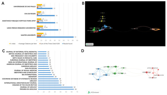
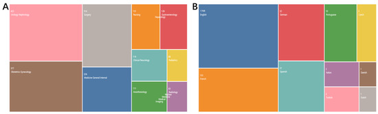
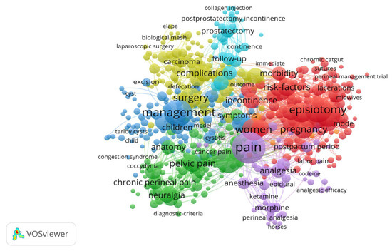
Background: Perineal pain is a painful neuropathic condition, which does not have a standard diagnostic or treatment approach. As such, we sought to evaluate the global scientific output of research into perineal pain and explore trends from 1981 to 2021 using bibliometric methods. Methods: Articles on perineal pain were retrieved from the Web of Science (WoS) database. We analyzed the content and quality of publications from within the specified timeframe. We also utilized VOSviewer to mine and cluster data from retrieved articles. Results: A total of 1917 articles were collected. The number of related papers published increased year by year. Articles were most frequently published by authors in the United States and France. Although the US remains at the center of this field, publications from China have become more frequent in recent years. We also found that French academic institutions dominate the field of perineal pain, and Jean-Jacques Labat from Nantes Universite is the most published author in the field. “Episiotomy”, “pain”, “management”, “prostatectomy”, “pelvic pain”, and “complication” were frequently cited as keywords. Conclusion: The increasing number of publications each year indicates that perineal pain has gained more attention as an important research topic.
Full article
(This article belongs to the Special Issue The Path to Personalized Pain Management)
►
Show Figures

Figure 1


