16 pages, 8886 KiB  
Article
Hydrophilic Shell Matrix Proteins of Nautilus pompilius and the Identification of a Core Set of Conchiferan Domains
by Davin H. E. Setiamarga 1,2,3,*, Kazuki Hirota 1,2, Masa-aki Yoshida 4, Yusuke Takeda 3,5, Keiji Kito 6, Makiko Ishikawa 2,7, Keisuke Shimizu 2,8, Yukinobu Isowa 2,9, Kazuho Ikeo 10, Takenori Sasaki 3 and Kazuyoshi Endo 2
1 Department of Applied Chemistry and Biochemistry, National Institute of Technology (KOSEN), Wakayama College, Gobo 644-0023, Japan
2 Graduate School of Sciences, The University of Tokyo, Bunkyo-ku, Tokyo 113-0033, Japan
3 The University Museum, The University of Tokyo, Tokyo 113-0033, Japan
4 Marine Biological Science Section, Education and Research Center for Biological Resources, Faculty of Life and Environmental Science, Shimane University, Unnan 685-0024, Japan
5 Graduate School of Science, Hokkaido University, Sapporo 060-0810, Japan
6 Department of Life Sciences, School of Agriculture, Meiji University, Kawasaki 214-8571, Japan
7 Faculty of Animal Health Technology, Yamazaki University of Animal Health Technology, Hachiouji 192-0364, Japan
8 Graduate School of Agriculture and Life Sciences, The University of Tokyo, Yayoi, Tokyo 113-8657, Japan
9 Shimoda Marine Research Center, University of Tsukuba, Shimoda 415-0025, Japan
10 Center for Information Biology, National Institute of Genetics, Mishima 411-8540, Japan
Genes 2021, 12(12), 1925; https://doi.org/10.3390/genes12121925 - 29 Nov 2021
Cited by 9 | Viewed by 4589
Abstract
Despite being a member of the shelled mollusks (Conchiferans), most members of extant cephalopods have lost their external biomineralized shells, except for the basally diverging Nautilids. Here, we report the result of our study to identify major Shell Matrix Proteins and their domains [...] Read more.
Despite being a member of the shelled mollusks (Conchiferans), most members of extant cephalopods have lost their external biomineralized shells, except for the basally diverging Nautilids. Here, we report the result of our study to identify major Shell Matrix Proteins and their domains in the Nautilid Nautilus pompilius, in order to gain a general insight into the evolution of Conchiferan Shell Matrix Proteins. In order to do so, we performed a multiomics study on the shell of N. pompilius, by conducting transcriptomics of its mantle tissue and proteomics of its shell matrix. Analyses of obtained data identified 61 distinct shell-specific sequences. Of the successfully annotated 27 sequences, protein domains were predicted in 19. Comparative analysis of Nautilus sequences with four Conchiferans for which Shell Matrix Protein data were available (the pacific oyster, the pearl oyster, the limpet and the Euhadra snail) revealed that three proteins and six protein domains were conserved in all Conchiferans. Interestingly, when the terrestrial Euhadra snail was excluded, another five proteins and six protein domains were found to be shared among the four marine Conchiferans. Phylogenetic analyses indicated that most of these proteins and domains were probably present in the ancestral Conchiferan, but employed in shell formation later and independently in most clades. Even though further studies utilizing deeper sequencing techniques to obtain genome and full-length sequences, and functional analyses, must be carried out in the future, our results here provide important pieces of information for the elucidation of the evolution of Conchiferan shells at the molecular level. Full article
(This article belongs to the Special Issue The Evolution of Invertebrate Animals)
Show Figures

Figure 1

12 pages, 1427 KiB  
Article
Genome-Wide Selective Signatures Reveal Candidate Genes Associated with Hair Follicle Development and Wool Shedding in Sheep
by Zhihui Lei 1,2,†, Weibo Sun 1,†, Tingting Guo 1, Jianye Li 1, Shaohua Zhu 2, Zengkui Lu 1, Guoyan Qiao 1, Mei Han 1, Hongchang Zhao 1, Bohui Yang 1, Liping Zhang 2, Jianbin Liu 1, Chao Yuan 1 and Yaojing Yue 1,*
1 Lanzhou Institute of Husbandry and Pharmaceutical Sciences, Chinese Academy of Agricultural Sciences, Lanzhou 730050, China
2 College of Animal Science and Technology, Gansu Agricultural University, Lanzhou 730070, China
Zhihui Lei and Weibo Sun both authors contributed equally to this work.
Genes 2021, 12(12), 1924; https://doi.org/10.3390/genes12121924 - 29 Nov 2021
Cited by 18 | Viewed by 3630
Abstract
Hair follicle development and wool shedding in sheep are poorly understood. This study investigated the population structures and genetic differences between sheep with different wool types to identify candidate genes related to these traits. We used Illumina ovine SNP 50K chip genotyping data [...] Read more.
Hair follicle development and wool shedding in sheep are poorly understood. This study investigated the population structures and genetic differences between sheep with different wool types to identify candidate genes related to these traits. We used Illumina ovine SNP 50K chip genotyping data of 795 sheep populations comprising 27 breeds with two wool types, measuring the population differentiation index (Fst), nucleotide diversity (θπ ratio), and extended haplotype homozygosity among populations (XP-EHH) to detect the selective signatures of hair sheep and fine-wool sheep. The top 5% of the Fst and θπ ratio values, and values of XP-EHH < −2 were considered strongly selected SNP sites. Annotation showed that the PRX, SOX18, TGM3, and TCF3 genes related to hair follicle development and wool shedding were strongly selected. Our results indicated that these methods identified important genes related to hair follicle formation, epidermal differentiation, and hair follicle stem cell development, and provide a meaningful reference for further study on the molecular mechanisms of economically important traits in sheep. Full article
(This article belongs to the Section Animal Genetics and Genomics)
Show Figures

Figure 1

11 pages, 1118 KiB  
Article
LTBP3 Frameshift Variant in British Shorthair Cats with Complex Skeletal Dysplasia
by Gabriela Rudd Garces 1,2,†, Anna Knebel 3,†, Kirsten Hülskötter 4,5,†, Vidhya Jagannathan 1, Theresa Störk 4, Marion Hewicker-Trautwein 4, Tosso Leeb 1,*,‡ and Holger A. Volk 3,5,‡
1 Institute of Genetics, Vetsuisse Faculty, University of Bern, 3001 Bern, Switzerland
2 Institute of Veterinary Genetics “Ing. Fernando Noel Dulout”, National University of La Plata, La Plata 1900, Argentina
3 Department of Small Animal Medicine and Surgery, University of Veterinary Medicine Hannover, 30559 Hannover, Germany
4 Department of Pathology, University of Veterinary Medicine Hannover, 30559 Hannover, Germany
5 Center for Systems Neuroscience Hannover (ZSN), 30559 Hannover, Germany
These authors contributed equally to this work.
These authors are senior authors.
Genes 2021, 12(12), 1923; https://doi.org/10.3390/genes12121923 - 29 Nov 2021
Cited by 2 | Viewed by 4551
Abstract
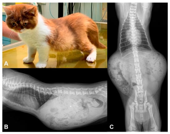
We investigated a highly inbred family of British Shorthair cats in which two offspring were affected by deteriorating paraparesis due to complex skeletal malformations. Radiographs of both affected kittens revealed vertebral deformations with marked stenosis of the vertebral canal from T11 to L3. [...] Read more.
We investigated a highly inbred family of British Shorthair cats in which two offspring were affected by deteriorating paraparesis due to complex skeletal malformations. Radiographs of both affected kittens revealed vertebral deformations with marked stenosis of the vertebral canal from T11 to L3. Additionally, compression of the spinal cord, cerebellar herniation, coprostasis and hypogangliosis were found. The pedigree suggested monogenic autosomal recessive inheritance of the trait. We sequenced the genome of an affected kitten and compared the data to 62 control genomes. This search yielded 55 private protein-changing variants of which only one was located in a likely functional candidate gene, LTBP3, encoding latent transforming growth factor β binding protein 3. This variant, c.158delG or p.(Gly53Alafs*16), represents a 1 bp frameshift deletion predicted to truncate 95% of the open reading frame. LTBP3 is a known key regulator of transforming growth factor β (TGF-β) and is involved in bone morphogenesis and remodeling. Genotypes at the LTBP3:c.158delG variant perfectly co-segregated with the phenotype in the investigated family. The available experimental data together with current knowledge on LTBP3 variants and their functional impact in human patients and mice suggest LTBP3:c.158delG as a candidate causative variant for the observed skeletal malformations in British Shorthair cats. To the best of our knowledge, this study represents the first report of LTBP3-related complex skeletal dysplasia in domestic animals. Full article
(This article belongs to the Special Issue Animal Domestication and Breeding)
Show Figures

Figure 1

15 pages, 2169 KiB  
Article
Identification and Characterization of a Novel Recurrent ERCC6 Variant in Patients with a Severe Form of Cockayne Syndrome B
by Khouloud Zayoud 1,2, Ichraf Kraoua 3, Asma Chikhaoui 1, Nadège Calmels 4,5, Sami Bouchoucha 6, Cathy Obringer 5, Clément Crochemore 7, Dorra Najjar 1, Sinda Zarrouk 8, Najoua Miladi 9, Vincent Laugel 5, Miria Ricchetti 7, Ilhem Turki 3 and Houda Yacoub-Youssef 1,*
1 Laboratory of Biomedical Genomics and Oncogenetics (LR16IPT05), Institut Pasteur de Tunis, Université Tunis El Manar, El Manar I, Tunis 1002, Tunisia
2 Faculté des Sciences de Bizerte, Bizerte 7000, Tunisia
3 LR18SP04 and Department of Child and Adolescent Neurology, National Institute Mongi Ben Hmida of Neurology, Tunis 1007, Tunisia
4 Laboratoires de Diagnostic Génétique, Institut de Génétique Médicale d’Alsace, Nouvel Hôpital Civil, Hôpitaux Universitaires de Strasbourg, 67000 Strasbourg, France
5 Laboratoire de Génétique Médicale, INSERM U1112, Institut de génétique médicale d’Alsace, CRBS, 67000 Strasbourg, France
6 Service Orthopédie, Hôpital d’enfant Béchir Hamza, Tunis 1000, Tunisia
7 Institut Pasteur, Team Stability of Nuclear and Mitochondrial DNA, Stem Cells and Development, UMR 3738 CNRS, 25-28 rue du Dr. Roux, 75015 Paris, France
8 Genomics Platform, Institut Pasteur de Tunis (IPT), Tunis-Belvédère, Tunis 1002, Tunisia
9 Maghreb Medical Center, El Manar III, Tunis 9000, Tunisia
Genes 2021, 12(12), 1922; https://doi.org/10.3390/genes12121922 - 29 Nov 2021
Cited by 9 | Viewed by 3274
Abstract
Cockayne syndrome (CS) is a rare disease caused by mutations in ERCC6/CSB or ERCC8/CSA. We report here the clinical, genetic, and functional analyses of three unrelated patients mutated in ERCC6/CSB with a severe phenotype. After clinical [...] Read more.
Cockayne syndrome (CS) is a rare disease caused by mutations in ERCC6/CSB or ERCC8/CSA. We report here the clinical, genetic, and functional analyses of three unrelated patients mutated in ERCC6/CSB with a severe phenotype. After clinical examination, two patients were investigated via next generation sequencing, targeting seventeen Nucleotide Excision Repair (NER) genes. All three patients harbored a novel, c.3156dup, homozygous mutation located in exon 18 of ERCC6/CSB that affects the C-terminal region of the protein. Sanger sequencing confirmed the mutation and the parental segregation in the three families, and Western blots showed a lack of the full-length protein. NER functional impairment was shown by reduced recovery of RNA synthesis with proficient unscheduled DNA synthesis after UV-C radiations in patient-derived fibroblasts. Despite sharing the same mutation, the clinical spectrum was heterogeneous among the three patients, and only two patients displayed clinical photosensitivity. This novel ERCC6 variant in Tunisian patients suggests a founder effect and has implications for setting-up prenatal diagnosis/genetic counselling in North Africa, where this disease is largely undiagnosed. This study reveals one of the rare cases of CS clinical heterogeneity despite the same mutation. Moreover, the occurrence of an identical homozygous mutation, which either results in clinical photosensitivity or does not, strongly suggests that this classic CS symptom relies on multiple factors. Full article
(This article belongs to the Section Human Genomics and Genetic Diseases)
Show Figures

Figure 1

17 pages, 3745 KiB  
Article
Weaving Mitochondrial DNA and Y-Chromosome Variation in the Panamanian Genetic Canvas
by Nicola Rambaldi Migliore 1,†, Giulia Colombo 1,†, Marco Rosario Capodiferro 1, Lucia Mazzocchi 1, Ana Maria Chero Osorio 1, Alessandro Raveane 1,2, Maribel Tribaldos 3, Ugo Alessandro Perego 1,4, Tomás Mendizábal 5,6, Alejandro García Montón 7, Gianluca Lombardo 1, Viola Grugni 1, Maria Garofalo 1,8, Luca Ferretti 1, Cristina Cereda 8, Stella Gagliardi 8, Richard Cooke 6,9, Nicole Smith-Guzmán 6,9, Anna Olivieri 1, Bethany Aram 7, Antonio Torroni 1, Jorge Motta 3, Ornella Semino 1,*,‡ and Alessandro Achilli 1,*,‡add Show full author list remove Hide full author list
1 Department of Biology and Biotechnology “L. Spallanzani”, University of Pavia, 27100 Pavia, Italy
2 Laboratory of Hematology-Oncology, European Institute of Oncology IRCCS, 20141 Milan, Italy
3 Gorgas Memorial Institute for Health Studies, Panama City 0816-02593, Panama
4 Department of Math and Science, Southeastern Community College, West Burlington, IA 52655, USA
5 Center for Historical, Anthropological and Cultural Research—AIP, Panama City 0816-07812, Panama
6 Smithsonian Tropical Research Institute, Panama City 0843-03092, Panama
7 Departamento de Geografía, Historia y Filosofía, Universidad Pablo de Olavide, 41013 Seville, Spain
8 Genomic and Post-Genomic Unit, IRCCS Mondino Foundation, 27100 Pavia, Italy
9 Sistema Nacional de Investigadores, Secretaría Nacional de Ciencia y Tecnología, Ciudad del Saber, Clayton 0816-02852, Panama
These authors contributed equally to this paper.
add Show full affiliation list remove Hide full affiliation list
Genes 2021, 12(12), 1921; https://doi.org/10.3390/genes12121921 - 29 Nov 2021
Cited by 4 | Viewed by 8585
Abstract
The Isthmus of Panama was a crossroads between North and South America during the continent’s first peopling (and subsequent movements) also playing a pivotal role during European colonization and the African slave trade. Previous analyses of uniparental systems revealed significant sex biases in [...] Read more.
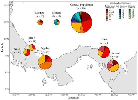
The Isthmus of Panama was a crossroads between North and South America during the continent’s first peopling (and subsequent movements) also playing a pivotal role during European colonization and the African slave trade. Previous analyses of uniparental systems revealed significant sex biases in the genetic history of Panamanians, as testified by the high proportions of Indigenous and sub-Saharan mitochondrial DNAs (mtDNAs) and by the prevalence of Western European/northern African Y chromosomes. Those studies were conducted on the general population without considering any self-reported ethnic affiliations. Here, we compared the mtDNA and Y-chromosome lineages of a new sample collection from 431 individuals (301 males and 130 females) belonging to either the general population, mixed groups, or one of five Indigenous groups currently living in Panama. We found different proportions of paternal and maternal lineages in the Indigenous groups testifying to pre-contact demographic events and genetic inputs (some dated to Pleistocene times) that created genetic structure. Then, while the local mitochondrial gene pool was marginally involved in post-contact admixtures, the Indigenous Y chromosomes were differentially replaced, mostly by lineages of western Eurasian origin. Finally, our new estimates of the sub-Saharan contribution, on a more accurately defined general population, reduce an apparent divergence between genetic and historical data. Full article
(This article belongs to the Special Issue The Peopling of the Americas: A Genetic Perspective)
Show Figures

Figure 1

12 pages, 2883 KiB  
Article
The Adenoviral E1B-55k Protein Present in HEK293 Cells Mediates Abnormal Accumulation of Key WNT Signaling Proteins in Large Cytoplasmic Aggregates
by Petter Angell Olsen 1,2,* and Stefan Krauss 1,2
1 Hybrid Technology Hub-Centre of Excellence, Institute of Basic Medical Sciences, University of Oslo, 0317 Oslo, Norway
2 Unit for Cell Signaling, Department of Immunology and Transfusion Medicine, Oslo University Hospital, P.O. Box 4950, Nydalen, 0424 Oslo, Norway
Genes 2021, 12(12), 1920; https://doi.org/10.3390/genes12121920 - 29 Nov 2021
Cited by 5 | Viewed by 3417
Abstract
HEK293 cells are one of the most widely used cell lines in research, and HEK293 cells are frequently used as an in vitro model for studying the WNT signaling pathway. The HEK293 cell line was originally established by transfection of human embryonic kidney [...] Read more.
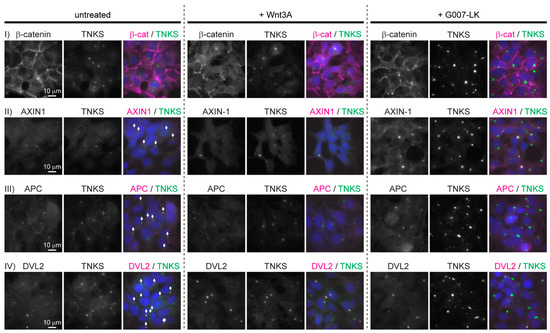
HEK293 cells are one of the most widely used cell lines in research, and HEK293 cells are frequently used as an in vitro model for studying the WNT signaling pathway. The HEK293 cell line was originally established by transfection of human embryonic kidney cells with sheared adenovirus 5 DNA, and it is known that that HEK293 cells stably express the adenoviral E1A and E1B-55k proteins. Here, we show that HEK293 cells display an unexpected distribution of key components of the WNT/β-catenin signaling pathway where AXIN1, APC, DVL2 and tankyrase are all co-localized in large spherical cytoplasmic aggregates. The cytoplasmic aggregates are enclosed by a narrow layer of the adenoviral E1B-55k protein. The reduction of E1B-55k protein levels leads to the disappearance of the cytoplasmic aggregates thus corroborating an essential role of the E1B-55k protein in mediating the formation of the aggregates. Furthermore, HEK293 cells with reduced E1B-55k protein levels display reduced levels of transcriptional activation of WNT/β-catenin signaling upon stimulation by the Wnt3A agonist. The demonstrated influence of the E1B-55k protein on the cellular localization of WNT/β-catenin signaling components and on transcriptional regulation of WNT/β-catenin signaling asks for caution in the interpretation of data derived from the HEK293 cell line. Full article
(This article belongs to the Special Issue Signaling Pathway of Cancer)
Show Figures

Figure 1

18 pages, 1621 KiB  
Review
Role and Regulation of the RECQL4 Family during Genomic Integrity Maintenance
by Thong T. Luong and Kara A. Bernstein *
Department of Pharmacology and Chemical Biology, UPMC Hillman Cancer Center, University of Pittsburgh School of Medicine, 5117 Centre Avenue, Pittsburgh, PA 15213, USA
Genes 2021, 12(12), 1919; https://doi.org/10.3390/genes12121919 - 29 Nov 2021
Cited by 21 | Viewed by 4802
Abstract
RECQL4 is a member of the evolutionarily conserved RecQ family of 3’ to 5’ DNA helicases. RECQL4 is critical for maintaining genomic stability through its functions in DNA repair, recombination, and replication. Unlike many DNA repair proteins, RECQL4 has unique functions in many [...] Read more.
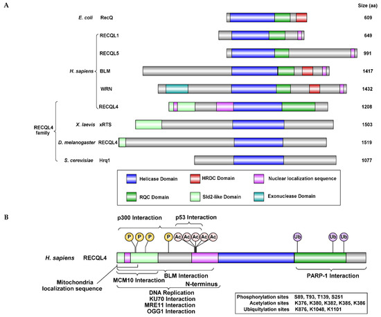
RECQL4 is a member of the evolutionarily conserved RecQ family of 3’ to 5’ DNA helicases. RECQL4 is critical for maintaining genomic stability through its functions in DNA repair, recombination, and replication. Unlike many DNA repair proteins, RECQL4 has unique functions in many of the central DNA repair pathways such as replication, telomere, double-strand break repair, base excision repair, mitochondrial maintenance, nucleotide excision repair, and crosslink repair. Consistent with these diverse roles, mutations in RECQL4 are associated with three distinct genetic diseases, which are characterized by developmental defects and/or cancer predisposition. In this review, we provide an overview of the roles and regulation of RECQL4 during maintenance of genome homeostasis. Full article
(This article belongs to the Special Issue Mechanisms of DNA Damage, Repair and Mutagenesis)
Show Figures

Figure 1

6 pages, 682 KiB  
Article
Should Patients with Kearns-Sayre Syndrome and Corneal Endothelial Failure Be Genotyped for a TCF4 Trinucleotide Repeat, Commonly Associated with Fuchs Endothelial Corneal Dystrophy?
by Lubica Dudakova 1, Pavlina Skalicka 1,2, Alice E. Davidson 3,4, Amanda N. Sadan 3, Monika Chylova 1, Helena Jahnova 1, Nicole Anteneova 1, Marketa Tesarova 1, Tomas Honzik 1 and Petra Liskova 1,2,3,*
1 Department of Paediatrics and Inherited Metabolic Disorders, First Faculty of Medicine, Charles University and General University Hospital in Prague, 128 08 Prague, Czech Republic
2 Department of Ophthalmology, First Faculty of Medicine, Charles University and General University Hospital in Prague, 128 08 Prague, Czech Republic
3 UCL Institute of Ophthalmology, London EC1V 9EL, UK
4 Moorfields Eye Hospital, London EC1V 2PD, UK
Genes 2021, 12(12), 1918; https://doi.org/10.3390/genes12121918 - 29 Nov 2021
Viewed by 2450
Abstract
The aim of this study was to describe the ocular phenotype in a case with Kearns-Sayre syndrome (KSS) spectrum and to determine if corneal endothelial cell dysfunction could be attributed to other known distinct genetic causes. Herein, genomic DNA was extracted from blood [...] Read more.
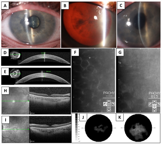
The aim of this study was to describe the ocular phenotype in a case with Kearns-Sayre syndrome (KSS) spectrum and to determine if corneal endothelial cell dysfunction could be attributed to other known distinct genetic causes. Herein, genomic DNA was extracted from blood and exome sequencing was performed. Non-coding gene regions implicated in corneal endothelial dystrophies were screened by Sanger sequencing. In addition, a repeat expansion situated within an intron of TCF4 (termed CTG18.1) was genotyped using the short tandem repeat assay. The diagnosis of KSS spectrum was based on the presence of ptosis, chronic progressive external ophthalmoplegia, pigmentary retinopathy, hearing loss, and muscle weakness, which were further supported by the detection of ~6.5 kb mtDNA deletion. At the age of 33 years, the proband’s best corrected visual acuity was reduced to 0.04 in the right eye and 0.2 in the left eye. Rare ocular findings included marked corneal oedema with central corneal thickness of 824 and 844 µm in the right and left eye, respectively. No pathogenic variants in the genes, which are associated with corneal endothelial dystrophies, were identified. Furthermore, the CTG18.1 genotype was 12/33, which exceeds a previously determined critical threshold for toxic RNA foci appearance in corneal endothelial cells. Full article
(This article belongs to the Special Issue Genetic Research in Metabolic Diseases)
Show Figures

Figure 1

7 pages, 2449 KiB  
Brief Report
BRCA1/2 NGS Somatic Testing in Clinical Practice: A Short Report
by Francesco Pepe 1,†, Pasquale Pisapia 1,†, Gianluca Russo 1, Mariantonia Nacchio 1, Pierlorenzo Pallante 2, Elena Vigliar 1, Carmine De Angelis 3, Luigi Insabato 4, Claudio Bellevicine 1, Sabino De Placido 3, Giancarlo Troncone 1,* and Umberto Malapelle 1
1 Department of Public Health, University of Naples Federico II, 80131 Naples, Italy
2 Institute for Experimental Endocrinology and Oncology (IEOS) “G. Salvatore”, National Research Council (CNR), 80131 Naples, Italy
3 Department of Clinical Medicine and Surgery, University of Naples Federico II, 80131 Naples, Italy
4 Department of Advanced Biomedical Sciences, School of Medicine, University of Naples Federico II, 80131 Naples, Italy
These Authors contributed equally as first authors.
Genes 2021, 12(12), 1917; https://doi.org/10.3390/genes12121917 - 28 Nov 2021
Cited by 4 | Viewed by 3181
Abstract
High-grade serous ovarian carcinoma (HGSOC) is the most common subtype of all ovarian carcinomas. HGSOC harboring BRCA1/2 germline or somatic mutations are sensitive to the poly (adenosine diphosphate-ribose) polymerase inhibitors (PARPi). Therefore, detecting these mutations is crucial to identifying patients for [...] Read more.
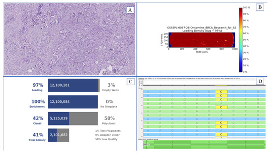
High-grade serous ovarian carcinoma (HGSOC) is the most common subtype of all ovarian carcinomas. HGSOC harboring BRCA1/2 germline or somatic mutations are sensitive to the poly (adenosine diphosphate-ribose) polymerase inhibitors (PARPi). Therefore, detecting these mutations is crucial to identifying patients for PARPi-targeted treatment. In the clinical setting, next generation sequencing (NGS) has proven to be a reliable diagnostic approach BRCA1/2 molecular evaluation. Here, we review the results of our BRCA1/2 NGS analysis obtained in a year and a half of diagnostic routine practice. BRCA1/2 molecular NGS records of HGSOC patients were retrieved from our institutional archive covering the period from January 2020 to September 2021. NGS analysis was performed on the Ion S5™ System (Thermo Fisher Scientific, Waltham, MA, USA) with the Oncomine™ BRCA Research Assay panel (Thermo Fisher Scientific). Variants were classified as pathogenic or likely pathogenic according to the guidelines of the American College of Medical Genetics and Genomics by using the inspection of Evidence-based Network for the Interpretation of Germline Mutant Alleles (ENIGMA) and ClinVar (NCBI) databases. Sixty-five HGSOC patient samples were successfully analyzed. Overall, 11 (16.9%) out of 65 cases harbored a pathogenic alteration in BRCA1/2, in particular, six BRCA1 and five BRCA2 pathogenic variations. This study confirms the efficiency and high sensitivity of NGS analysis in detecting BRCA1/2 germline or somatic variations in patients with HGSOC. Full article
(This article belongs to the Special Issue Deciphering Epigenetic Signature in Human Health and Disease)
Show Figures

Figure 1

11 pages, 1878 KiB  
Article
The Decisive Case-Control Study Elaborates the Null Association between ADAMTS5 rs226794 and Osteoarthritis in Asians: A Case-Control Study and Meta-Analysis
by Chung-Cheng Kao 1, Hsiang-En Hsu 2, Yi-Chou Chen 3,4, Ming-Yu Tu 5, Su-Wen Chuang 2,6 and Sui-Lung Su 2,*
1 Tri-Service General Hospital Songshan Branch, National Defense Medical Center, Taipei 105309, Taiwan
2 School of Public Health, National Defense Medical Center, Taipei 114201, Taiwan
3 Department of Orthopedics, Taoyuan General Hospital, Ministry of Health and Welfare, Taoyuan 325208, Taiwan
4 Institute of Medical Sciences, National Defense Medical Center, Taipei 114201, Taiwan
5 Department of Orthopedics, Kaohsiung Armed Forces General Hospital Gangshan Branch, Kaohsiung 820004, Taiwan
6 Graduate Institute of Life Sciences, National Defense Medical Center, Taipei 114201, Taiwan
Genes 2021, 12(12), 1916; https://doi.org/10.3390/genes12121916 - 28 Nov 2021
Cited by 3 | Viewed by 2297
Abstract
Background: Osteoarthritis is an important health issue for the elderly. Many studies indicate that genetics is an important risk factor for osteoarthritis, and a disintegrin and metalloproteinase with thrombospondin motifs 5 (ADAMTS5) is one gene that is most frequently implicated. Many [...] Read more.
Background: Osteoarthritis is an important health issue for the elderly. Many studies indicate that genetics is an important risk factor for osteoarthritis, and a disintegrin and metalloproteinase with thrombospondin motifs 5 (ADAMTS5) is one gene that is most frequently implicated. Many recent studies have examined the relationship between a polymorphism in the ADAMTS5 gene (rs226794) and the risk for developing osteoarthritis without definitive results. Objective: In this case-control study, we examined the correlation between the ADAMTS5 gene polymorphism, rs226794, and knee osteoarthritis. We used a meta-analysis and trial sequential analysis to determine whether ADAMTS5 rs226794 expression increases susceptibility to osteoarthritis. Methods: This study consisted of two parts: a case-control study and a meta-analysis. The case-control study included subjects who underwent knee radiography at the Health Examination Center of the Tri Service General Hospital from 2015 to 2019. The Kellgren–Lawrence (KL) grading system was used as diagnostic criteria. Patients with unsuccessful gene sequencing were excluded. There were 606 subjects in the knee osteoarthritis group (KL ≥ 2) and 564 in the control group (KL < 2). Gene sequencing was performed using iPLEX Gold to determine the association between the gene polymorphism of ADAMTS5 rs226794 and knee osteoarthritis. For the meta-analysis, databases such as PubMed, Embase, and Cochrane were queried to identify studies that examined the relationship between ADAMTS5 rs226794 and osteoarthritis. Next, the findings of the meta-analysis were incorporated with the results of the case-control study and samples from the published studies to estimate the association between the genetic polymorphism and osteoarthritis using an odds ratio and a 95% confidence interval. Results: We found a non-significant association between the G allele and knee OA (crude-OR: 0.93 (95% CI: 0.79–1.10) and adjusted-OR: 1.02 (95% CI: 0.76–1.36) in the allele model) in the present study, and the analysis of other genetic models revealed a similar trend. After including five published studies and our case-control study, the results with 2866 Asians indicated a conclusively null association between ADAMTS5 rs226794 and knee OA) OR: 1.09 (95% CI: 0.93–1.26). The results for Caucasians also revealed a null association (OR: 1.21 (95% CI: 0.81–1.82)). Conclusions: This study indicates that the gene polymorphism, ADAMTS5 rs226794, is not significantly associated with knee osteoarthritis. Additionally, assuming that the cumulative sample size in the allele model is sufficient, we confirmed that the G allele is not a risk factor for osteoarthritis. This study integrated all available evidence to arrive at this conclusion, and it suggests that no additional studies are necessary. Full article
(This article belongs to the Section Human Genomics and Genetic Diseases)
Show Figures

Figure 1

9 pages, 681 KiB  
Article
Exome Sequencing Identifies a Novel FBN1 Variant in a Pakistani Family with Marfan Syndrome That Includes Left Ventricle Diastolic Dysfunction
by Nadia Farooqi 1, Louise A. Metherell 2, Isabelle Schrauwen 3, Anushree Acharya 3, Qayum Khan 1, Liz M. Nouel Saied 3, Yasir Ali 1, Hamed A. El-Serehy 4, Fazal Jalil 1,* and Suzanne M. Leal 3,5,*
1 Department of Biotechnology, Faculty of Chemical and Life Sciences, Abdul Wali Khan University, Mardan 23200, Pakistan
2 Centre for Endocrinology, William Harvey Research Institute, Charterhouse Square Campus, Barts and The London School of Medicine and Dentistry, Queen Mary University of London, London EC1M 6BQ, UK
3 Center for Statistical Genetics, Gertrude H. Sergievsky Center, Department of Neurology, Columbia University Medical Center, New York, NY 10032, USA
4 Department of Zoology, College of Science, King Saud University, Riyadh I1451, Saudi Arabia
5 Taub Institute for Alzheimer’s Disease and the Aging Brain, Columbia University Medical Center, New York, NY 10032, USA
Genes 2021, 12(12), 1915; https://doi.org/10.3390/genes12121915 - 28 Nov 2021
Cited by 1 | Viewed by 3144
Abstract
Introduction: Cardiomyopathies are diseases of the heart muscle and are important causes of heart failure. Dilated cardiomyopathy (DCM) is a common form of cardiomyopathy that can be acquired, syndromic or non-syndromic. The current study was conducted to explore the genetic defects in a [...] Read more.
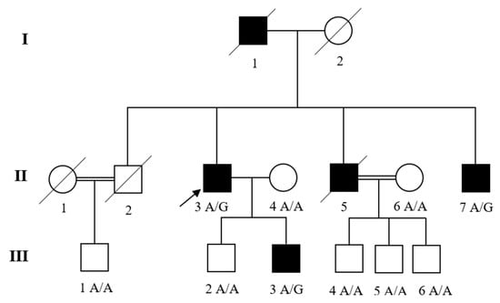
Introduction: Cardiomyopathies are diseases of the heart muscle and are important causes of heart failure. Dilated cardiomyopathy (DCM) is a common form of cardiomyopathy that can be acquired, syndromic or non-syndromic. The current study was conducted to explore the genetic defects in a Pakistani family with cardiac disease and features of Marfan’s syndrome (MFS). Methods: A family with left ventricle (LV) diastolic dysfunction and MFS phenotype was assessed in Pakistan. The clinical information and blood samples from the patients were collected after physical, cardiovascular, and ophthalmologic examinations. An affected individual (proband) was subjected to whole-exome sequencing (WES). The findings were further validated through Sanger sequencing in the family. Results: Through WES and sanger validation, we identified a novel variant NM_000138.4; c.1402A>G in the Fibrillin-1 (FBN1) gene that segregates with LV diastolic dysfunction and MFS. Furthermore, bioinformatic evaluation suggested that the novel variant is deleterious and disease-causing. Conclusions: This study identified for the first time a novel FBN1 variant in a family with LV diastolic dysfunction and MFS in Pakistan. Full article
(This article belongs to the Special Issue Cardiovascular Disease Risk Factors, Genetics, and Prevention)
Show Figures

Figure 1

10 pages, 1310 KiB  
Article
TNFA and IL10 Polymorphisms and IL-6 and IL-10 Levels Influence Disease Severity in Influenza A(H1N1)pdm09 Virus Infected Patients
by Kalichamy Alagarasu 1, Himanshu Kaushal 1, Pooja Shinde 1, Mahadeo Kakade 1, Urmila Chaudhary 1, Vikram Padbidri 2, Shashikala A. Sangle 3, Sonali Salvi 3, Ashish R. Bavdekar 4, Pradeep D’costa 4 and Manohar Lal Choudhary 1,*
1 ICMR-National Institute of Virology, Pune 411001, India
2 Jehangir Hospital Research Center, Pune 411001, India
3 Department of Medicine, BJ Medical College, Pune 411001, India
4 KEM Hospital Research Center, Pune 411001, India
Genes 2021, 12(12), 1914; https://doi.org/10.3390/genes12121914 - 28 Nov 2021
Cited by 16 | Viewed by 2558
Abstract
Cytokines are key modulators of immune response, and dysregulated production of proinflammatory and anti-inflammatory cytokines contributes to the pathogenesis of influenza A(H1N1)pdm09 virus infection. Cytokine production is impacted by single nucleotide polymorphisms (SNPs) in the genes coding for them. In the present study, [...] Read more.
Cytokines are key modulators of immune response, and dysregulated production of proinflammatory and anti-inflammatory cytokines contributes to the pathogenesis of influenza A(H1N1)pdm09 virus infection. Cytokine production is impacted by single nucleotide polymorphisms (SNPs) in the genes coding for them. In the present study, SNPs in the IL6, TNFA, IFNG, IL17A, IL10, and TGFB were investigated for their association with disease severity and fatality in influenza A(H1N1)pdm09-affected patients with mild disease (n = 293) and severe disease (n = 86). Among those with severe disease, 41 patients had fatal outcomes. In a subset of the patients, levels of IL-2, IL-4, IL-6, IL-10, TNF, IFN-γ, and IL-17 were assayed in the plasma for their association with severe disease. The frequency of TNFA rs1800629 G/A allele was significantly higher in severe cases and survived severe cases group compared to that of those with mild infection (OR with 95% for mild vs. severe cases 2.95 (1.52–5.73); mild vs. survived severe cases 4.02 (1.84–8.82)). IL10 rs1800896-rs1800872 G-C haplotype was significantly lower (OR with 95% 0.34 (0.12–0.95)), while IL10 rs1800896-rs1800872 G-A haplotype was significantly higher (OR with 95% 12.11 (2.23–76.96)) in fatal cases group compared to that of the mild group. IL-6 and IL-10 levels were significantly higher in fatal cases compared to that of survived severe cases. IL-6 levels had greater discriminatory power than IL-10 to predict progression to fatal outcome in influenza A(H1N1)pdm09 virus-infected patients. To conclude, the present study reports the association of TNFA and IL10 SNPs with severe disease in Influenza A(H1N1)pdm09 virus-infected subjects. Furthermore, IL-6 levels can be a potential biomarker for predicting fatal outcomes in Influenza A(H1N1)pdm09 virus infected subjects. Full article
(This article belongs to the Section Molecular Genetics and Genomics)
Show Figures

Graphical abstract

18 pages, 2083 KiB  
Article
The Effects of Sequence Length and Composition of Random Sequence Peptides on the Growth of E. coli Cells
by Johana F. Castro and Diethard Tautz *
Max Planck Institute for Evolutionary Biology, August-Thienemann Strasse 2, 24306 Plön, Germany
Genes 2021, 12(12), 1913; https://doi.org/10.3390/genes12121913 - 28 Nov 2021
Cited by 12 | Viewed by 3099
Abstract
We study the potential for the de novo evolution of genes from random nucleotide sequences using libraries of E. coli expressing random sequence peptides. We assess the effects of such peptides on cell growth by monitoring frequency changes in individual clones in a [...] Read more.
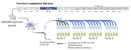
We study the potential for the de novo evolution of genes from random nucleotide sequences using libraries of E. coli expressing random sequence peptides. We assess the effects of such peptides on cell growth by monitoring frequency changes in individual clones in a complex library through four serial passages. Using a new analysis pipeline that allows the tracing of peptides of all lengths, we find that over half of the peptides have consistent effects on cell growth. Across nine different experiments, around 16% of clones increase in frequency and 36% decrease, with some variation between individual experiments. Shorter peptides (8–20 residues), are more likely to increase in frequency, longer ones are more likely to decrease. GC content, amino acid composition, intrinsic disorder, and aggregation propensity show slightly different patterns between peptide groups. Sequences that increase in frequency tend to be more disordered with lower aggregation propensity. This coincides with the observation that young genes with more disordered structures are better tolerated in genomes. Our data indicate that random sequences can be a source of evolutionary innovation, since a large fraction of them are well tolerated by the cells or can provide a growth advantage. Full article
(This article belongs to the Special Issue How Do New Genes Originate and Evolve?)
Show Figures

Figure 1

16 pages, 2369 KiB  
Article
Genome-Wide Analysis of DNA Methylation in Buccal Cells of Children Conceived through IVF and ICSI
by Bastien Ducreux 1, Jean Frappier 1,2, Céline Bruno 1,2, Abiba Doukani 3, Magali Guilleman 1,2, Emmanuel Simon 1,4, Aurélie Martinaud 1,2, Déborah Bourc’his 5, Julie Barberet 1,2 and Patricia Fauque 1,2,*
1 Equipe Génétique des Anomalies du Développement (GAD), Université Bourgogne Franche-Comté, INSERM UMR1231, 2 Rue Angélique Ducoudray, F-21000 Dijon, France
2 Laboratoire de Biologie de la Reproduction—CECOS, CHU Dijon Bourgogne, 14 Rue Gaffarel, F-21000 Dijon, France
3 Faculté de Médecine, Sorbonne Université, UMS 37 PASS Plateforme P3S, 91, Bd de l’hôpital, F-75634 Paris, France
4 Service de Gynécologie-Obstétrique, CHU Dijon Bourgogne, 14 Rue Gaffarel, F-21000 Dijon, France
5 Institut Curie, PSL University, CNRS, INSERM, 26 Rue d’Ulm, F-75248 Paris, France
Genes 2021, 12(12), 1912; https://doi.org/10.3390/genes12121912 - 28 Nov 2021
Cited by 10 | Viewed by 3272
Abstract
Early life periconceptional exposures during assisted reproductive technology (ART) procedures could alter the DNA methylation profiles of ART children, notably in imprinted genes and repetitive elements. At the genome scale, DNA methylation differences have been reported in ART conceptions at birth, but it [...] Read more.
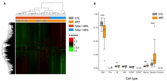
Early life periconceptional exposures during assisted reproductive technology (ART) procedures could alter the DNA methylation profiles of ART children, notably in imprinted genes and repetitive elements. At the genome scale, DNA methylation differences have been reported in ART conceptions at birth, but it is still unclear if those differences remain at childhood. Here, we performed an epigenome-wide DNA methylation association study using Illumina InfiniumEPIC BeadChip to assess the effects of the mode of conception on the methylome of buccal cells from 7- to 8-year-old children (48 children conceived after ART or naturally (control, CTL)) and according to the embryo culture medium in which they were conceived. We identified 127 differentially methylated positions (DMPs) and 16 differentially methylated regions (DMRs) (FDR < 0.05) with low delta beta differences between the two groups (ART vs. CTL). DMPs were preferentially located inside promoter proximal regions and CpG islands and were mostly hypermethylated with ART. We highlighted that the use of distinct embryo culture medium was not associated with DNA methylation differences in childhood. Overall, we bring additional evidence that children conceived via ART display limited genome-wide DNA methylation variation compared with those conceived naturally. Full article
(This article belongs to the Special Issue Epigenetic Safety after Assisted Reproductive Technologies)
Show Figures

Figure 1

8 pages, 731 KiB  
Article
WDR13: A Novel Gene Implicated in Non-Syndromic Intellectual Disability
by Sylwia Rzońca-Niewczas 1,*, Jolanta Wierzba 2, Ewa Kaczorowska 3, Milena Poryszewska 1, Joanna Kosińska 4, Piotr Stawiński 4, Rafał Płoski 4 and Jerzy Bal 1
1 Department of Medical Genetics, Institute of Mother and Child, 01-211 Warsaw, Poland
2 Department of Internal and Pediatric Nursing, Faculty of Health Sciences with Institute of Maritime and Tropical Medicine, Medical University of Gdansk, 80-210 Gdansk, Poland
3 Department of Biology and Medical Genetics, Medical University of Gdansk, 80-211 Gdansk, Poland
4 Department of Medical Genetics, Warsaw Medical University, 02-106 Warsaw, Poland
Genes 2021, 12(12), 1911; https://doi.org/10.3390/genes12121911 - 28 Nov 2021
Viewed by 2354
Abstract
Investigating novel genetic variants involved in intellectual disability (ID) development is essential. X-linked intellectual disability (XLID) accounts for over 10% of all cases of ID in males. XLID genes are involved in many cellular pathways and processes. Some of them are not specific [...] Read more.
Investigating novel genetic variants involved in intellectual disability (ID) development is essential. X-linked intellectual disability (XLID) accounts for over 10% of all cases of ID in males. XLID genes are involved in many cellular pathways and processes. Some of them are not specific to the development and functioning of the neural system. The implementation of exome sequencing simplifies the search for novel variants, especially those less expected. Here, we describe a nonsense variant of the XLID gene, WDR13. The mutation c.757C>T (p.Arg253Ter) was uncovered by X-chromosome exome sequencing in males with a familial form of intellectual disability. Quantitative PCR (qPCR) analysis showed that variant c.757C>T caused a significant decrease in WDR13 expression in the patient's fibroblast. Moreover, it dysregulated other genes linked to intellectual disability, such as FMR1, SYN1, CAMK2A, and THOC2. The obtained results indicate the pathogenic nature of the detected variant and suggest that the WDR13 gene interacts with other genes essential for the functioning of the nervous system, especially the synaptic plasticity process. Full article
(This article belongs to the Section Human Genomics and Genetic Diseases)
Show Figures

Figure 1