24 pages, 1299 KiB  
Review
Importance, Applications and Features of Assays Measuring SARS-CoV-2 Neutralizing Antibodies
by Pia Gattinger 1, Anna Ohradanova-Repic 2 and Rudolf Valenta 1,3,4,5,*
1 Department of Pathophysiology and Allergy Research, Center for Pathophysiology, Infectiology and Immunology, Medical University of Vienna, 1090 Vienna, Austria
2 Institute for Hygiene and Applied Immunology, Center for Pathophysiology, Infectiology and Immunology, Medical University of Vienna, 1090 Vienna, Austria
3 Karl Landsteiner University, 3500 Krems an der Donau, Austria
4 Laboratory for Immunopathology, Department of Clinical Immunology and Allergology, Sechenov First Moscow State Medical University, 119435 Moscow, Russia
5 NRC Institute of Immunology FMBA of Russia, 115478 Moscow, Russia
Int. J. Mol. Sci. 2023, 24(6), 5352; https://doi.org/10.3390/ijms24065352 - 10 Mar 2023
Cited by 18 | Viewed by 5254
Abstract
More than three years ago, the Severe Acute Respiratory Syndrome Coronavirus 2 (SARS-CoV-2) caused the unforeseen COVID-19 pandemic with millions of deaths. In the meantime, SARS-CoV-2 has become endemic and is now part of the repertoire of viruses causing seasonal severe respiratory infections. [...] Read more.
More than three years ago, the Severe Acute Respiratory Syndrome Coronavirus 2 (SARS-CoV-2) caused the unforeseen COVID-19 pandemic with millions of deaths. In the meantime, SARS-CoV-2 has become endemic and is now part of the repertoire of viruses causing seasonal severe respiratory infections. Due to several factors, among them the development of SARS-CoV-2 immunity through natural infection, vaccination and the current dominance of seemingly less pathogenic strains belonging to the omicron lineage, the COVID-19 situation has stabilized. However, several challenges remain and the possible new occurrence of highly pathogenic variants remains a threat. Here we review the development, features and importance of assays measuring SARS-CoV-2 neutralizing antibodies (NAbs). In particular we focus on in vitro infection assays and molecular interaction assays studying the binding of the receptor binding domain (RBD) with its cognate cellular receptor ACE2. These assays, but not the measurement of SARS-CoV-2-specific antibodies per se, can inform us of whether antibodies produced by convalescent or vaccinated subjects may protect against the infection and thus have the potential to predict the risk of becoming newly infected. This information is extremely important given the fact that a considerable number of subjects, in particular vulnerable persons, respond poorly to the vaccination with the production of neutralizing antibodies. Furthermore, these assays allow to determine and evaluate the virus-neutralizing capacity of antibodies induced by vaccines and administration of plasma-, immunoglobulin preparations, monoclonal antibodies, ACE2 variants or synthetic compounds to be used for therapy of COVID-19 and assist in the preclinical evaluation of vaccines. Both types of assays can be relatively quickly adapted to newly emerging virus variants to inform us about the magnitude of cross-neutralization, which may even allow us to estimate the risk of becoming infected by newly appearing virus variants. Given the paramount importance of the infection and interaction assays we discuss their specific features, possible advantages and disadvantages, technical aspects and not yet fully resolved issues, such as cut-off levels predicting the degree of in vivo protection. Full article
Show Figures

Figure 1

12 pages, 1557 KiB  
Article
Baseline Susceptibility, Cross-Resistance, and Sublethal Effects of Broflanilide, a Novel Meta-Diamide Pesticide, in Spodoptera litura
by Yunyi Li 1,2,†, Cheng Qu 2,†, Qinghe Zhang 2, Liping Zhang 1,2,*, Chen Luo 2 and Ran Wang 2,*
1 Institute of Cotton Research, Shanxi Agricultural University, Yuncheng 044000, China
2 Institute of Plant Protection, Beijing Academy of Agriculture and Forestry Sciences, Beijing 100097, China
These authors contributed equally to this work.
Int. J. Mol. Sci. 2023, 24(6), 5351; https://doi.org/10.3390/ijms24065351 - 10 Mar 2023
Cited by 15 | Viewed by 3141
Abstract
Spodoptera litura is a damaging and notorious insect pest of agricultural crops that has developed resistance to various insecticides. Broflanilide is a novel pesticide with a unique mode of action that displays high efficiency against lepidopterous larvae. We here determined the baseline susceptibility [...] Read more.
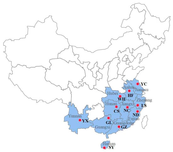
Spodoptera litura is a damaging and notorious insect pest of agricultural crops that has developed resistance to various insecticides. Broflanilide is a novel pesticide with a unique mode of action that displays high efficiency against lepidopterous larvae. We here determined the baseline susceptibility of a laboratory strain of S. litura to broflanilide and 10 other popular insecticides. Furthermore, we measured susceptibility and cross-resistance using three common insecticides in 11 field-collected S. litura populations. Broflanilide caused the highest toxicity among all tested insecticides, with the laboratory strain and all field-collected populations showing high susceptibility. Moreover, no cross-resistance was detected between broflanilide and the other tested insecticides. We subsequently evaluated the sublethal effects of broflanilide and found that treatment with the 25% lethal concentration (LC25) prolonged the development duration in the larvae, reduced the pupation rate and pupae weight, and decreased egg hatchability. Finally, the activities of three detoxifying enzymes were measured in S. litura after treatment with the LC25 dose. The results suggested that enhanced cytochrome P450 monooxygenase (P450) activity could be involved in broflanilide detoxification. Overall, these findings demonstrate the strong toxicity and significant sublethal effects of broflanilide in S. litura and suggest that increased P450 activity may be associated with broflanilide detoxification. Full article
(This article belongs to the Special Issue Pesticides Exposure and Toxicity)
Show Figures

Figure 1

23 pages, 1206 KiB  
Review
Bottom-Up Proteomics: Advancements in Sample Preparation
by Van-An Duong and Hookeun Lee *
College of Pharmacy, Gachon University, Incheon 21936, Republic of Korea
Int. J. Mol. Sci. 2023, 24(6), 5350; https://doi.org/10.3390/ijms24065350 - 10 Mar 2023
Cited by 82 | Viewed by 16531
Abstract
Liquid chromatography–tandem mass spectrometry (LC–MS/MS)-based proteomics is a powerful technique for profiling proteomes of cells, tissues, and body fluids. Typical bottom-up proteomic workflows consist of the following three major steps: sample preparation, LC–MS/MS analysis, and data analysis. LC–MS/MS and data analysis techniques have [...] Read more.
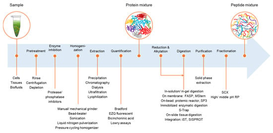
Liquid chromatography–tandem mass spectrometry (LC–MS/MS)-based proteomics is a powerful technique for profiling proteomes of cells, tissues, and body fluids. Typical bottom-up proteomic workflows consist of the following three major steps: sample preparation, LC–MS/MS analysis, and data analysis. LC–MS/MS and data analysis techniques have been intensively developed, whereas sample preparation, a laborious process, remains a difficult task and the main challenge in different applications. Sample preparation is a crucial stage that affects the overall efficiency of a proteomic study; however, it is prone to errors and has low reproducibility and throughput. In-solution digestion and filter-aided sample preparation are the typical and widely used methods. In the past decade, novel methods to improve and facilitate the entire sample preparation process or integrate sample preparation and fractionation have been reported to reduce time, increase throughput, and improve reproducibility. In this review, we have outlined the current methods used for sample preparation in proteomics, including on-membrane digestion, bead-based digestion, immobilized enzymatic digestion, and suspension trapping. Additionally, we have summarized and discussed current devices and methods for integrating different steps of sample preparation and peptide fractionation. Full article
(This article belongs to the Special Issue Advances in Mass Spectrometry-Based Proteomics)
Show Figures

Figure 1

27 pages, 2334 KiB  
Review
The Roles of Secreted Wnt Ligands in Cancer
by Johannes Werner 1,2,†, Kim E. Boonekamp 1,†, Tianzuo Zhan 3,4,5,‡ and Michael Boutros 1,5,*,‡
1 Division of Signaling and Functional Genomics, German Cancer Research Center (DKFZ), Medical Faculty Mannheim, Heidelberg University, 69120 Heidelberg, Germany
2 Medical Faculty Heidelberg, Heidelberg University, 69120 Heidelberg, Germany
3 Department of Medicine II, Medical Faculty Mannheim, Mannheim University Hospital, Heidelberg University, D-68167 Mannheim, Germany
4 Mannheim Cancer Center, Medical Faculty Mannheim, Heidelberg University, D-68167 Mannheim, Germany
5 DKFZ-Hector Cancer Institute, University Medical Center Mannheim, D-68167 Mannheim, Germany
These authors contributed equally to this work.
These authors also contributed equally to this work.
Int. J. Mol. Sci. 2023, 24(6), 5349; https://doi.org/10.3390/ijms24065349 - 10 Mar 2023
Cited by 21 | Viewed by 4345
Abstract
Wnt ligands are secreted signaling proteins that display a wide range of biological effects. They play key roles in stimulating Wnt signaling pathways to facilitate processes such as tissue homeostasis and regeneration. Dysregulation of Wnt signaling is a hallmark of many cancers and [...] Read more.
Wnt ligands are secreted signaling proteins that display a wide range of biological effects. They play key roles in stimulating Wnt signaling pathways to facilitate processes such as tissue homeostasis and regeneration. Dysregulation of Wnt signaling is a hallmark of many cancers and genetic alterations in various Wnt signaling components, which result in ligand-independent or ligand-dependent hyperactivation of the pathway that have been identified. Recently, research is focusing on the impact of Wnt signaling on the interaction between tumor cells and their micro-environment. This Wnt-mediated crosstalk can act either in a tumor promoting or suppressing fashion. In this review, we comprehensively outline the function of Wnt ligands in different tumor entities and their impact on key phenotypes, including cancer stemness, drug resistance, metastasis, and immune evasion. Lastly, we elaborate approaches to target Wnt ligands in cancer therapy. Full article
(This article belongs to the Special Issue Wnt/β-Catenin Signaling in Health and Disease)
Show Figures

Figure 1

21 pages, 5153 KiB  
Article
Induction of Antimicrobial Protein S100A15 Expression by Oral Microbial Pathogens Is Toll-like Receptors-Dependent Activation of c-Jun-N-Terminal Kinase (JNK), p38, and NF-κB Pathways
by Denis Selimovic 1,2, Naji Kharouf 1,3, Florence Carrouel 4, Sofie-Yasmin Hassan 5, Thomas W. Flanagan 6, Sarah-Lilly Hassan 5, Mosaad Megahed 7, Youssef Haikel 1,3, Simeon Santourlidis 8 and Mohamed Hassan 1,3,9,*
1 Institut National de la Santé et de la Recherche Médicale, University of Strasbourg, 67000 Strasbourg, France
2 Department of Restorative Dentistry, Endodontology and Biomaterials, Faculty of Dentistry, University of Tours, 37000 Tours, France
3 Department of Operative Dentistry and Endodontics, Dental Faculty, University of Strasbourg, 67000 Strasbourg, France
4 Health, Systemic, Process, UR 4129 Research Unit, University Claude Bernard Lyon 1, University of Lyon, 69008 Lyon, France
5 Department of Chemistry, Faculty of Science, Heinrich-Heine University Duesseldorf, 40225 Duesseldorf, Germany
6 Department of Pharmacology and Experimental Therapeutics, LSU Health Sciences Center, New Orleans, LA 70112, USA
7 Clinic of Dermatology, University Hospital of Aachen, 52074 Aachen, Germany
8 Institute of Cell Therapeutics and Diagnostics, University Medical Center of Duesseldorf, 40225 Duesseldorf, Germany
9 Research Laboratory of Surgery-Oncology, Department of Surgery, Tulane University School of Medicine, New Orleans, LA 70112, USA
Int. J. Mol. Sci. 2023, 24(6), 5348; https://doi.org/10.3390/ijms24065348 - 10 Mar 2023
Cited by 3 | Viewed by 2224
Abstract
The antimicrobial protein S100A15 belongs to the S100 family, which is differentially expressed in a variety of normal and pathological tissues. Although the function of S100A15 protein has been discussed in several studies, its induction and regulation in oral mucosa, so far, are [...] Read more.
The antimicrobial protein S100A15 belongs to the S100 family, which is differentially expressed in a variety of normal and pathological tissues. Although the function of S100A15 protein has been discussed in several studies, its induction and regulation in oral mucosa, so far, are largely unknown. In this study, we demonstrate that S100A15 is induced by the stimulation of oral mucosa with gram or gram+ bacterial pathogens, as well as with the purified membrane components, namely lipopolysaccharides (LPS) and lipoteichoic acid (LTA). The stimulation of the human gingival fibroblast (GF) and the human mouth epidermal carcinoma (KB) cell lines with either gram or gram+ bacterial pathogens or their purified membrane components (LPS and LTA) results in the activation of NF-κB, apoptosis-regulating kinase1 (ASK1), and MAP kinase signaling pathways including, c-Jun N-terminal kinase (JNK) and p38 together with their physiological substrates AP-1 and ATF-2, respectively. Inhibition of S100A15 by antibodies-mediated Toll-like receptor 4 (TLR4) or Toll-like receptor 2 (TLR2) neutralization reveals the induction of S100A15 protein by LPS/gram bacterial pathogens to be TLR4- dependent mechanism, whereas induction by LTA/gram+ bacterial pathogens to be TLR2- dependent mechanism. Pre-treatment of GF and KB cells with JNK (SP600125), p38 (SB-203580), or NF-κB (Bay11-7082) specific inhibitors further demonstrates the importance of JNK, p38 and NF-κB pathways in the regulation of gram/gram+ bacterial pathogen-induced S100A15 expression. Our data provide evidence that S100A15 is induced in cancer and non-cancer oral mucosa-derived cell lines by gram/gram+ bacterial pathogens and provide insight into the molecular mechanisms by which gram and gram+ bacterial pathogens induce S100A15 expression in the oral mucosa. Full article
(This article belongs to the Section Biochemistry)
Show Figures

Figure 1

21 pages, 15070 KiB  
Article
VPg Impact on Ryegrass Mottle Virus Serine-like 3C Protease Proteolysis and Structure
by Gints Kalnins 1,†, Rebeka Ludviga 2,†, Ieva Kalnciema 3, Gunta Resevica 3, Vilija Zeltina 3, Janis Bogans 4, Kaspars Tars 1, Andris Zeltins 3 and Ina Balke 2,*
1 Structural Biology Group, Latvian Biomedical Research and Study Centre, Ratsupites Street 1, k-1, LV-1067 Riga, Latvia
2 Plant Virus Protein Research Group, Latvian Biomedical Research and Study Centre, Ratsupites Street 1, k-1, LV-1067 Riga, Latvia
3 Plant Virology Group, Latvian Biomedical Research and Study Centre, Ratsupites Street 1, k-1, LV-1067 Riga, Latvia
4 Biotechnology Core Facility, Latvian Biomedical Research and Study Centre, Ratsupites Street 1, k-1, LV-1067 Riga, Latvia
These authors contributed equally to this work.
Int. J. Mol. Sci. 2023, 24(6), 5347; https://doi.org/10.3390/ijms24065347 - 10 Mar 2023
Cited by 1 | Viewed by 1876
Abstract
Sobemoviruses encode serine-like 3C proteases (Pro) that participate in the processing and maturation of other virus-encoded proteins. Its cis and trans activity is mediated by the naturally unfolded virus-genome-linked protein (VPg). Nuclear magnetic resonance studies show a Pro–VPg complex interaction and VPg tertiary [...] Read more.
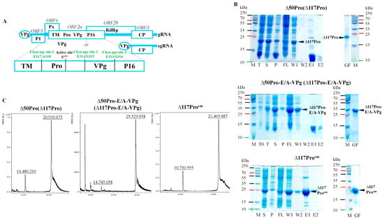
Sobemoviruses encode serine-like 3C proteases (Pro) that participate in the processing and maturation of other virus-encoded proteins. Its cis and trans activity is mediated by the naturally unfolded virus-genome-linked protein (VPg). Nuclear magnetic resonance studies show a Pro–VPg complex interaction and VPg tertiary structure; however, information regarding structural changes of the Pro–VPg complex during interaction is lacking. Here, we solved a full Pro–VPg 3D structure of ryegrass mottle virus (RGMoV) that demonstrates the structural changes in three different conformations due to VPg interaction with Pro. We identified a unique site of VPg interaction with Pro that was not observed in other sobemoviruses, and observed different conformations of the Pro β2 barrel. This is the first report of a full plant Pro crystal structure with its VPg cofactor. We also confirmed the existence of an unusual previously unmapped cleavage site for sobemovirus Pro in the transmembrane domain: E/A. We demonstrated that RGMoV Pro in cis activity is not regulated by VPg and that in trans, VPg can also mediate Pro in free form. Additionally, we observed Ca2+ and Zn2+ inhibitory effects on the Pro cleavage activity. Full article
(This article belongs to the Collection Feature Papers in “Molecular Biology”)
Show Figures

Figure 1

17 pages, 4573 KiB  
Article
Hepatocyte-Specific Triggering of Hepatic Stellate Cell Profibrotic Activation by Apoptotic Bodies: The Role of Hepatoma-Derived Growth Factor, HIV, and Ethanol
by Moses New-Aaron 1,2, Siva Sankar Koganti 2,3, Murali Ganesan 2,3, Sharma Kanika 4, Vikas Kumar 4, Weimin Wang 4, Edward Makarov 4, Kusum K. Kharbanda 2,3, Larisa Y. Poluektova 5 and Natalia A. Osna 1,2,3,5,*
1 Department of Environmental Health, Occupational Health and Toxicology, College of Public Health, University of Nebraska Medical Center, Omaha, NE 68198, USA
2 Research Service, Veterans Affairs Nebraska-Western Iowa Health Care System, Omaha, NE 68105, USA
3 Department of Internal Medicine, University of Nebraska Medical Center, Omaha, NE 68105, USA
4 Department of Genetics Cell Biology & Anatomy, University of Nebraska Medical Center, Omaha, NE 68105, USA
5 Department of Pharmacology and Experimental Neuroscience, University of Nebraska Medical Center, Omaha, NE 68105, USA
Int. J. Mol. Sci. 2023, 24(6), 5346; https://doi.org/10.3390/ijms24065346 - 10 Mar 2023
Cited by 8 | Viewed by 2547
Abstract
Liver disease is one of the leading comorbidities in HIV infection. The risk of liver fibrosis development is potentiated by alcohol abuse. In our previous studies, we reported that hepatocytes exposed to HIV and acetaldehyde undergo significant apoptosis, and the engulfment of apoptotic [...] Read more.
Liver disease is one of the leading comorbidities in HIV infection. The risk of liver fibrosis development is potentiated by alcohol abuse. In our previous studies, we reported that hepatocytes exposed to HIV and acetaldehyde undergo significant apoptosis, and the engulfment of apoptotic bodies (ABs) by hepatic stellate cells (HSC) potentiates their pro-fibrotic activation. However, in addition to hepatocytes, under the same conditions, ABs can be generated from liver-infiltrating immune cells. The goal of this study is to explore whether lymphocyte-derived ABs trigger HSC profibrotic activation as strongly as hepatocyte-derived ABs. ABs were generated from Huh7.5-CYP2E1 (RLW) cells and Jurkat cells treated with HIV+acetaldehyde and co-culture with HSC to induce their pro-fibrotic activation. ABs cargo was analyzed by proteomics. ABs generated from RLW, but not from Jurkat cells activated fibrogenic genes in HSC. This was driven by the expression of hepatocyte-specific proteins in ABs cargo. One of these proteins is Hepatocyte-Derived Growth Factor, for which suppression attenuates pro-fibrotic activation of HSC. In mice humanized with only immune cells but not human hepatocytes, infected with HIV and fed ethanol, liver fibrosis was not observed. We conclude that HIV+ABs of hepatocyte origin promote HSC activation, which potentially may lead to liver fibrosis progression. Full article
Show Figures

Figure 1

21 pages, 4009 KiB  
Article
Novel Synthetic Derivative of Renieramycin T Right-Half Analog Induces Apoptosis and Inhibits Cancer Stem Cells via Targeting the Akt Signal in Lung Cancer Cells
by Korrakod Petsri 1,2, Masashi Yokoya 3, Satapat Racha 1,2,4, Sunisa Thongsom 1,2, Chorpaka Thepthanee 1,2, Bhurichaya Innets 1,2, Zin Zin Ei 1,2, Daiki Hotta 3, Hongbin Zou 5 and Pithi Chanvorachote 1,2,*
1 Center of Excellence in Cancer Cell and Molecular Biology, Faculty of Pharmaceutical Sciences, Chulalongkorn University, Bangkok 10330, Thailand
2 Department of Pharmacology and Physiology, Faculty of Pharmaceutical Sciences, Chulalongkorn University, Bangkok 10330, Thailand
3 Department of Pharmaceutical Chemistry, Meiji Pharmaceutical University, 2-522-1, Noshio, Kiyose, Tokyo 204-8588, Japan
4 Interdisciplinary Program in Pharmacology, Graduate School, Chulalongkorn University, Bangkok 10330, Thailand
5 College of Pharmaceutical Sciences, Zhejiang University, Hangzhou 310058, China
Int. J. Mol. Sci. 2023, 24(6), 5345; https://doi.org/10.3390/ijms24065345 - 10 Mar 2023
Cited by 7 | Viewed by 2510
Abstract
Akt is a key regulatory protein of cancer stem cells (CSCs) and is responsible for cancer aggressiveness and metastasis. Targeting Akt is beneficial for the development of cancer drugs. renieramycin T (RT) has been reported to have Mcl-1 targeting activity, and the study [...] Read more.
Akt is a key regulatory protein of cancer stem cells (CSCs) and is responsible for cancer aggressiveness and metastasis. Targeting Akt is beneficial for the development of cancer drugs. renieramycin T (RT) has been reported to have Mcl-1 targeting activity, and the study of the structure-activity relationships (SARs) demonstrated that cyanide and the benzene ring are essential for its effects. In this study, novel derivatives of the RT right-half analog with cyanide and the modified ring were synthesized to further investigate the SARs for improving the anticancer effects of RT analogs and evaluate CSC-suppressing activity through Akt inhibition. Among the five derivatives, a compound with a substituted thiazole structure (DH_25) exerts the most potent anticancer activity in lung cancer cells. It has the ability to induce apoptosis, which is accompanied by an increase in PARP cleavage, a decrease in Bcl-2, and a diminishment of Mcl-1, suggesting that residual Mcl-1 inhibitory effects exist even after modifying the benzene ring to thiazole. Furthermore, DH_25 is found to induce CSC death, as well as a decrease in CSC marker CD133, CSC transcription factor Nanog, and CSC-related oncoprotein c-Myc. Notably, an upstream member of these proteins, Akt and p-Akt, are also downregulated, indicating that Akt can be a potential target of action. Computational molecular docking showing a high-affinity interaction between DH_25 and an Akt at the allosteric binding site supports that DH_25 can bind and inhibit Akt. This study has revealed a novel SAR and CSC inhibitory effect of DH_25 via Akt inhibition, which may encourage further development of RT compounds for cancer therapy. Full article
(This article belongs to the Section Molecular Pharmacology)
Show Figures

Figure 1

13 pages, 2337 KiB  
Article
The Role of Toll-like Receptor 2 (TLR2) in the Development and Progression of Hashimoto’s Disease (HD): A Case Study on Female Patients in Poland
by Maria Klatka 1, Agnieszka Polak 2, Paulina Mertowska 3,*, Sebastian Mertowski 3, Jakub Łyczba 3, Anna Hymos 3, Izabela Korona-Głowniak 4 and Ewelina Grywalska 3
1 Department of Pediatric Endocrinology and Diabetology, Medical University of Lublin, 20-093 Lublin, Poland
2 Department of Endocrinology, Medical University of Lublin, 20-093 Lublin, Poland
3 Department of Experimental Immunology, Medical University of Lublin, 20-093 Lublin, Poland
4 Department of Pharmaceutical Microbiology, Medical University of Lublin, 20-093 Lublin, Poland
Int. J. Mol. Sci. 2023, 24(6), 5344; https://doi.org/10.3390/ijms24065344 - 10 Mar 2023
Cited by 2 | Viewed by 2343
Abstract
Chronic lymphocytic thyroiditis, commonly known as HD, is one of the most common thyroid disorders. Due to the diverse factors affecting the etiopathogenesis of this disease (hormonal disorders and genetic and environmental factors), as well as the direct involvement of the immune system, [...] Read more.
Chronic lymphocytic thyroiditis, commonly known as HD, is one of the most common thyroid disorders. Due to the diverse factors affecting the etiopathogenesis of this disease (hormonal disorders and genetic and environmental factors), as well as the direct involvement of the immune system, scientists are increasingly willing to undertake research aimed at explaining the impact of the loss of immune tolerance and reactivity of autoantigens on the development of the disease. One of the directions of research in recent years is the role of the innate immune response, particularly Toll-like receptors (TLRs), in the pathogenesis of HD. The purpose of this study was to determine the importance of Toll-like receptor 2 (TLR2) expression on selected populations of immune cells, namely, monocytes (MONs) and dendritic cells (DCs), in the course of HD. Particular attention was paid to the analysis of TLR2’s correlation with clinical parameters and the possibility its use as a potential biomarker molecule in the diagnostic process. Based on the obtained results, we found a statistically significant increase in the percentage of all analyzed populations of immune cells, i.e., mDC BDCA-1+CD19−, pDC BDCA-1+CD123, classical MONs CD14+CD16−, and non-classical MONs CD14+CD16+ showing on their surface TLR2 expression in patients diagnosed with HD compared to the healthy volunteers. Moreover, in the study group, we noted a more than 6-fold increase in the concentration of the soluble form of TLR2 in plasma compared to healthy patients. In addition, the correlation analysis showed significant positive correlations between the level of TLR2 expression on selected subpopulations of immune cells and biochemical indicators of thyroid function. Based on the obtained results, we can assume that TLR2 may be involved in the immunopathogenesis of HD. Full article
(This article belongs to the Special Issue Thyroid Autoimmunity and Autoimmune Hypothyroidism)
Show Figures

Figure 1

18 pages, 17186 KiB  
Article
Genome-Wide Analysis of Long Noncoding RNAs in Porcine Intestine during Weaning Stress
by Shujie Liu, Xin Tao, Bo Deng, Yongming Li and Ziwei Xu *
Institute of Animal Husbandry and Veterinary Science, Zhejiang Academy of Agricultural Sciences, 298 Desheng Middle Road, Hangzhou 310021, China
Int. J. Mol. Sci. 2023, 24(6), 5343; https://doi.org/10.3390/ijms24065343 - 10 Mar 2023
Cited by 1 | Viewed by 1701
Abstract
Long noncoding RNAs (lncRNAs) play crucial roles in various biological processes, and they are considered to be closely associated with the pathogenesis of intestinal diseases. However, the role and expression of lncRNAs in intestinal damage during weaning stress remain unknown. Herein, we investigated [...] Read more.
Long noncoding RNAs (lncRNAs) play crucial roles in various biological processes, and they are considered to be closely associated with the pathogenesis of intestinal diseases. However, the role and expression of lncRNAs in intestinal damage during weaning stress remain unknown. Herein, we investigated the expression profiles of jejunal tissue from weaning piglets at 4 and 7 d after weaning (groups W4 and W7, respectively) and from suckling piglets on the same days (groups S4 and S7, respectively). Genome-wide analysis of lncRNAs was also performed using RNA sequencing technology. A total of 1809 annotated lncRNAs and 1612 novel lncRNAs were obtained from the jejunum of piglets. In W4 vs. S4, a total of 331 lncRNAs showed significant differential expression, and a total of 163 significantly differentially expressed lncRNAs (DElncRNAs) was identified in W7 vs. S7. Biological analysis indicated that DElncRNAs were involved in intestinal diseases, inflammation, and immune functions, and were mainly enriched in the Jak-STAT signaling pathway, inflammatory bowel disease, T cell receptor signaling pathway, B cell receptor signaling pathway and intestinal immune network for IgA production. Moreover, we found that lnc_000884 and target gene KLF5 were significantly upregulated in the intestine of weaning piglets. The overexpression of lnc_000884 also significantly promoted the proliferation and depressed apoptosis of IPEC-J2 cells. This result suggested that lnc_000884 may contribute to repairing intestinal damage. Our study identified the characterization and expression profile of lncRNAs in the small intestine of weaning piglets and provided new insights into the molecular regulation of intestinal damage during weaning stress. Full article
(This article belongs to the Section Molecular Biology)
Show Figures

Figure 1

18 pages, 1169 KiB  
Review
Endocrine-Disrupting Chemicals and Disease Endpoints
by Changhwan Ahn 1 and Eui-Bae Jeung 2,*
1 Laboratory of Veterinary Physiology, College of Veterinary Medicine, Jeju National University, Jeju 63243, Republic of Korea
2 Laboratory of Veterinary Biochemistry and Molecular Biology, College of Veterinary Medicine, Chungbuk National University, Cheongju 28644, Republic of Korea
Int. J. Mol. Sci. 2023, 24(6), 5342; https://doi.org/10.3390/ijms24065342 - 10 Mar 2023
Cited by 105 | Viewed by 17070
Abstract
Endocrine-disrupting chemicals (EDCs) have significant impacts on biological systems, and have been shown to interfere with physiological systems, especially by disrupting the hormone balance. During the last few decades, EDCs have been shown to affect reproductive, neurological, and metabolic development and function and [...] Read more.
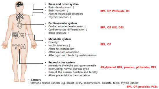
Endocrine-disrupting chemicals (EDCs) have significant impacts on biological systems, and have been shown to interfere with physiological systems, especially by disrupting the hormone balance. During the last few decades, EDCs have been shown to affect reproductive, neurological, and metabolic development and function and even stimulate tumor growth. EDC exposure during development can disrupt normal development patterns and alter susceptibility to disease. Many chemicals have endocrine-disrupting properties, including bisphenol A, organochlorines, polybrominated flame retardants, alkylphenols, and phthalates. These compounds have gradually been elucidated as risk factors for many diseases, such as reproductive, neural, and metabolic diseases and cancers. Endocrine disruption has been spread to wildlife and species that are connected to the food chains. Dietary uptake represents an important source of EDC exposure. Although EDCs represent a significant public health concern, the relationship and specific mechanism between EDCs and diseases remain unclear. This review focuses on the disease-EDC relationship and the disease endpoints associated with endocrine disruption for a better understanding of the relationship between EDCs-disease and elucidates the development of new prevention/treatment opportunities and screening methods. Full article
(This article belongs to the Special Issue Advances in Endocrine Disruptors 2.0)
Show Figures

Figure 1

16 pages, 3791 KiB  
Article
Production of Silver Nano-Inks and Surface Coatings for Anti-Microbial Food Packaging and Its Ecological Impact
by N. Arul Manikandan 1, Ronan McCann 2,3,4, Dimitrios Kakavas 1, Keith D. Rochfort 5, Sithara P. Sreenilayam 2,3, Godze Alkan 6, Tom Stornetta 1, Allan Robert McGivern 1, Konstantinos Grintzalis 1, Bernd Friedrich 6, Greg Foley 1, Dermot Brabazon 2,3,* and Brian Freeland 1,2,3
1 School of Biotechnology, Dublin City University, Glasnevin, D09 K20V Dublin, Ireland
2 I-Form, Advanced Manufacturing Research Centre, School of Mechanical and Manufacturing Engineering, Dublin City University, Glasnevin, D09 NA55 Dublin, Ireland
3 Advanced Processing Technology Research Centre, School of Mechanical and Manufacturing Engineering, Dublin City University, Glasnevin, D09 NA55 Dublin, Ireland
4 Department of Science, School of Science and Computing, South East Technological University, Cork Road, X91 K0EK Waterford City, Ireland
5 School of Nursing, Psychotherapy and Community Health, Dublin City University, Glasnevin, D09 NA55 Dublin, Ireland
6 Institute of Process Metallurgy and Metal Recycling (IME), RWTH Aachen University, Intzestraße 3, 52056 Aachen, Germany
Int. J. Mol. Sci. 2023, 24(6), 5341; https://doi.org/10.3390/ijms24065341 - 10 Mar 2023
Cited by 10 | Viewed by 3683
Abstract
Food spoilage is an ongoing global issue that contributes to rising carbon dioxide emissions and increased demand for food processing. This work developed anti-bacterial coatings utilising inkjet printing of silver nano-inks onto food-grade polymer packaging, with the potential to enhance food safety and [...] Read more.
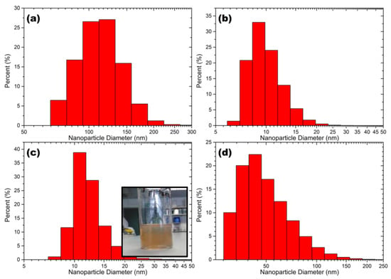
Food spoilage is an ongoing global issue that contributes to rising carbon dioxide emissions and increased demand for food processing. This work developed anti-bacterial coatings utilising inkjet printing of silver nano-inks onto food-grade polymer packaging, with the potential to enhance food safety and reduce food spoilage. Silver nano-inks were synthesised via laser ablation synthesis in solution (LaSiS) and ultrasound pyrolysis (USP). The silver nanoparticles (AgNPs) produced using LaSiS and USP were characterised using transmission electron microscopy (TEM), Fourier transform infrared (FTIR) spectroscopy, UV-Vis spectrophotometry and dynamic light scattering (DLS) analysis. The laser ablation technique, operated under recirculation mode, produced nanoparticles with a small size distribution with an average diameter ranging from 7–30 nm. Silver nano-ink was synthesised by blending isopropanol with nanoparticles dispersed in deionised water. The silver nano-inks were printed on plasma-cleaned cyclo-olefin polymer. Irrespective of the production methods, all silver nanoparticles exhibited strong antibacterial activity against E. coli with a zone of inhibition exceeding 6 mm. Furthermore, silver nano-inks printed cyclo-olefin polymer reduced the bacterial cell population from 1235 (±45) × 106 cell/mL to 960 (±110) × 106 cell/mL. The bactericidal performance of silver-coated polymer was comparable to that of the penicillin-coated polymer, wherein a reduction in bacterial population from 1235 (±45) × 106 cell/mL to 830 (±70) × 106 cell/mL was observed. Finally, the ecotoxicity of the silver nano-ink printed cyclo-olefin polymer was tested with daphniids, a species of water flea, to simulate the release of coated packaging into a freshwater environment. Full article
(This article belongs to the Special Issue Silver Nanomaterials for Biological Applications)
Show Figures

Figure 1

13 pages, 2754 KiB  
Article
Ligand-Induced Activation of GPR110 (ADGRF1) to Improve Visual Function Impaired by Optic Nerve Injury
by Heung-Sun Kwon 1, Karl Kevala 1, Haohua Qian 2, Mones Abu-Asab 3, Samarjit Patnaik 4, Juan Marugan 4 and Hee-Yong Kim 1,*
1 Laboratory of Molecular Signaling, NIAAA, National Institutes of Health, 5625 Fishers Lane Room 3S-02, Rockville, MD 20892-9410, USA
2 Visual Function Core, NEI, National Institutes of Health, Bethesda, MD 20892-0616, USA
3 Electron Microscopy Laboratory, Biological Imaging Core, NEI, National Institutes of Health, Bethesda, MD 20850-2510, USA
4 Division of Pre-Clinical Innovation, NCATS, National Institutes of Health, Rockville, MD 20817, USA
Int. J. Mol. Sci. 2023, 24(6), 5340; https://doi.org/10.3390/ijms24065340 - 10 Mar 2023
Cited by 4 | Viewed by 2385
Abstract
It is extremely difficult to achieve functional recovery after axonal injury in the adult central nervous system. The activation of G-protein coupled receptor 110 (GPR110, ADGRF1) has been shown to stimulate neurite extension in developing neurons and after axonal injury in adult mice. [...] Read more.
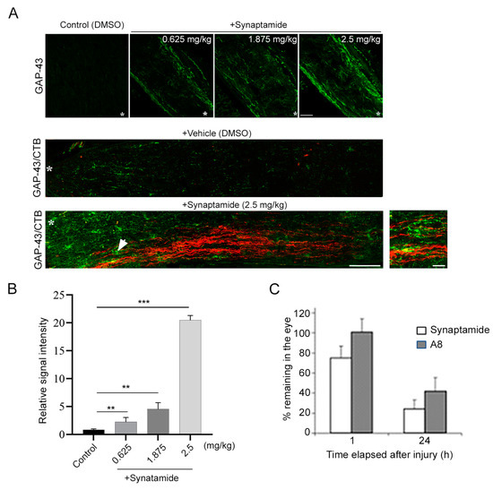
It is extremely difficult to achieve functional recovery after axonal injury in the adult central nervous system. The activation of G-protein coupled receptor 110 (GPR110, ADGRF1) has been shown to stimulate neurite extension in developing neurons and after axonal injury in adult mice. Here, we demonstrate that GPR110 activation partially restores visual function impaired by optic nerve injury in adult mice. Intravitreal injection of GPR110 ligands, synaptamide and its stable analogue dimethylsynaptamide (A8) after optic nerve crush significantly reduced axonal degeneration and improved axonal integrity and visual function in wild-type but not gpr110 knockout mice. The retina obtained from the injured mice treated with GPR110 ligands also showed a significant reduction in the crush-induced loss of retinal ganglion cells. Our data suggest that targeting GPR110 may be a viable strategy for functional recovery after optic nerve injury. Full article
Show Figures

Figure 1

22 pages, 1444 KiB  
Review
Role of Terpenophenolics in Modulating Inflammation and Apoptosis in Cardiovascular Diseases: A Review
by Muhamad Adib Abdul Ghani 1, Azizah Ugusman 2, Jalifah Latip 3,* and Satirah Zainalabidin 1,*
1 Programme of Biomedical Sciences, Centre of Toxicology and Health Risk Study, Faculty of Health Sciences, Universiti Kebangsaan Malaysia, Kuala Lumpur 50300, Malaysia
2 Department of Physiology, Faculty of Medicine, Universiti Kebangsaan Malaysia, Kuala Lumpur 56000, Malaysia
3 Department of Chemical Sciences, Faculty of Science and Technology, Universiti Kebangsaan Malaysia, Bangi 43600, Malaysia
Int. J. Mol. Sci. 2023, 24(6), 5339; https://doi.org/10.3390/ijms24065339 - 10 Mar 2023
Cited by 31 | Viewed by 4364
Abstract
One in every three deaths worldwide is caused by cardiovascular diseases (CVDs), estimating a total of 17.9 million deaths annually. By 2030, it is expected that more than 24 million people will die from CVDs related complications. The most common CVDs are coronary [...] Read more.
One in every three deaths worldwide is caused by cardiovascular diseases (CVDs), estimating a total of 17.9 million deaths annually. By 2030, it is expected that more than 24 million people will die from CVDs related complications. The most common CVDs are coronary heart disease, myocardial infarction, stroke, and hypertension. A plethora of studies has shown inflammation causing both short-term and long-term damage to the tissues in many organ systems, including the cardiovascular system. In parallel to inflammation processes, it has been discovered that apoptosis, a mode of programmed cell death, may also contribute to CVD development due to the loss of cardiomyocytes. Terpenophenolic compounds are comprised of terpenes and natural phenols as secondary metabolites by plants and are commonly found in the genus Humulus and Cannabis. A growing body of evidence has shown that terpenophenolic compounds exhibit protective properties against inflammation and apoptosis within the cardiovascular system. This review highlights the current evidence elucidating the molecular actions of terpenophenolic compounds in protecting the cardiovascular system, i.e., bakuchiol, ferruginol, carnosic acid, carnosol, carvacrol, thymol and hinokitiol. The potential of these compounds is discussed as the new nutraceutical drugs that may help to decrease the burden of cardiovascular disorders. Full article
(This article belongs to the Special Issue Nutraceuticals in Cardiovascular and Metabolic Diseases)
Show Figures

Figure 1

22 pages, 10999 KiB  
Article
Knockout Mutants of OsPUB7 Generated Using CRISPR/Cas9 Revealed Abiotic Stress Tolerance in Rice
by Me-Sun Kim 1,†, Seo-Rin Ko 1,†, Yu Jin Jung 2, Kwon-Kyoo Kang 2, Yung-Jo Lee 3 and Yong-Gu Cho 1,*
1 Department of Crop Science, College of Agriculture and Life & Environment Sciences, Chungbuk National University, Cheongju 28644, Republic of Korea
2 Division of Horticultural Biotechnology, Hankyong National University, Anseong 17579, Republic of Korea
3 Institute of Korean Prehistory, Cheongju 28763, Republic of Korea
These authors contributed equally to this work.
Int. J. Mol. Sci. 2023, 24(6), 5338; https://doi.org/10.3390/ijms24065338 - 10 Mar 2023
Cited by 26 | Viewed by 3897
Abstract
Plants produce and accumulate stress-resistant substances when exposed to abiotic stress, which involves a protein conversion mechanism that breaks down stress-damaged proteins and supplies usable amino acids. Eukaryotic protein turnover is mostly driven by the ubiquitination pathway. Among the three enzymes required for [...] Read more.
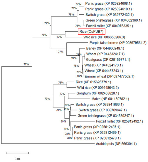
Plants produce and accumulate stress-resistant substances when exposed to abiotic stress, which involves a protein conversion mechanism that breaks down stress-damaged proteins and supplies usable amino acids. Eukaryotic protein turnover is mostly driven by the ubiquitination pathway. Among the three enzymes required for protein degradation, E3 ubiquitin ligase plays a pivotal role in most cells, as it determines the specificity of ubiquitination and selects target proteins for degradation. In this study, to investigate the function of OsPUB7 (Plant U-box gene in Oryza sativa), we constructed a CRISPR/Cas9 vector, generated OsPUB7 gene-edited individuals, and evaluated resistance to abiotic stress using gene-edited lines. A stress-tolerant phenotype was observed as a result of drought and salinity stress treatment in the T2 OsPUB7 gene-edited null lines (PUB7-GE) lacking the T-DNA. In addition, although PUB7-GE did not show any significant change in mRNA expression analysis, it showed lower ion leakage and higher proline content than the wild type (WT). Protein–protein interaction analysis revealed that the expression of the genes (OsPUB23, OsPUB24, OsPUB66, and OsPUB67) known to be involved in stress increased in PUB7-GE and this, by forming a 1-node network with OsPUB66 and OsPUB7, acted as a negative regulator of drought and salinity stress. This result provides evidence that OsPUB7 will be a useful target for both breeding and future research on drought tolerance/abiotic stress in rice. Full article
(This article belongs to the Special Issue Genetic Analysis Based on CRISPR/Cas9 Technology)
Show Figures

Figure 1