Int. J. Mol. Sci. 2023, 24(4), 3067; https://doi.org/10.3390/ijms24043067 - 4 Feb 2023
Cited by 39 | Viewed by 6199
Abstract
Acute liver injury (ALI) is a globally important public health issue that, when severe, rapidly progresses to acute liver failure, seriously compromising the life safety of patients. The pathogenesis of ALI is defined by massive cell death in the liver, which triggers a
[...] Read more.
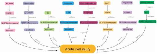
Acute liver injury (ALI) is a globally important public health issue that, when severe, rapidly progresses to acute liver failure, seriously compromising the life safety of patients. The pathogenesis of ALI is defined by massive cell death in the liver, which triggers a cascade of immune responses. Studies have shown that the aberrant activation of the nod-like receptor protein 3 (NLRP3) inflammasome plays an important role in various types of ALI and that the activation of the NLRP3 inflammasome causes various types of programmed cell death (PCD), and these cell death effectors can in turn regulate NLRP3 inflammasome activation. This indicates that NLRP3 inflammasome activation is inextricably linked to PCD. In this review, we summarize the role of NLRP3 inflammasome activation and PCD in various types of ALI (APAP, liver ischemia reperfusion, CCl4, alcohol, Con A, and LPS/D-GalN induced ALI) and analyze the underlying mechanisms to provide references for future relevant studies.
Full article
(This article belongs to the Special Issue Mechanism of Cellular Signaling, Dysfunction and Drug Effects on Liver Diseases)
►
Show Figures

Figure 1


