22 pages, 7479 KiB  
Article
Selective Calpain Inhibition Improves Functional and Histopathological Outcomes in a Canine Spinal Cord Injury Model
by Elsayed Metwally, Hatim A. Al-Abbadi, Mohamed A. Hashem, Yasmina K. Mahmoud, Eman A. Ahmed, Ahmed I. Maaty, Ibrahim E. Helal and Mahmoud F. Ahmed
Int. J. Mol. Sci. 2022, 23(19), 11772; https://doi.org/10.3390/ijms231911772 - 4 Oct 2022
Cited by 5 | Viewed by 3326
Abstract
Calpain activation has been implicated in various pathologies, including neurodegeneration. Thus, calpain inhibition could effectively prevent spinal cord injury (SCI) associated with neurodegeneration. In the current study, a dog SCI model was used to evaluate the therapeutic potential of a selective calpain inhibitor [...] Read more.
Calpain activation has been implicated in various pathologies, including neurodegeneration. Thus, calpain inhibition could effectively prevent spinal cord injury (SCI) associated with neurodegeneration. In the current study, a dog SCI model was used to evaluate the therapeutic potential of a selective calpain inhibitor (PD150606) in combination with methylprednisolone sodium succinate (MPSS) as an anti-inflammatory drug. SCI was experimentally induced in sixteen mongrel dogs through an epidural balloon compression technique. The dogs were allocated randomly into four groups: control, MPSS, PD150606, and MPSS+PD150606. Clinical evaluation, serum biochemical, somatosensory evoked potentials, histopathological, and immunoblotting analyses were performed to assess treated dogs during the study. The current findings revealed that the combined administration of MPSS+PD150606 demonstrated considerably lower neuronal loss and microglial cell infiltration than the other groups, with a significant improvement in the locomotor score. The increased levels of inflammatory markers (GFAP and CD11) and calcium-binding proteins (Iba1 and S100) were significantly reduced in the combination group and to a lesser extent in MPSS or PD150606 treatment alone. Interestingly, the combined treatment effectively inhibited the calpain-induced cleavage of p35, limited cdk5 activation, and inhibited tau phosphorylation. These results suggest that early MPSS+PD150606 therapy after acute SCI may prevent subsequent neurodegeneration via calpain inhibition. Full article
(This article belongs to the Special Issue Neurodegeneration 2022: From Genetics to Molecules)
Show Figures

Figure 1

12 pages, 3276 KiB  
Article
The First Evidence of Bacterial Foci in the Hair Part and Dermal Papilla of Scalp Hair Follicles: A Pilot Comparative Study in Alopecia Areata
by Fabio Rinaldi, Daniela Pinto, Elisa Borsani, Stefania Castrezzati, Amedeo Amedei and Rita Rezzani
Int. J. Mol. Sci. 2022, 23(19), 11956; https://doi.org/10.3390/ijms231911956 - 8 Oct 2022
Cited by 11 | Viewed by 3306
Abstract
The role of the microbiome in hair follicle (HF) growth represents a growing field of research. Here, we studied the bacterial population in the scalp hair follicles of subjects with alopecia areata (AA). Two Healthy and two AA subjects, respectively (20–60 years old), [...] Read more.
The role of the microbiome in hair follicle (HF) growth represents a growing field of research. Here, we studied the bacterial population in the scalp hair follicles of subjects with alopecia areata (AA). Two Healthy and two AA subjects, respectively (20–60 years old), were enrolled and studied regarding the microbial community in the subepidermal scalp compartments by means of a 4-mm biopsy punch. Samples were examined by 16S sequencing, histochemical staining (Gram’s method), and transmission electron microscopy (TEM). Bacterial foci were observed in the AA subjects’ follicles with both the two adopted complementary approaches (electron microscopy and Gram staining). Significant (p < 0.05) differences were also found in the three-layer biopsy samples (p < 0.05) regarding the bacterial population. In particular, in the deep epidermis and dermis levels, a significant (p < 0.05) lower abundance of Firmicutes and a higher abundance of Proteobacteria were found in AA samples compared to the healthy control. Firmicutes also showed a significant (p < 0.05) lower abundance in hypodermis in AA subjects. In addition, Enterobacteriaceae and the genera Streptococcus, Gemella, Porphyromonas, and Granulicatella were relatively more abundant in AA groups at the deep epidermis level. The Staphylococcus and Flavobacterium genera were significantly less abundant in AA samples than in controls in all three-layer biopsy samples (p < 0.05). In contrast, Veillonella and Neisseriaceae were relatively more abundant in the healthy control group compared to the AA sample. Therefore, higher alpha diversity was observed in all three-layer biopsy samples of AA patients compared to the control. In conclusion, our data suggest that tAA could be defined as a “hair disease associated with dysregulated microbiome-immunity axis of hair follicles”. Full article
(This article belongs to the Section Molecular Pathology, Diagnostics, and Therapeutics)
Show Figures

Figure 1

13 pages, 1189 KiB  
Article
A Physiologically Based Pharmacokinetic Model to Predict the Impact of Metabolic Changes Associated with Metabolic Associated Fatty Liver Disease on Drug Exposure
by Elise M. Newman and Andrew Rowland
Int. J. Mol. Sci. 2022, 23(19), 11751; https://doi.org/10.3390/ijms231911751 - 4 Oct 2022
Cited by 10 | Viewed by 3306
Abstract
Metabolic associated fatty liver disease (MAFLD) is the most common chronic liver disease, with an estimated prevalence of between 20 and 30% worldwide. Observational data supported by in vitro and pre-clinical animal models of MAFLD suggest meaningful differences in drug disposition in MAFLD [...] Read more.
Metabolic associated fatty liver disease (MAFLD) is the most common chronic liver disease, with an estimated prevalence of between 20 and 30% worldwide. Observational data supported by in vitro and pre-clinical animal models of MAFLD suggest meaningful differences in drug disposition in MAFLD patients. This study aimed to build a physiologically based pharmacokinetic (PBPK) model reflecting observed changes in physiological and molecular parameters relevant to drug disposition that are associated with MAFLD. A comprehensive literature review and meta-analysis was conducted to identify all studies describing in vivo physiological changes along with in vitro and pre-clinical model changes in CYP 1A2, 2C9, 2C19, 2D6 and 3A4 protein abundance associated with MAFLD. A MAFLD population profile was constructed in Simcyp (version 19.1) by adapting demographic and physiological covariates from the Sim-Healthy population profile based on a meta-analysis of observed data from the published literature. Simulations demonstrated that single dose and steady state area under the plasma concentration time curve (AUC) for caffeine, clozapine, omeprazole, metoprolol, dextromethorphan and midazolam, but not s-warfarin or rosiglitazone, were increased by >20% in the MAFLD population compared to the healthy control population. These findings indicate that MAFLD patients are likely to be experience meaningfully higher exposure to drugs that are primarily metabolized by CYP 1A2, 2C19, 2D6 and 3A4, but not CYP2C9. Closer monitoring of MAFLD patients using drugs primarily cleared by CYP 1A2, 2C19 and 3A4 is warranted as reduced metabolic activity and increased drug exposure are likely to result in an increased incidence of toxicity in this population. Full article
Show Figures

Figure 1

18 pages, 1052 KiB  
Review
Molecular Advances in MAFLD—A Link between Sphingolipids and Extracellular Matrix in Development and Progression to Fibrosis
by Adrian Kołakowski, Sylwia Dziemitko, Aleksandra Chmielecka, Hubert Żywno, Wiktor Bzdęga, Tomasz Charytoniuk, Adrian Chabowski and Karolina Konstantynowicz-Nowicka
Int. J. Mol. Sci. 2022, 23(19), 11380; https://doi.org/10.3390/ijms231911380 - 27 Sep 2022
Cited by 5 | Viewed by 3289
Abstract
Metabolic-Associated Fatty Liver Disease (MAFLD) is a major cause of liver diseases globally and its prevalence is expected to grow in the coming decades. The main cause of MAFLD development is changed in the composition of the extracellular matrix (ECM). Increased production of [...] Read more.
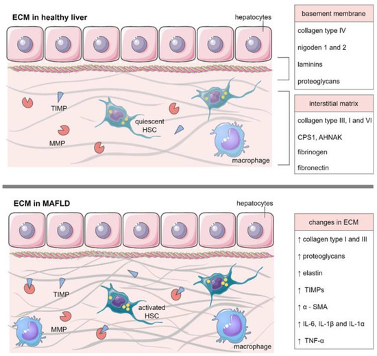
Metabolic-Associated Fatty Liver Disease (MAFLD) is a major cause of liver diseases globally and its prevalence is expected to grow in the coming decades. The main cause of MAFLD development is changed in the composition of the extracellular matrix (ECM). Increased production of matrix molecules and inflammatory processes lead to progressive fibrosis, cirrhosis, and ultimately liver failure. In addition, increased accumulation of sphingolipids accompanied by increased expression of pro-inflammatory cytokines in the ECM is closely related to lipogenesis, MAFLD development, and its progression to fibrosis. In our work, we will summarize all information regarding the role of sphingolipids e.g., ceramide and S1P in MAFLD development. These sphingolipids seem to have the most significant effect on macrophages and, consequently, HSCs which trigger the entire cascade of overproduction matrix molecules, especially type I and III collagen, proteoglycans, elastin, and also tissue inhibitors of metalloproteinases, which as a result cause the development of liver fibrosis. Full article
(This article belongs to the Special Issue Molecular Advances in MAFLD)
Show Figures

Figure 1

13 pages, 2955 KiB  
Article
KITLG Promotes Glomerular Endothelial Cell Injury in Diabetic Nephropathy by an Autocrine Effect
by Jiun-Chi Huang, Szu-Chia Chen, Wei-An Chang, Wei-Wen Hung, Ping-Hsun Wu, Ling-Yu Wu, Jer-Ming Chang, Ya-Ling Hsu and Yi-Chun Tsai
Int. J. Mol. Sci. 2022, 23(19), 11723; https://doi.org/10.3390/ijms231911723 - 3 Oct 2022
Cited by 4 | Viewed by 3287
Abstract
Diabetic nephropathy (DN) is an increasing threat to human health. The impact of hyperglycemia or its metabolites, advanced glycation end-products (AGEs), on glomerular endothelial cells (GECs) and their pathophysiologic mechanisms are not well explored. Our results reveal that AGEs increased the expression and [...] Read more.
Diabetic nephropathy (DN) is an increasing threat to human health. The impact of hyperglycemia or its metabolites, advanced glycation end-products (AGEs), on glomerular endothelial cells (GECs) and their pathophysiologic mechanisms are not well explored. Our results reveal that AGEs increased the expression and secretion of the KIT ligand (KITLG) in GECs. Both AGEs and KITLG promoted endothelial-to-mesenchymal transition (EndoMT) in GECs and further increased the permeability of GECs through the AKT/extracellular-signal-regulated kinase pathway. Inhibition of KITLG’s effects by imatinib prevented AGE-medicated EndoMT in GECs, supporting the belief that KITLG is a critical factor for GEC injury. We found higher KITLG levels in the GECs and urine of db/db mice compared with db/m mice, and urinary KITLG levels were positively correlated with the urinary albumin-to-creatinine ratio (ACR). Furthermore, type 2 diabetic patients had higher urinary KITLG levels than normal individuals, as well as urinary KITLG levels that were positively correlated with urinary ACR and negatively correlated with the estimated glomerular filtration rate. KITLG plays a pathogenic role in GEC injury in DN and might act as a biomarker of DN progression. Full article
(This article belongs to the Special Issue Advances in the Pathogenesis of Diabetic Kidney Disease)
Show Figures

Figure 1

15 pages, 1768 KiB  
Article
SP1 and NFY Regulate the Expression of PNPT1, a Gene Encoding a Mitochondrial Protein Involved in Cancer
by Ignacio Ventura, Fernando Revert, Francisco Revert-Ros, Lucía Gómez-Tatay, Jesús A. Prieto-Ruiz and José Miguel Hernández-Andreu
Int. J. Mol. Sci. 2022, 23(19), 11399; https://doi.org/10.3390/ijms231911399 - 27 Sep 2022
Cited by 4 | Viewed by 3279
Abstract
The Polyribonucleotide nucleotidyltransferase 1 gene (PNPT1) encodes polynucleotide phosphorylase (PNPase), a 3′-5′ exoribonuclease involved in mitochondrial RNA degradation and surveillance and RNA import into the mitochondrion. Here, we have characterized the PNPT1 promoter by in silico analysis, luciferase reporter assays, electrophoretic [...] Read more.
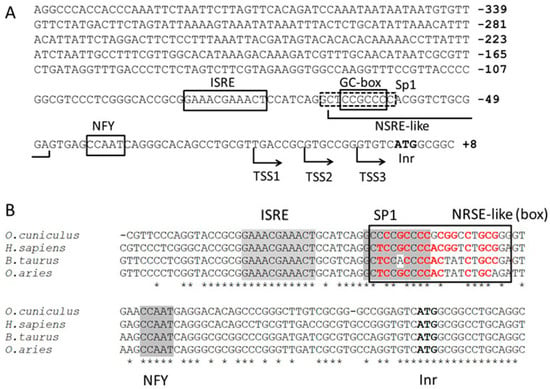
The Polyribonucleotide nucleotidyltransferase 1 gene (PNPT1) encodes polynucleotide phosphorylase (PNPase), a 3′-5′ exoribonuclease involved in mitochondrial RNA degradation and surveillance and RNA import into the mitochondrion. Here, we have characterized the PNPT1 promoter by in silico analysis, luciferase reporter assays, electrophoretic mobility shift assays (EMSA), chromatin immunoprecipitation (ChIP), siRNA-based mRNA silencing and RT-qPCR. We show that the Specificity protein 1 (SP1) transcription factor and Nuclear transcription factor Y (NFY) bind the PNPT1 promoter, and have a relevant role regulating the promoter activity, PNPT1 expression, and mitochondrial activity. We also found in Kaplan–Meier survival curves that a high expression of either PNPase, SP1 or NFY subunit A (NFYA) is associated with a poor prognosis in liver cancer. In summary, our results show the relevance of SP1 and NFY in PNPT1 expression, and point to SP1/NFY and PNPase as possible targets in anti-cancer therapy. Full article
Show Figures

Figure 1

20 pages, 7436 KiB  
Review
Oligonucleotide-Recognizing Topoisomerase Inhibitors (OTIs): Precision Gene Editors for Neurodegenerative Diseases?
by Ben D. Bax, Dmitry Sutormin, Neil Q. McDonald, Glenn A. Burley and Tatyana Shelkovnikova
Int. J. Mol. Sci. 2022, 23(19), 11541; https://doi.org/10.3390/ijms231911541 - 29 Sep 2022
Viewed by 3234
Abstract
Topoisomerases are essential enzymes that recognize and modify the topology of DNA to allow DNA replication and transcription to take place. Topoisomerases are divided into type I topoisomerases, that cleave one DNA strand to modify DNA topology, and type II, that cleave both [...] Read more.
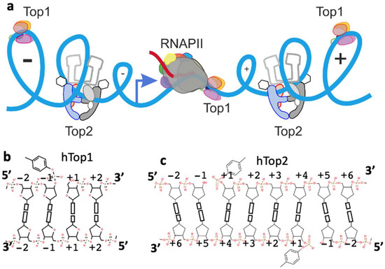
Topoisomerases are essential enzymes that recognize and modify the topology of DNA to allow DNA replication and transcription to take place. Topoisomerases are divided into type I topoisomerases, that cleave one DNA strand to modify DNA topology, and type II, that cleave both DNA strands. Topoisomerases normally rapidly religate cleaved-DNA once the topology has been modified. Topoisomerases do not recognize specific DNA sequences, but actively cleave positively supercoiled DNA ahead of transcription bubbles or replication forks, and negative supercoils (or precatenanes) behind, thus allowing the unwinding of the DNA-helix to proceed (during both transcription and replication). Drugs that stabilize DNA-cleavage complexes with topoisomerases produce cytotoxic DNA damage and kill fast-dividing cells; they are widely used in cancer chemotherapy. Oligonucleotide-recognizing topoisomerase inhibitors (OTIs) have given drugs that stabilize DNA-cleavage complexes specificity by linking them to either: (i) DNA duplex recognizing triplex forming oligonucleotide (TFO-OTIs) or DNA duplex recognizing pyrrole-imidazole-polyamides (PIP-OTIs) (ii) or by conventional Watson–Crick base pairing (WC-OTIs). This converts compounds from indiscriminate DNA-damaging drugs to highly specific targeted DNA-cleaving OTIs. Herein we propose simple strategies to enable DNA-duplex strand invasion of WC-OTIs giving strand-invading SI-OTIs. This will make SI-OTIs similar to the guide RNAs of CRISPR/Cas9 nuclease bacterial immune systems. However, an important difference between OTIs and CRISPR/Cas9, is that OTIs do not require the introduction of foreign proteins into cells. Recent successful oligonucleotide therapeutics for neurodegenerative diseases suggest that OTIs can be developed to be highly specific gene editing agents for DNA lesions that cause neurodegenerative diseases. Full article
(This article belongs to the Special Issue DNA Replication Stress and Chromosomal Instability 2.0)
Show Figures

Figure 1

15 pages, 2923 KiB  
Article
Proof-of-Principle Study Suggesting Potential Anti-Inflammatory Activity of Butyrate and Propionate in Periodontal Cells
by Ana Flávia Piquera Santos, Lara Cristina Cunha Cervantes, Layla Panahipour, Francisley Ávila Souza and Reinhard Gruber
Int. J. Mol. Sci. 2022, 23(19), 11006; https://doi.org/10.3390/ijms231911006 - 20 Sep 2022
Cited by 14 | Viewed by 3233
Abstract
Short-chain fatty acids (SCFAs) are potent immune modulators present in the gingival crevicular fluid. It is therefore likely that SCFAs exert a role in periodontal health and disease. To better understand how SCFAs can module inflammation, we screened acetic acid, propionic acid, and [...] Read more.
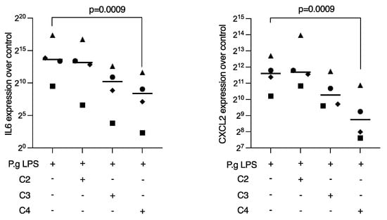
Short-chain fatty acids (SCFAs) are potent immune modulators present in the gingival crevicular fluid. It is therefore likely that SCFAs exert a role in periodontal health and disease. To better understand how SCFAs can module inflammation, we screened acetic acid, propionic acid, and butyric acid for their potential ability to lower the inflammatory response of macrophages, gingival fibroblasts, and oral epithelial cells in vitro. To this end, RAW 264.7 and primary macrophages were exposed to LPSs from Porphyromonas gingivalis (P. gingivalis) with and without the SCFAs. Moreover, gingival fibroblasts and HSC2 oral epithelial cells were exposed to IL1β and TNFα with and without the SCFAs. We report here that butyrate was effective in reducing the lipopolysaccharide (LPS)-induced expression of IL6 and chemokine (C-X-C motif) ligand 2 (CXCL2) in the RAW 264.7 and primary macrophages. Butyrate also reduced the IL1β and TNFα-induced expression of IL8, chemokine (C-X-C motif) ligand 1 (CXCL1), and CXCL2 in gingival fibroblasts. Likewise, butyrate lowered the induced expression of CXCL1 and CXCL2, but not IL8, in HSC2 cells. Butyrate further caused a reduction of p65 nuclear translocation in RAW 264.7 macrophages, gingival fibroblasts, and HSC2 cells. Propionate and acetate partially lowered the inflammatory response in vitro but did not reach the level of significance. These findings suggest that not only macrophages, but also gingival fibroblasts and oral epithelial cells are susceptive to the anti-inflammatory activity of butyrate. Full article
(This article belongs to the Special Issue Molecular Mechanisms of Periodontal Disease 3.0)
Show Figures

Figure 1

13 pages, 956 KiB  
Article
Evaluation of Dried Blood and Cerebrospinal Fluid Filter Paper Spots for Storing and Transporting Clinical Material for the Molecular Diagnosis of Invasive Meningococcal Disease
by Brenda A. Kwambana-Adams, Stephen A. Clark, Nicole Tay, Schadrac Agbla, Chrispin Chaguza, Eunice W. Kagucia, Ray Borrow and Robert S. Heyderman
Int. J. Mol. Sci. 2022, 23(19), 11879; https://doi.org/10.3390/ijms231911879 - 6 Oct 2022
Cited by 2 | Viewed by 3209
Abstract
To improve the storage and transport of clinical specimens for the diagnosis of Neisseria meningitidis (Nm) infections in resource-limited settings, we have evaluated the performance of dried blood spot (DBS) and dried cerebrospinal fluid spot (DCS) assays. DBS and DCS were prepared on [...] Read more.
To improve the storage and transport of clinical specimens for the diagnosis of Neisseria meningitidis (Nm) infections in resource-limited settings, we have evaluated the performance of dried blood spot (DBS) and dried cerebrospinal fluid spot (DCS) assays. DBS and DCS were prepared on filter paper from liquid specimens previously tested for Nm in the United Kingdom. Nm was detected and genogrouped by real-time PCR performed on crude genomic DNA extracted from the DBS (n = 226) and DCS (n = 226) specimens. Targeted whole-genome sequencing was performed on a subset of specimens, DBS (n = 4) and DCS (n = 6). The overall agreement between the analysis of liquid and dried specimens was (94.2%; 95% CI 90.8–96.7) for blood and (96.4%; 95% CI 93.5–98.0) for cerebrospinal fluid. Relative to liquid specimens as the reference, the DBS and DCS assays had sensitivities of (89.1%; 95% CI 82.7–93.8) and (94.2%; 95% CI 88.9–97.5), respectively, and both assays had specificities above 98%. A genogroup was identified by dried specimen analysis for 81.9% of the confirmed meningococcal infections. Near full-length Nm genome sequences (>86%) were obtained for all ten specimens tested which allowed determination of the sequence type, clonal complex, presence of antimicrobial resistance and other meningococcal genotyping. Dried blood and CSF filter spot assays offer a practical alternative to liquid specimens for the molecular and genomic characterisation of invasive meningococcal diseases in low-resource settings. Full article
Show Figures

Figure 1

43 pages, 9099 KiB  
Review
Magnetic Micellar Nanovehicles: Prospects of Multifunctional Hybrid Systems for Precision Theranostics
by Margarida S. Miranda, Ana F. Almeida, Manuela E. Gomes and Márcia T. Rodrigues
Int. J. Mol. Sci. 2022, 23(19), 11793; https://doi.org/10.3390/ijms231911793 - 4 Oct 2022
Cited by 10 | Viewed by 3207
Abstract
Hybrid nanoarchitectures such as magnetic polymeric micelles (MPMs) are among the most promising nanotechnology-enabled materials for biomedical applications combining the benefits of polymeric micelles and magnetic nanoparticles within a single bioinstructive system. MPMs are formed by the self-assembly of polymer amphiphiles above the [...] Read more.
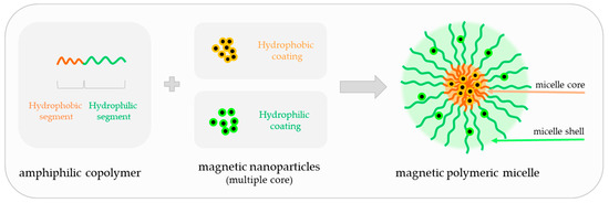
Hybrid nanoarchitectures such as magnetic polymeric micelles (MPMs) are among the most promising nanotechnology-enabled materials for biomedical applications combining the benefits of polymeric micelles and magnetic nanoparticles within a single bioinstructive system. MPMs are formed by the self-assembly of polymer amphiphiles above the critical micelle concentration, generating a colloidal structure with a hydrophobic core and a hydrophilic shell incorporating magnetic particles (MNPs) in one of the segments. MPMs have been investigated most prominently as contrast agents for magnetic resonance imaging (MRI), as heat generators in hyperthermia treatments, and as magnetic-susceptible nanocarriers for the delivery and release of therapeutic agents. The versatility of MPMs constitutes a powerful route to ultrasensitive, precise, and multifunctional diagnostic and therapeutic vehicles for the treatment of a wide range of pathologies. Although MPMs have been significantly explored for MRI and cancer therapy, MPMs are multipurpose functional units, widening their applicability into less expected fields of research such as bioengineering and regenerative medicine. Herein, we aim to review published reports of the last five years about MPMs concerning their structure and fabrication methods as well as their current and foreseen expectations for advanced biomedical applications. Full article
(This article belongs to the Special Issue Nanotechnology in Cancer Treatment 3.0)
Show Figures

Figure 1

18 pages, 3562 KiB  
Article
Polo-Like Kinase 2 Plays an Essential Role in Cytoprotection against MG132-Induced Proteasome Inhibition via Phosphorylation of Serine 19 in HSPB5
by Shuji Ueda, Moeka Nishihara, Yuuki Hioka, Ken-ichi Yoshino, Soichiro Yamada, Minoru Yamanoue and Yasuhito Shirai
Int. J. Mol. Sci. 2022, 23(19), 11257; https://doi.org/10.3390/ijms231911257 - 24 Sep 2022
Cited by 3 | Viewed by 3158
Abstract
Protein homeostasis, including protein folding, refolding, and degradation, is thought to decline with aging. HSPB5 (also known as αB-crystallin) prevents target protein aggregation as a molecular chaperone and exhibits a cytoprotective function against various cell stresses. To elucidate the effect of HSPB5 on [...] Read more.
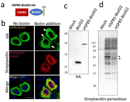
Protein homeostasis, including protein folding, refolding, and degradation, is thought to decline with aging. HSPB5 (also known as αB-crystallin) prevents target protein aggregation as a molecular chaperone and exhibits a cytoprotective function against various cell stresses. To elucidate the effect of HSPB5 on endoplasmic reticulum (ER) stress, we searched for novel binding proteins of HSPB5 using the proximity-dependent biotin labeling method. Proteins presumed to interact with HSPB5 in cells treated with the proteasome inhibitor MG132 were identified by a reversible biotin-binding capacity method combining tamavidin2-REV magnetic beads and mass spectrometry. We discovered a new binding protein for HSPB5, polo-like kinase 2 (PLK2), which is an apoptosis-related enzyme. The expression of PLK2 was upregulated by MG132 treatment, and it was co-localized with HSPB5 near the ER in L6 muscle cells. Inhibition of PLK2 decreased ER stress-induced phosphorylation of serine 19 in HSPB5 and increased apoptosis by activation of caspase 3 under ER stress. Overexpression of HSPB5 (WT) suppressed the ER stress-induced caspase 3 activity, but this was not observed with phospho-deficient HSPB5 (3A) mutants. These results clarify the role of HSPB5 phosphorylation during ER stress and suggest that the PLK2/HSPB5 pathway plays an essential role in cytoprotection against proteasome inhibition-induced ER stress. Full article
(This article belongs to the Special Issue Molecular Research of Aging Stress Response)
Show Figures

Figure 1

14 pages, 2454 KiB  
Article
Hepatic Sam68 Regulates Systemic Glucose Homeostasis and Insulin Sensitivity
by Aijun Qiao, Wenxia Ma, Ying Jiang, Chaoshan Han, Baolong Yan, Junlan Zhou and Gangjian Qin
Int. J. Mol. Sci. 2022, 23(19), 11469; https://doi.org/10.3390/ijms231911469 - 29 Sep 2022
Cited by 2 | Viewed by 3152
Abstract
Hepatic glucose production (HGP) is an important component of glucose homeostasis, and deregulated HGP, particularly through gluconeogenesis, contributes to hyperglycemia and pathology of type-2 diabetes (T2D). It has been shown that the gluconeogenic gene expression is governed primarily by the transcription factor cAMP-response [...] Read more.
Hepatic glucose production (HGP) is an important component of glucose homeostasis, and deregulated HGP, particularly through gluconeogenesis, contributes to hyperglycemia and pathology of type-2 diabetes (T2D). It has been shown that the gluconeogenic gene expression is governed primarily by the transcription factor cAMP-response element (CRE)-binding protein (CREB) and its coactivator, CREB-regulated transcriptional coactivator 2 (CRTC2). Recently, we have discovered that Sam68, an adaptor protein and Src kinase substrate, potently promotes hepatic gluconeogenesis by promoting CRTC2 stability; however, the detailed mechanisms remain unclear. Here we show that in response to glucagon, Sam68 increases CREB/CRTC2 transactivity by interacting with CRTC2 in the CREB/CRTC2 complex and occupying the CRE motif of promoters, leading to gluconeogenic gene expression and glucose production. In hepatocytes, glucagon promotes Sam68 nuclear import, whereas insulin elicits its nuclear export. Furthermore, ablation of Sam68 in hepatocytes protects mice from high-fat diet (HFD)-induced hyperglycemia and significantly increased hepatic and peripheral insulin sensitivities. Thus, hepatic Sam68 potentiates CREB/CRTC2-mediated glucose production, contributes to the pathogenesis of insulin resistance, and may serve as a therapeutic target for T2D. Full article
Show Figures

Figure 1

7 pages, 840 KiB  
Article
Familial Linkage and Association of the NR3C1 Gene with Type 2 Diabetes and Depression Comorbidity
by Mutaz Amin, Shumail Syed, Rongling Wu, Teodor Tudorel Postolache and Claudia Gragnoli
Int. J. Mol. Sci. 2022, 23(19), 11951; https://doi.org/10.3390/ijms231911951 - 8 Oct 2022
Cited by 8 | Viewed by 3148
Abstract
Impairment in the hypothalamic-pituitary-adrenal (HPA) axis and cortisol pathway may be major contributing factors to the common pathogenesis of major depressive disorders (MDD) and type 2 diabetes (T2D). A significant player in the neuroendocrine HPA axis and cortisol response is the glucocorticoid receptor [...] Read more.
Impairment in the hypothalamic-pituitary-adrenal (HPA) axis and cortisol pathway may be major contributing factors to the common pathogenesis of major depressive disorders (MDD) and type 2 diabetes (T2D). A significant player in the neuroendocrine HPA axis and cortisol response is the glucocorticoid receptor (GR), which is encoded by the nuclear receptor subfamily 3 group C member (NR3C1) gene. Variants in the NR3C1 gene have been reported in patients with MDD and obesity and found to confer reduced risk for quantitative metabolic traits and T2D in Cushing syndrome; variants have not been reported in T2D and MDD-T2D comorbid patients. We studied 212 original Italian families with a rich family history for T2D and tested 24 single nucleotide polymorphisms (SNPs) in the NR3C1 gene for linkage to and linkage disequilibrium (LD) with T2D and MDD across different inheritance models. We identified a total of 6 novel SNPs significantly linked/in LD to/with T2D (rs6196, rs10482633, rs13186836, rs13184611, rs10482681 and rs258751) and 1 SNP (rs10482668) significantly linked to/in LD with both T2D and MDD. These findings expand understanding of the role that NR3C1 variants play in modulating the risk of T2D-MDD comorbidity. Replication and functional studies are needed to confirm these findings. Full article
(This article belongs to the Section Molecular Pathology, Diagnostics, and Therapeutics)
Show Figures

Figure 1

12 pages, 1116 KiB  
Review
Pyroptosis and Insulin Resistance in Metabolic Organs
by Huiting Wei and Di Cui
Int. J. Mol. Sci. 2022, 23(19), 11638; https://doi.org/10.3390/ijms231911638 - 1 Oct 2022
Cited by 7 | Viewed by 3137
Abstract
Skeletal muscle serves as the optimal effective organ to balance glucose homeostasis, but insulin resistance (IR) in skeletal muscle breaks this balance by impeding glucose uptake and causes metabolic disorders. IR in skeletal muscle is caused by multiple factors, and it has been [...] Read more.
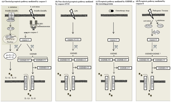
Skeletal muscle serves as the optimal effective organ to balance glucose homeostasis, but insulin resistance (IR) in skeletal muscle breaks this balance by impeding glucose uptake and causes metabolic disorders. IR in skeletal muscle is caused by multiple factors, and it has been reported that systemic low-grade inflammation is related to skeletal muscle IR, though its molecular mechanisms need to be ulteriorly studied. Pyroptosis is a novel inflammatory-mediated type of cell death. It has recently been reported that pyroptosis is associated with a decline in insulin sensitivity in skeletal muscle. The appropriate occurrence of pyroptosis positively eliminates pathogenic factors, whereas its excessive activation may aggravate inflammatory responses and expedite disease progression. The relationship between pyroptosis and IR in skeletal muscle and its underlined mechanism need to be further illustrated. The role of pyroptosis during the process of IR alleviation induced by non-drug interventions, such as exercise, also needs to be clarified. In this paper, we review and describe the molecular mechanisms of pyroptosis and further comb the roles of its relevant key factors in skeletal muscle IR, aiming to propose a novel theoretical basis for the relationship between pyroptosis and muscle IR and provide new research targets for the improvement of IR-related diseases. Full article
(This article belongs to the Special Issue Oxidative Stress and Skeletal Muscle Function)
Show Figures

Figure 1

13 pages, 1747 KiB  
Article
Carvedilol Selectively Stimulates βArrestin2-Dependent SERCA2a Activity in Cardiomyocytes to Augment Contractility
by Jennifer Maning, Victoria L. Desimine, Celina M. Pollard, Jennifer Ghandour and Anastasios Lymperopoulos
Int. J. Mol. Sci. 2022, 23(19), 11315; https://doi.org/10.3390/ijms231911315 - 26 Sep 2022
Cited by 12 | Viewed by 3136
Abstract
Heart failure (HF) carries the highest mortality in the western world and β-blockers [β-adrenergic receptor (AR) antagonists] are part of the cornerstone pharmacotherapy for post-myocardial infarction (MI) chronic HF. Cardiac β1AR-activated βarrestin2, a G protein-coupled receptor (GPCR) adapter protein, promotes Sarco(endo)plasmic [...] Read more.
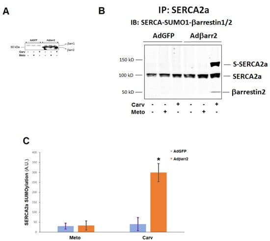
Heart failure (HF) carries the highest mortality in the western world and β-blockers [β-adrenergic receptor (AR) antagonists] are part of the cornerstone pharmacotherapy for post-myocardial infarction (MI) chronic HF. Cardiac β1AR-activated βarrestin2, a G protein-coupled receptor (GPCR) adapter protein, promotes Sarco(endo)plasmic reticulum Ca2+-ATPase (SERCA)2a SUMO (small ubiquitin-like modifier)-ylation and activity, thereby directly increasing cardiac contractility. Given that certain β-blockers, such as carvedilol and metoprolol, can activate βarrestins and/or SERCA2a in the heart, we investigated the effects of these two agents on cardiac βarrestin2-dependent SERCA2a SUMOylation and activity. We found that carvedilol, but not metoprolol, acutely induces βarrestin2 interaction with SERCA2a in H9c2 cardiomyocytes and in neonatal rat ventricular myocytes (NRVMs), resulting in enhanced SERCA2a SUMOylation. However, this translates into enhanced SERCA2a activity only in the presence of the β2AR-selective inverse agonist ICI 118,551 (ICI), indicating an opposing effect of carvedilol-occupied β2AR subtype on carvedilol-occupied β1AR-stimulated, βarrestin2-dependent SERCA2a activation. In addition, the amplitude of fractional shortening of NRVMs, transfected to overexpress βarrestin2, is acutely enhanced by carvedilol, again in the presence of ICI only. In contrast, metoprolol was without effect on NRVMs’ shortening amplitude irrespective of ICI co-treatment. Importantly, the pro-contractile effect of carvedilol was also observed in human induced pluripotent stem cell (hIPSC)-derived cardiac myocytes (CMs) overexpressing βarrestin2, and, in fact, it was present even without concomitant ICI treatment of human CMs. Metoprolol with or without concomitant ICI did not affect contractility of human CMs, either. In conclusion, carvedilol, but not metoprolol, stimulates βarrestin2-mediated SERCA2a SUMOylation and activity through the β1AR in cardiac myocytes, translating into direct positive inotropy. However, this unique βarrestin2-dependent pro-contractile effect of carvedilol may be opposed or masked by carvedilol-bound β2AR subtype signaling. Full article
(This article belongs to the Special Issue Role of G-Protein Coupled Receptors in Cardiovascular Diseases)
Show Figures

Figure 1