by
Laia Bertran 1,† , Marta Portillo-Carrasquer 1,†, Andrea Barrientos-Riosalido 1, Carmen Aguilar 1, David Riesco 2, Salomé Martínez 3, Amada Culebradas 4, Margarita Vives 4, Fàtima Sabench 4, Daniel Del Castillo 4, Cristóbal Richart 1,2 and Teresa Auguet 1,2,*
, Marta Portillo-Carrasquer 1,†, Andrea Barrientos-Riosalido 1, Carmen Aguilar 1, David Riesco 2, Salomé Martínez 3, Amada Culebradas 4, Margarita Vives 4, Fàtima Sabench 4, Daniel Del Castillo 4, Cristóbal Richart 1,2 and Teresa Auguet 1,2,*
1
Grup de Recerca GEMMAIR (AGAUR)—Medicina Aplicada (URV), Departament de Medicina i Cirurgia, Universitat Rovira i Virgili (URV), Institut d’Investigació Sanitària Pere Virgili (IISPV), 43007 Tarragona, Spain
2
Servei Medicina Interna, Hospital Universitari de Tarragona Joan XXIII, Mallafré Guasch, 4, 43007 Tarragona, Spain
3
Servei Anatomia Patològica, Hospital Universitari de Tarragona Joan XXIII, Mallafré Guasch, 4, 43007 Tarragona, Spain
4
Servei de Cirurgia, Hospital Sant Joan de Reus, Departament de Medicina i Cirurgia, Universitat Rovira i Virgili (URV), IISPV, Avinguda Doctor Josep Laporte, 2, 43204 Reus, Spain
†
These authors contributed equally to this work.
Int. J. Mol. Sci. 2022, 23(17), 9871; https://doi.org/10.3390/ijms23179871 - 30 Aug 2022
Cited by 5 | Viewed by 3659
Abstract
Secreted frizzled-related protein 5 (SFRP5) is an anti-inflammatory adipocytokine secreted by adipocytes that seems to be linked with nonalcoholic fatty liver disease (NAFLD). We aimed to evaluate the role of the SFRP5-wingless-MMTV integration site family member 5a (WNT5A) pathway, closely related to adipogenesis,
[...] Read more.
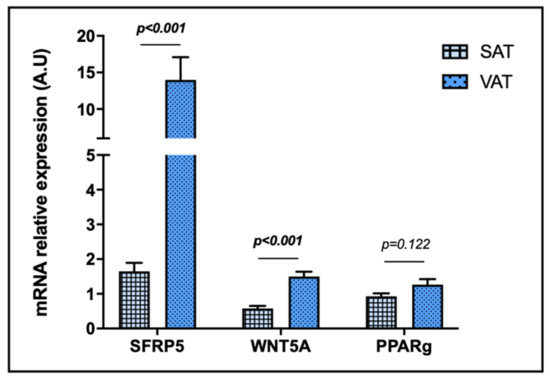
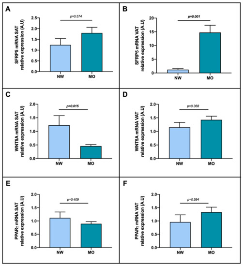
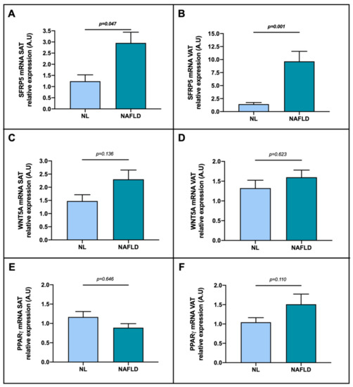
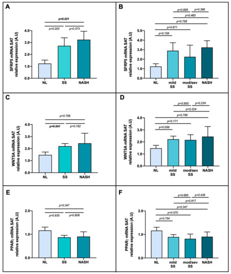
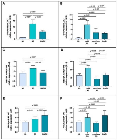
Secreted frizzled-related protein 5 (SFRP5) is an anti-inflammatory adipocytokine secreted by adipocytes that seems to be linked with nonalcoholic fatty liver disease (NAFLD). We aimed to evaluate the role of the SFRP5-wingless-MMTV integration site family member 5a (WNT5A) pathway, closely related to adipogenesis, in subcutaneous (SAT) and visceral adipose tissues (VAT) and its relationship with obesity-related NAFLD. Our cohort was composed of 60 women with morbid obesity (MO), who underwent hypocaloric diet, subclassified according to their hepatic histopathology and 15 women with normal weight. We observed increased SFRP5 mRNA expression in VAT and lower WNT5A expression in SAT in MO compared to normal weight. We found elevated SFRP5 expression in nonalcoholic steatohepatitis (NASH) in SAT and in mild simple steatosis (SS) and NASH in VAT. We observed higher WNT5A expression in SS compared to normal liver in SAT, and a peak of WNT5A expression in mild SS. To conclude, we reported increased SFRP5 mRNA expression in SAT and VAT of NAFLD-related to obesity subjects, suggesting an implication of the SFRP5-WNT5A pathway in NAFLD pathogenesis, probably due to the adipose tissue-liver axis. Since the mechanisms by which this potential interaction takes place remain elusive, more research in this field is needed.
Full article
(This article belongs to the Special Issue Nutrition and Metabolism in Health and Disease: From Gene to Organism)
▼
Show Figures
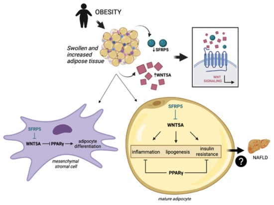
Figure 1


