Int. J. Mol. Sci. 2022, 23(16), 8997; https://doi.org/10.3390/ijms23168997 - 12 Aug 2022
Cited by 15 | Viewed by 3864
Abstract
This study aimed to discuss the role of 12/15-lipoxygenase (12/15-LOX) regulation involved in diabetes cognitive dysfunction. First, Mini Mental State Examination (MMSE) test was used to evaluate cognitive ability in diabetic patients and normal controls. The plasma test showed that the plasma level
[...] Read more.
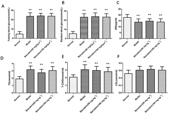
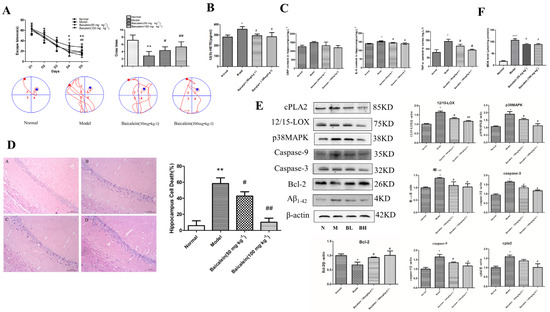
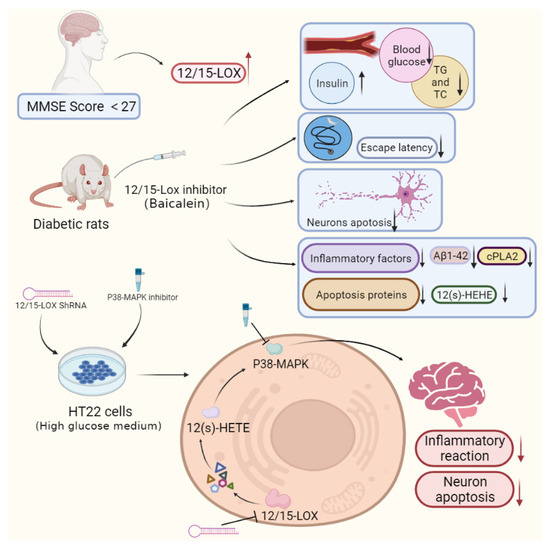
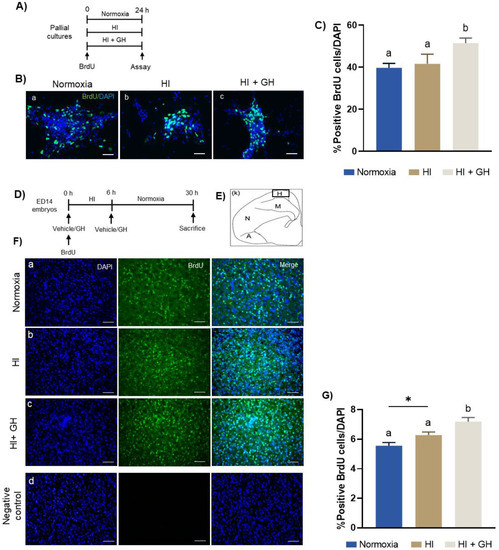
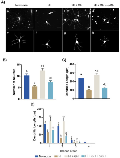
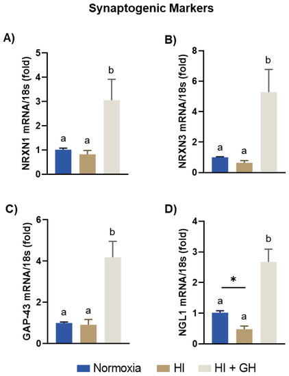
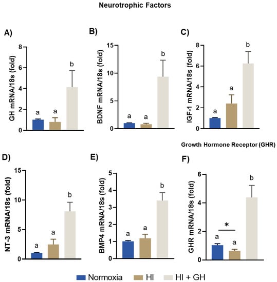
This study aimed to discuss the role of 12/15-lipoxygenase (12/15-LOX) regulation involved in diabetes cognitive dysfunction. First, Mini Mental State Examination (MMSE) test was used to evaluate cognitive ability in diabetic patients and normal controls. The plasma test showed that the plasma level of 12/15-LOX in patients with MMSE scores below 27 was significantly increased compared with that of the normal group. Second, 12/15-LOX inhibitor was administered to diabetic rats. Behavioral tests, biochemistry, enzyme-linked immunosorbent assays, and Western blotting were used in this study. We found that the levels of fasting and random blood glucose increased rapidly in diabetic rats, the levels of triglycerides and total cholesterol in the diabetic group increased, and insulin levels decreased significantly. In the Morris water maze test, the escape latency was prolonged, and the crossing times decreased in the diabetic group. Under the microscope, the apoptosis of hippocampal neurons in diabetic rats increased significantly. The levels of TNF-α, IL-6 and 12-hydroxyindoleic acid (12(S)-HETE) significantly increased, and the protein expression of 12/15-LOX, p38 MAPK, Aβ1-42, caspase-3, caspase-9 and cPLA2 increased, while that of Bcl-2 decreased. However, the use of 12/15-LOX inhibitor reversed these results. Third, 12/15-LOX shRNA and p38MAPK inhibitor were administered to HT22 cells in high-glucose medium. The results of the cell experiment were consistent with those of the animal experiment. Our results indicated that the 12/15-LOX pathway participates in diabetic brain damage by activating p38MAPK to promote inflammation and neuronal apoptosis, and intervention 12/15-LOX can improve diabetic cognitive dysfunction.
Full article
(This article belongs to the Special Issue Molecular Signals and Genetic Regulations of Neurological Disorders)
►
Show Figures

Figure 1


