Int. J. Mol. Sci. 2021, 22(22), 12417; https://doi.org/10.3390/ijms222212417 - 17 Nov 2021
Cited by 18 | Viewed by 3349
Abstract
During angiogenesis, cell adhesion molecules expressed on the endothelial cell surface promote the growth and survival of newly forming vessels. Hence, elucidation of the signaling pathways activated by cell-to-matrix adhesion may assist in the discovery of new targets to be used in antiangiogenic
[...] Read more.
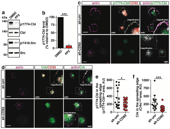
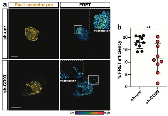
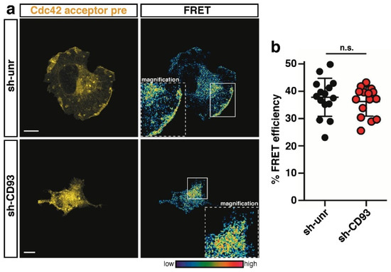
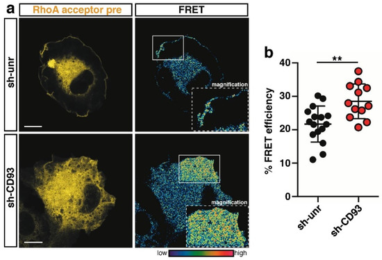
During angiogenesis, cell adhesion molecules expressed on the endothelial cell surface promote the growth and survival of newly forming vessels. Hence, elucidation of the signaling pathways activated by cell-to-matrix adhesion may assist in the discovery of new targets to be used in antiangiogenic therapy. In proliferating endothelial cells, the single-pass transmembrane glycoprotein CD93 has recently emerged as an important endothelial cell adhesion molecule regulating vascular maturation. In this study, we unveil a signaling pathway triggered by CD93 that regulates actin cytoskeletal dynamics responsible of endothelial cell adhesion. We show that the Src-dependent phosphorylation of CD93 and the adaptor protein Cbl leads to the recruitment of Crk, which works as a downstream integrator in the CD93-mediated signaling. Moreover, confocal microscopy analysis of FRET-based biosensors shows that CD93 drives the coordinated activation of Rac1 and RhoA at the cell edge of spreading cells, thus promoting the establishment of cell polarity and adhesion required for cell motility.
Full article
(This article belongs to the Special Issue Cell and Molecular Interactions in Blood Vessels 2021)
►
Show Figures
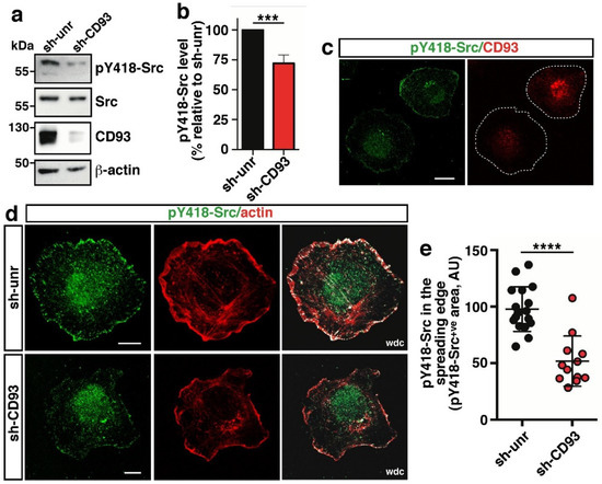
Figure 1


