Int. J. Mol. Sci. 2016, 17(2), 70; https://doi.org/10.3390/ijms17020070 - 2 Feb 2016
Cited by 12 | Viewed by 6341
Abstract
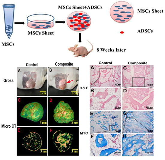
To determine the effect of adipose-derived stem cells (ADSCs) added to bone marrow-derived mesenchymal stem cell (MSC) sheets on bone formation at an ectopic site. We isolated MSCs and ADSCs from the same rabbits. We then prepared MSC sheets for implantation with or
[...] Read more.
To determine the effect of adipose-derived stem cells (ADSCs) added to bone marrow-derived mesenchymal stem cell (MSC) sheets on bone formation at an ectopic site. We isolated MSCs and ADSCs from the same rabbits. We then prepared MSC sheets for implantation with or without ADSCs subcutaneously in the backs of severe combined immunodeficiency (SCID) mice. We assessed bone formation at eight weeks after implantation by micro-computed tomography and histological analysis. In osteogenic medium, MSCs grew to form multilayer sheets containing many calcium nodules. MSC sheets without ADSCs formed bone-like tissue; although neo-bone and cartilage-like tissues were sparse and unevenly distributed by eight weeks after implantation. In comparison, MSC sheets with ADSCs promoted better bone regeneration as evidenced by the greater density of bone, increased mineral deposition, obvious formation of blood vessels, large number of interconnected ossified trabeculae and woven bone structures, and greater bone volume/total volume within the composite constructs. Our results indicate that although sheets of only MSCs have the potential to form tissue engineered bone at an ectopic site, the addition of ADSCs can significantly increase the osteogenic potential of MSC sheets. Thus, the combination of MSC sheets with ADSCs may be regarded as a promising therapeutic strategy to stimulate bone regeneration.
Full article
(This article belongs to the Section Biochemistry)
►
Show Figures
Graphical abstract


