14 pages, 1542 KiB  
Article
Exogenous Carbon Monoxide Decreases Sepsis-Induced Acute Kidney Injury and Inhibits NLRP3 Inflammasome Activation in Rats
by Peng Wang 1,2,†, Jian Huang 3,†, Yi Li 1,2, Ruiming Chang 1, Haidong Wu 1, Jiali Lin 1,2 and Zitong Huang 1,2,*
1 Department of Emergency Medicine, Sun Yat-sen Memorial Hospital of Sun Yat-sen University, Guangzhou 510120, China
2 Institute of Cardiopulmonary Cerebral Resuscitation, Sun Yat-sen University, Guangzhou 510120, China
3 Department of Nephrology, the Fourth Affiliated Hospital of Guangzhou Medical University, Guangzhou 510120, China
These authors contributed equally to this work.
Int. J. Mol. Sci. 2015, 16(9), 20595-20608; https://doi.org/10.3390/ijms160920595 - 31 Aug 2015
Cited by 67 | Viewed by 6950
Abstract
Carbon monoxide (CO) has shown various physiological effects including anti-inflammatory activity in several diseases, whereas the therapeutic efficacy of CO on sepsis-induced acute kidney injury (AKI) has not been reported as of yet. The purpose of the present study was to explore the [...] Read more.
Carbon monoxide (CO) has shown various physiological effects including anti-inflammatory activity in several diseases, whereas the therapeutic efficacy of CO on sepsis-induced acute kidney injury (AKI) has not been reported as of yet. The purpose of the present study was to explore the effects of exogenous CO on sepsis-induced AKI and nucleotide-binding domain-like receptor protein 3 (NLRP3) inflammasome activation in rats. Male rats were subjected to cecal ligation and puncture (CLP) to induce sepsis and AKI. Exogenous CO delivered from CO-releasing molecule 2 (CORM-2) was used intraperitoneally as intervention after CLP surgery. Therapeutic effects of CORM-2 on sepsis-induced AKI were assessed by measuring serum creatinine (Scr) and blood urea nitrogen (BUN), kidney histology scores, apoptotic cell scores, oxidative stress, levels of cytokines TNF-α and IL-1β, and NLRP3 inflammasome expression. CORM-2 treatment protected against the sepsis-induced AKI as evidenced by reducing serum Scr/BUN levels, apoptotic cells scores, increasing survival rates, and decreasing renal histology scores. Furthermore, treatment with CORM-2 significantly reduced TNF-α and IL-1β levels and oxidative stress. Moreover, CORM-2 treatment significantly decreased NLRP3 inflammasome protein expressions. Our study provided evidence that CORM-2 treatment protected against sepsis-induced AKI and inhibited NLRP3 inflammasome activation, and suggested that CORM-2 could be a potential therapeutic candidate for treating sepsis-induced AKI. Full article
(This article belongs to the Section Molecular Pathology, Diagnostics, and Therapeutics)
Show Figures

Figure 1

19 pages, 1326 KiB  
Article
Impact of Chronic Hepatitis C Virus Genotype 1b Infection on Triglyceride Concentration in Serum Lipoprotein Fractions
by Tomohisa Nagano 1,*, Nobuyoshi Seki 1, Yoichi Tomita 1, Tomonori Sugita 1, Yuta Aida 1, Munenori Itagaki 1, Satoshi Sutoh 1, Hiroshi Abe 1, Akihito Tsubota 2 and Yoshio Aizawa 1
1 Department of Gastroenterology and Hepatology Internal Medicine, Jikei University Katsushika Medical Center, 6-41-2 Aoto, Katsushika-ku, Tokyo 125-8506, Japan
2 Core Research Facilities for Basic Science, Research Center for Medical Science, Jikei University School of Medicine, 3-25-8 Nishi-Shimbashi, Minato-ku, Tokyo 105-8461, Japan
Int. J. Mol. Sci. 2015, 16(9), 20576-20594; https://doi.org/10.3390/ijms160920576 - 31 Aug 2015
Cited by 14 | Viewed by 6766
Abstract
Reduced low-density lipoprotein (LDL) cholesterol level is a characteristic feature of dyslipidemia in chronic hepatitis C virus (HCV) infection. However, abnormality in serum triglyceride (TG) has not been fully investigated. To clarify the impact of HCV genotype 1b (G1b) infection and advanced fibrosis [...] Read more.
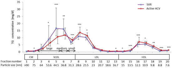
Reduced low-density lipoprotein (LDL) cholesterol level is a characteristic feature of dyslipidemia in chronic hepatitis C virus (HCV) infection. However, abnormality in serum triglyceride (TG) has not been fully investigated. To clarify the impact of HCV genotype 1b (G1b) infection and advanced fibrosis on serum TG profiles, TG concentrations in lipoprotein fractions were examined in fasting sera from 185 subjects with active or cleared HCV infection by high-performance liquid chromatography. Serum lipoproteins were fractionated into four classes: chylomicron, very low-density lipoprotein (VLDL), LDL, and high-density lipoprotein (HDL). Then, the significance of HCV G1b infection on TG levels in each lipoprotein fraction was determined using multiple regression models. We found that active HCV G1b infection was positively associated with high HDL-TG levels and low VLDL-TG levels, independent of other factors included in the regression model. In VLDL sub-fractions, active HCV infection was only found to be associated with low levels of large VLDL-TG. Similarly, advanced liver fibrosis in chronic HCV G1b infection was associated with high levels of LDL-TG, HDL-TG, and small VLDL-TG, independent of other clinical factors. These findings indicate that active HCV G1b infection and advanced fibrosis are closely associated with abnormal serum TG profiles. Full article
(This article belongs to the Special Issue Viral Hepatitis Research)
Show Figures

Graphical abstract

16 pages, 1293 KiB  
Review
Biomarkers of Chondrocyte Apoptosis and Autophagy in Osteoarthritis
by Giuseppe Musumeci 1,*, Paola Castrogiovanni 1, Francesca Maria Trovato 2, Annelie Martina Weinberg 3, Mohammad K. Al-Wasiyah 4,5, Mohammed H. Alqahtani 4 and Ali Mobasheri 5,6,7
1 Department of Biomedical and Biotechnological Sciences, Human Anatomy and Histology Section, School of Medicine, University of Catania, Catania 95123, Italy
2 Department of Clinical and Experimental Medicine, Internal Medicine Division, School of Medicine, University of Catania, Catania 95123, Italy
3 Department of Orthopaedic Surgery, Medical University of Graz, 8036 Graz, Austria
4 Aziziah Maternity and Children's Hospital, Jeddah 50204, Saudi Arabia
5 King Fahd Medical Research Center (KFMRC), King AbdulAziz University, Jeddah 21589, Saudi Arabia
6 The D-BOARD European Consortium for Biomarker Discovery, Department of Veterinary Preclinical Sciences, School of Veterinary Medicine, Faculty of Health and Medical Sciences, University of Surrey, Guildford GU2 7XH, UK
7 Arthritis Research UK Centre for Sport, Exercise and Osteoarthritis, Arthritis Research UK Pain Centre, Medical Research Council and Arthritis Research UK Centre for Musculoskeletal Ageing Research, University of Nottingham, Queen's Medical Centre, Nottingham NG7 2UH, UK
Int. J. Mol. Sci. 2015, 16(9), 20560-20575; https://doi.org/10.3390/ijms160920560 - 31 Aug 2015
Cited by 250 | Viewed by 18737
Abstract
Cell death with morphological and molecular features of apoptosis has been detected in osteoarthritic (OA) cartilage, which suggests a key role for chondrocyte death/survival in the pathogenesis of OA. Identification of biomarkers of chondrocyte apoptosis may facilitate the development of novel therapies that [...] Read more.
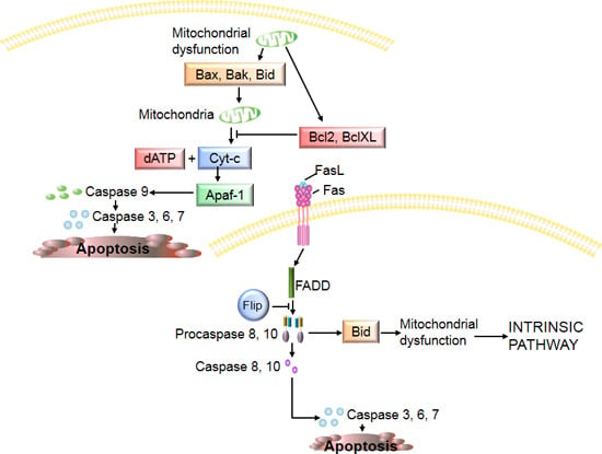
Cell death with morphological and molecular features of apoptosis has been detected in osteoarthritic (OA) cartilage, which suggests a key role for chondrocyte death/survival in the pathogenesis of OA. Identification of biomarkers of chondrocyte apoptosis may facilitate the development of novel therapies that may eliminate the cause or, at least, slow down the degenerative processes in OA. The aim of this review was to explore the molecular markers and signals that induce chondrocyte apoptosis in OA. A literature search was conducted in PubMed, Scopus, Web of Science and Google Scholar using the keywords chondrocyte death, apoptosis, osteoarthritis, autophagy and biomarker. Several molecules considered to be markers of chondrocyte apoptosis will be discussed in this brief review. Molecular markers and signalling pathways associated with chondroycte apoptosis may turn out to be therapeutic targets in OA and approaches aimed at neutralizing apoptosis-inducing molecules may at least delay the progression of cartilage degeneration in OA. Full article
(This article belongs to the Special Issue Apoptotic Chondrocytes and Osteoarthritis)
Show Figures

Graphical abstract

37 pages, 1663 KiB  
Review
Photodynamic Efficiency: From Molecular Photochemistry to Cell Death
by Isabel O. L. Bacellar 1, Tayana M. Tsubone 1, Christiane Pavani 2 and Mauricio S. Baptista 1,*
1 Instituto de Química, Universidade de São Paulo, São Paulo 05508-900, Brazil
2 Programa de Pós Graduação em Biofotônica Aplicada às Ciências da Saúde, Universidade Nove de Julho, São Paulo 01504-001, Brazil
Int. J. Mol. Sci. 2015, 16(9), 20523-20559; https://doi.org/10.3390/ijms160920523 - 31 Aug 2015
Cited by 341 | Viewed by 14948
Abstract
Photodynamic therapy (PDT) is a clinical modality used to treat cancer and infectious diseases. The main agent is the photosensitizer (PS), which is excited by light and converted to a triplet excited state. This latter species leads to the formation of singlet oxygen [...] Read more.
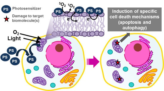
Photodynamic therapy (PDT) is a clinical modality used to treat cancer and infectious diseases. The main agent is the photosensitizer (PS), which is excited by light and converted to a triplet excited state. This latter species leads to the formation of singlet oxygen and radicals that oxidize biomolecules. The main motivation for this review is to suggest alternatives for achieving high-efficiency PDT protocols, by taking advantage of knowledge on the chemical and biological processes taking place during and after photosensitization. We defend that in order to obtain specific mechanisms of cell death and maximize PDT efficiency, PSes should oxidize specific molecular targets. We consider the role of subcellular localization, how PS photochemistry and photophysics can change according to its nanoenvironment, and how can all these trigger specific cell death mechanisms. We propose that in order to develop PSes that will cause a breakthrough enhancement in the efficiency of PDT, researchers should first consider tissue and intracellular localization, instead of trying to maximize singlet oxygen quantum yields in in vitro tests. In addition to this, we also indicate many open questions and challenges remaining in this field, hoping to encourage future research. Full article
(This article belongs to the Special Issue Advances in Photodynamic Therapy)
Show Figures

Graphical abstract

12 pages, 1265 KiB  
Communication
Supracolloidal Assemblies as Sacrificial Templates for Porous Silk-Based Biomaterials
by John G. Hardy 1,2,3,*, Chiara E. Ghezzi 3, Richard J. Saballos 1, David L. Kaplan 3,* and Christine E. Schmidt 1,2,*
1 J. Crayton Pruitt Family Department of Biomedical Engineering, University of Florida, Biomedical Sciences Building JG-53, P.O. Box 116131, Gainesville, FL 32611-6131, USA
2 Department of Biomedical Engineering, the University of Texas at Austin, Austin, TX 78712, USA
3 Department of Biomedical Engineering, Tufts University, Medford, MA 02155, USA
Int. J. Mol. Sci. 2015, 16(9), 20511-20522; https://doi.org/10.3390/ijms160920511 - 28 Aug 2015
Cited by 6 | Viewed by 6850
Abstract
Tissues in the body are hierarchically structured composite materials with tissue-specific properties. Urea self-assembles via hydrogen bonding interactions into crystalline supracolloidal assemblies that can be used to impart macroscopic pores to polymer-based tissue scaffolds. In this communication, we explain the solvent interactions governing [...] Read more.
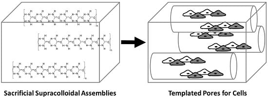
Tissues in the body are hierarchically structured composite materials with tissue-specific properties. Urea self-assembles via hydrogen bonding interactions into crystalline supracolloidal assemblies that can be used to impart macroscopic pores to polymer-based tissue scaffolds. In this communication, we explain the solvent interactions governing the solubility of urea and thereby the scope of compatible polymers. We also highlight the role of solvent interactions on the morphology of the resulting supracolloidal crystals. We elucidate the role of polymer-urea interactions on the morphology of the pores in the resulting biomaterials. Finally, we demonstrate that it is possible to use our urea templating methodology to prepare Bombyx mori silk protein-based biomaterials with pores that human dermal fibroblasts respond to by aligning with the long axis of the pores. This methodology has potential for application in a variety of different tissue engineering niches in which cell alignment is observed, including skin, bone, muscle and nerve. Full article
(This article belongs to the Special Issue Supramolecular Interactions)
Show Figures

Graphical abstract

19 pages, 1510 KiB  
Article
Influence of Parathyroid Hormone-Loaded PLGA Nanoparticles in Porous Scaffolds for Bone Regeneration
by Piergiorgio Gentile 1,†, Vijay Kumar Nandagiri 2,3,†, Ritesh Pabari 3, Jacqueline Daly 4, Chiara Tonda-Turo 2, Gianluca Ciardelli 2 and Zebunnissa Ramtoola 3,*
1 School of Clinical Dentistry, University of Sheffield, Sheffield S10 2TA, UK
2 Department of Mechanical and Aerospace Engineering, Politecnico di Torino, Turin 10129, Italy
3 School of Pharmacy, Royal College of Surgeons in Ireland, Dublin 2, Ireland
4 Division of Biology, Department of Anatomy, Royal College of Surgeons in Ireland, Dublin 2, Ireland
These authors contributed equally to this work.
Int. J. Mol. Sci. 2015, 16(9), 20492-20510; https://doi.org/10.3390/ijms160920492 - 28 Aug 2015
Cited by 29 | Viewed by 6810
Abstract
Biodegradable poly(lactide-co-glycolide) (PLGA) nanoparticles, containing human parathyroid hormone (PTH (1–34)), prepared by a modified double emulsion-solvent diffusion-evaporation method, were incorporated in porous freeze-dried chitosan-gelatin (CH-G) scaffolds. The PTH-loaded nanoparticles (NPTH) were characterised in terms of morphology, size, protein loading, release kinetics [...] Read more.
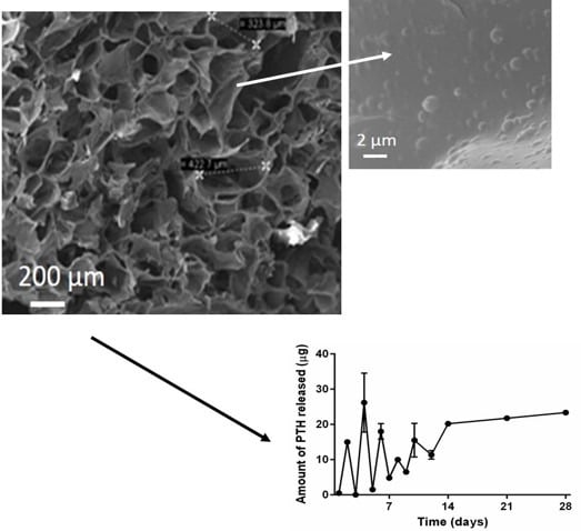
Biodegradable poly(lactide-co-glycolide) (PLGA) nanoparticles, containing human parathyroid hormone (PTH (1–34)), prepared by a modified double emulsion-solvent diffusion-evaporation method, were incorporated in porous freeze-dried chitosan-gelatin (CH-G) scaffolds. The PTH-loaded nanoparticles (NPTH) were characterised in terms of morphology, size, protein loading, release kinetics and in vitro assessment of biological activity of released PTH and cytocompatibility studies against clonal human osteoblast (hFOB) cells. Structural integrity of incorporated and released PTH from nanoparticles was found to be intact by using Tris-tricine SDS-PAGE. In vitro PTH release kinetics from PLGA nanoparticles were characterised by a burst release followed by a slow release phase for 3–4 weeks. The released PTH was biologically active as evidenced by the stimulated release of cyclic AMP from hFOB cells as well as increased mineralisation studies. in vitro and cell studies demonstrated that the PTH bioactivity was maintained during the fabrication of PLGA nanoparticles and upon release. Finally, a content of 33.3% w/w NPTHs was incorporated in CH-G scaffolds, showing an intermittent release during the first 10 days and, followed by a controlled release over 28 days of observation time. The increased expression of Alkaline Phosphatase levels on hFOB cells further confirmed the activity of intermittently released PTH from scaffolds. Full article
(This article belongs to the Special Issue Bioactive Nanoparticles)
Show Figures

Graphical abstract

24 pages, 1226 KiB  
Article
Evaluation of Appropriate Reference Genes for Reverse Transcription-Quantitative PCR Studies in Different Tissues of a Desert Poplar via Comparision of Different Algorithms
by Hou-Ling Wang 1,2,†, Lan Li 1,†, Sha Tang 3, Chao Yuan 1, Qianqian Tian 1, Yanyan Su 1, Hui-Guang Li 1, Lin Zhao 1, Weilun Yin 1,2, Rui Zhao 1,* and Xinli Xia 1,*
1 National Engineering Laboratory for Tree Breeding, College of Biological Sciences and Technology, Beijing Forestry University, Beijing 100083, China
2 The Key Laboratory for Silviculture and Conservation of Ministry Education, College of Forestry, Beijing Forestry University, Beijing 100083, China
3 Institute of Crop Sciences, Chinese Academy of Agricultural Sciences, Beijing 100081, China
These authors contributed equally to this work.
Int. J. Mol. Sci. 2015, 16(9), 20468-20491; https://doi.org/10.3390/ijms160920468 - 28 Aug 2015
Cited by 36 | Viewed by 7193
Abstract
Despite the unshakable status of reverse transcription-quantitative PCR in gene expression analysis, it has certain disadvantages, including that the results are highly dependent on the reference genes selected for data normalization. Since inappropriate endogenous control genes will lead to inaccurate target gene expression [...] Read more.
Despite the unshakable status of reverse transcription-quantitative PCR in gene expression analysis, it has certain disadvantages, including that the results are highly dependent on the reference genes selected for data normalization. Since inappropriate endogenous control genes will lead to inaccurate target gene expression profiles, the validation of suitable internal reference genes is essential. Given the increasing interest in functional genes and genomics of Populus euphratica, a desert poplar showing extraordinary adaptation to salt stress, we evaluated the expression stability of ten candidate reference genes in P. euphratica roots, stems, and leaves under salt stress conditions. We used five algorithms, namely, ΔCt, NormFinder, geNorm, GrayNorm, and a rank aggregation method (RankAggreg) to identify suitable normalizers. To support the suitability of the identified reference genes and to compare the relative merits of these different algorithms, we analyzed and compared the relative expression levels of nine P. euphratica functional genes in different tissues. Our results indicate that a combination of multiple reference genes recommended by GrayNorm algorithm (e.g., a combination of Actin, EF1α, GAPDH, RP, UBQ in root) should be used instead of a single reference gene. These results are valuable for research of gene identification in different P. euphratica tissues. Full article
(This article belongs to the Special Issue Abiotic Stress and Gene Networks in Plants)
Show Figures

Graphical abstract

19 pages, 993 KiB  
Article
Mechanistic and Kinetic Studies on the Homogeneous Gas-Phase Formation of PCTA/DTs from 2,4-Dichlorothiophenol and 2,4,6-Trichlorothiophenol
by Fei Xu, Xiangli Shi, Yunfeng Li and Qingzhu Zhang *
Environment Research Institute, Shandong University, Jinan 250100, China
Int. J. Mol. Sci. 2015, 16(9), 20449-20467; https://doi.org/10.3390/ijms160920449 - 28 Aug 2015
Cited by 12 | Viewed by 4728
Abstract
Polychlorinated thianthrene/dibenzothiophenes (PCTA/DTs) are sulfur analogues compounds to polychlorinated dibenzo-p-dioxin/dibenzofurans (PCDD/Fs). Chlorothiophenols (CTPs) are key precursors to form PCTA/DTs. 2,4-DCTP has the minimum number of Cl atoms to form 2,4,6,8-tetrachlorinated dibenzothiophenes (2,4,6,8-TeCDT), which is the most important and widely detected of the PCDTs. [...] Read more.
Polychlorinated thianthrene/dibenzothiophenes (PCTA/DTs) are sulfur analogues compounds to polychlorinated dibenzo-p-dioxin/dibenzofurans (PCDD/Fs). Chlorothiophenols (CTPs) are key precursors to form PCTA/DTs. 2,4-DCTP has the minimum number of Cl atoms to form 2,4,6,8-tetrachlorinated dibenzothiophenes (2,4,6,8-TeCDT), which is the most important and widely detected of the PCDTs. In this paper, quantum chemical calculations were carried out to investigate the homogeneous gas-phase formation of PCTA/DTs from 2,4-DCTP and 2,4,6-TCTP precursors at the MPWB1K/6-311+G(3df,2p)//MPWB1K/6-31+G(d,p) level. Several energetically feasible pathways were revealed to compare the formation potential of PCTA/DT products. The rate constants of the crucial elementary reactions were evaluated by the canonical variational transition-state (CVT) theory with the small curvature tunneling (SCT) correction over a wide temperature range of 600–1200 K. This study shows that pathways that ended with elimination of Cl step were dominant over pathways ended with elimination of the H step. The water molecule has a negative catalytic effect on the H-shift step and hinders the formation of PCDTs from 2,4-DCTP. This study, together with works already published from our group, clearly illustrates an increased propensity for the dioxin formation from CTPs over the analogous CPs. Full article
(This article belongs to the Section Physical Chemistry, Theoretical and Computational Chemistry)
Show Figures

Figure 1

18 pages, 1474 KiB  
Article
BMP9-Induced Survival Effect in Liver Tumor Cells Requires p38MAPK Activation
by María García-Álvaro 1, Annalisa Addante 1, Cesáreo Roncero 1, Margarita Fernández 1, Isabel Fabregat 2, Aránzazu Sánchez 1 and Blanca Herrera 1,*
1 Department of Biochemistry and Molecular Biology II, Faculty of Pharmacy, Complutense University of Madrid. San Carlos Clinical Hospital Health Research Institute (IdISSC), Plaza Ramón y Cajal S/N, Madrid 28040, Spain
2 Bellvitge Biomedical Research Institute (IDIBELL) and University of Barcelona (UB), L'Hospitalet de Llobregat, Barcelona 08908, Spain
Int. J. Mol. Sci. 2015, 16(9), 20431-20448; https://doi.org/10.3390/ijms160920431 - 28 Aug 2015
Cited by 24 | Viewed by 5836
Abstract
The study of bone morphogenetic proteins (BMPs) role in tumorigenic processes, and specifically in the liver, has gathered importance in the last few years. Previous studies have shown that BMP9 is overexpressed in about 40% of hepatocellular carcinoma (HCC) patients. In vitro data [...] Read more.
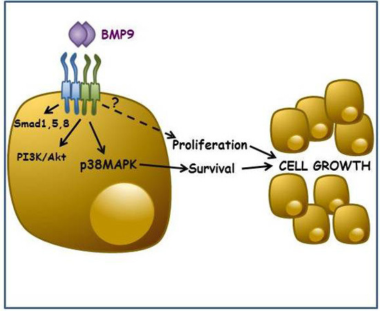
The study of bone morphogenetic proteins (BMPs) role in tumorigenic processes, and specifically in the liver, has gathered importance in the last few years. Previous studies have shown that BMP9 is overexpressed in about 40% of hepatocellular carcinoma (HCC) patients. In vitro data have also shown evidence that BMP9 has a pro-tumorigenic action, not only by inducing epithelial to mesenchymal transition (EMT) and migration, but also by promoting proliferation and survival in liver cancer cells. However, the precise mechanisms driving these effects have not yet been established. In the present work, we deepened our studies into the intracellular mechanisms implicated in the BMP9 proliferative and pro-survival effect on liver tumor cells. In HepG2 cells, BMP9 induces both Smad and non-Smad signaling cascades, specifically PI3K/AKT and p38MAPK. However, only the p38MAPK pathway contributes to the BMP9 growth-promoting effect on these cells. Using genetic and pharmacological approaches, we demonstrate that p38MAPK activation, although dispensable for the BMP9 proliferative activity, is required for the BMP9 protective effect on serum withdrawal-induced apoptosis. These findings contribute to a better understanding of the signaling pathways involved in the BMP9 pro-tumorigenic role in liver tumor cells. Full article
(This article belongs to the Special Issue Molecular Mechanisms of Human Liver Diseases)
Show Figures

Graphical abstract

14 pages, 5568 KiB  
Article
Photodynamic and Antibiotic Therapy in Combination to Fight Biofilms and Resistant Surface Bacterial Infections
by Federica Barra 1,†, Emanuela Roscetto 1,†, Amata A. Soriano 1, Adriana Vollaro 1, Ilaria Postiglione 1, Giovanna Maria Pierantoni 1,2, Giuseppe Palumbo 1,* and Maria Rosaria Catania 1
1 Department of Molecular Medicine and Medical Biotechnology, University of Naples Federico II, Naples 80131, Italy
2 Institute of Experimental Endocrinology and Oncology (IEOS), National Research Council (CNR), Naples 80131, Italy
These authors contributed equally to this work.
Int. J. Mol. Sci. 2015, 16(9), 20417-20430; https://doi.org/10.3390/ijms160920417 - 28 Aug 2015
Cited by 92 | Viewed by 7371
Abstract
Although photodynamic therapy (PDT), a therapeutic approach that involves a photosensitizer, light and O2, has been principally considered for the treatment of specific types of cancers, other applications exist, including the treatment of infections. Unfortunately, PDT does not always guarantee full [...] Read more.
Although photodynamic therapy (PDT), a therapeutic approach that involves a photosensitizer, light and O2, has been principally considered for the treatment of specific types of cancers, other applications exist, including the treatment of infections. Unfortunately, PDT does not always guarantee full success since it exerts lethal effects only in cells that have taken up a sufficient amount of photosensitizer and have been exposed to adequate light doses, conditions that are not always achieved. Based on our previous experience on the combination PDT/chemotherapy, we have explored the possibility of fighting bacteria that commonly crowd infected surfaces by combining PDT with an antibiotic, which normally does not harm the strain at low concentrations. To this purpose, we employed 5-aminolevulinic acid (5-ALA), a pro-drug that, once absorbed by proliferating bacteria, is converted into the natural photosensitizer Protoporphyrin IX (PpIX), followed by Gentamicin. Photoactivation generates reactive oxygen species (ROS) which damage or kill the cell, while Gentamicin, even at low doses, ends the work. Our experiments, in combination, have been highly successful against biofilms produced by several Gram positive bacteria (i.e., Staphylococcus aureus, Staphylococcus epidermidis, etc.). This original approach points to potentially new and wide applications in the therapy of infections of superficial wounds and sores. Full article
(This article belongs to the Special Issue Advances in Photodynamic Therapy)
Show Figures

Graphical abstract

25 pages, 1533 KiB  
Review
Abiotic Stresses: Insight into Gene Regulation and Protein Expression in Photosynthetic Pathways of Plants
by Mohammad-Zaman Nouri 1,*, Ali Moumeni 1 and Setsuko Komatsu 2,*
1 Rice Research Institute of Iran, Mazandaran Branch, Agricultural Research, Education and Extension Organization (AREEO), Amol 46191-91951, Iran
2 National Institute of Crop Science, National Agriculture and Food Research Organization, Tsukuba 305-8518, Japan
Int. J. Mol. Sci. 2015, 16(9), 20392-20416; https://doi.org/10.3390/ijms160920392 - 28 Aug 2015
Cited by 128 | Viewed by 10390
Abstract
Global warming and climate change intensified the occurrence and severity of abiotic stresses that seriously affect the growth and development of plants,especially, plant photosynthesis. The direct impact of abiotic stress on the activity of photosynthesis is disruption of all photosynthesis components such as [...] Read more.
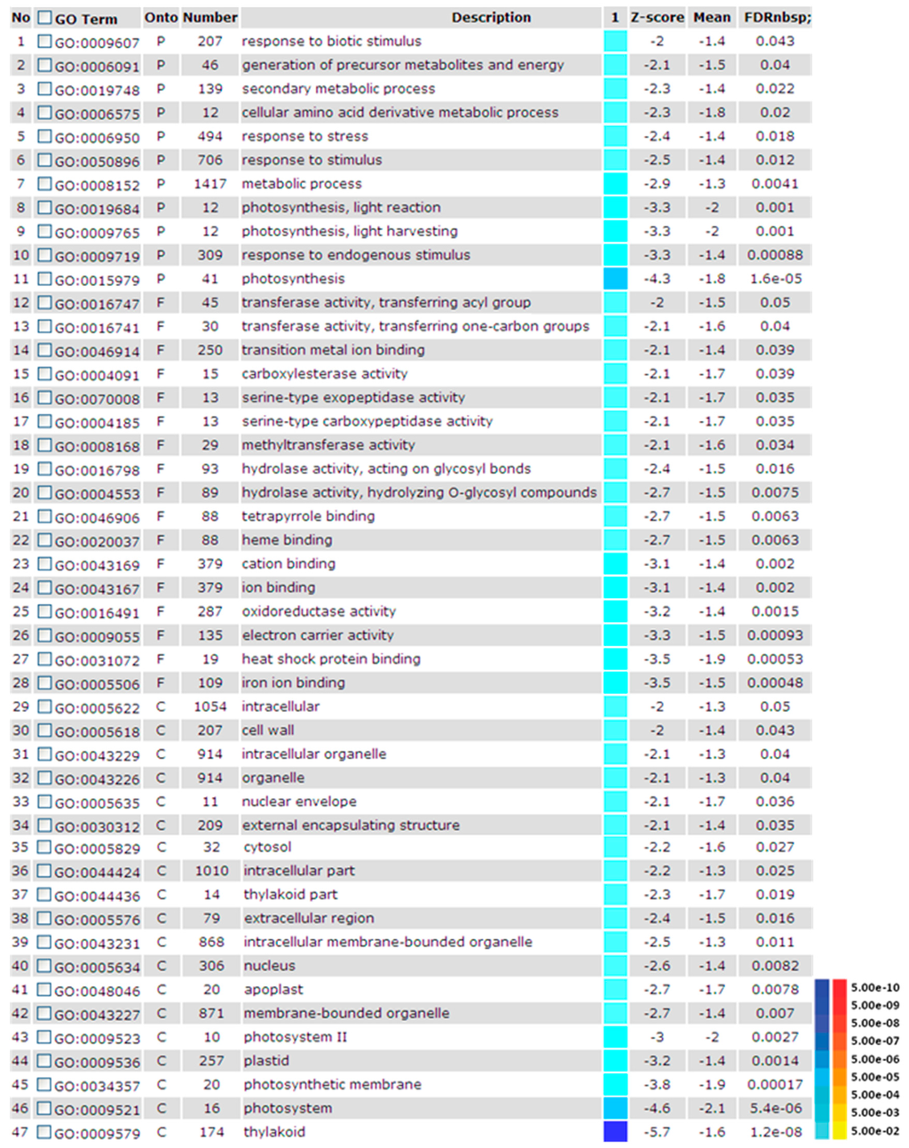
Global warming and climate change intensified the occurrence and severity of abiotic stresses that seriously affect the growth and development of plants,especially, plant photosynthesis. The direct impact of abiotic stress on the activity of photosynthesis is disruption of all photosynthesis components such as photosystem I and II, electron transport, carbon fixation, ATP generating system and stomatal conductance. The photosynthetic system of plants reacts to the stress differently, according to the plant type, photosynthetic systems (C3 or C4), type of the stress, time and duration of the occurrence and several other factors. The plant responds to the stresses by a coordinate chloroplast and nuclear gene expression. Chloroplast, thylakoid membrane, and nucleus are the main targets of regulated proteins and metabolites associated with photosynthetic pathways. Rapid responses of plant cell metabolism and adaptation to photosynthetic machinery are key factors for survival of plants in a fluctuating environment. This review gives a comprehensive view of photosynthesis-related alterations at the gene and protein levels for plant adaptation or reaction in response to abiotic stress. Full article
(This article belongs to the Special Issue Plant Proteomic Research)
Show Figures

Figure 1

17 pages, 1295 KiB  
Article
Mitochondrial Malfunctioning, Proteasome Arrest and Apoptosis in Cancer Cells by Focused Intracellular Generation of Oxygen Radicals
by Ilaria Postiglione 1,†, Angela Chiaviello 1,†, Federica Barra 1, Emanuela Roscetto 1, Amata A. Soriano 1, Maria Rosaria Catania 1, Giuseppe Palumbo 1,* and Giovanna Maria Pierantoni 1,2
1 Department of Molecular Medicine and Medical Biotechnology, University of Naples Federico II, Naples 80131, Italy
2 Institute of Experimental Endocrinology and Oncology (IEOS), National Research Council (CNR), Naples 80131, Italy
These authors contributed equally to this work.
Int. J. Mol. Sci. 2015, 16(9), 20375-20391; https://doi.org/10.3390/ijms160920375 - 28 Aug 2015
Cited by 2 | Viewed by 5872
Abstract
Photofrin/photodynamic therapy (PDT) at sub-lethal doses induced a transient stall in proteasome activity in surviving A549 (p53+/+) and H1299 (p53−/−) cells as indicated by the time-dependent decline/recovery of chymotrypsin-like activity. Indeed, within 3 h of incubation, Photofrin invaded the [...] Read more.
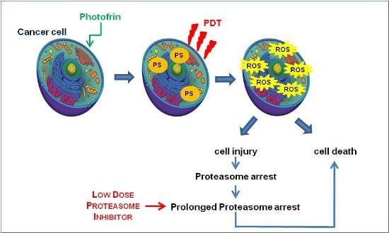
Photofrin/photodynamic therapy (PDT) at sub-lethal doses induced a transient stall in proteasome activity in surviving A549 (p53+/+) and H1299 (p53−/−) cells as indicated by the time-dependent decline/recovery of chymotrypsin-like activity. Indeed, within 3 h of incubation, Photofrin invaded the cytoplasm and localized preferentially within the mitochondria. Its light activation determined a decrease in mitochondrial membrane potential and a reversible arrest in proteasomal activity. A similar result is obtained by treating cells with Antimycin and Rotenone, indicating, as a common denominator of this effect, the ATP decrease. Both inhibitors, however, were more toxic to cells as the recovery of proteasomal activity was incomplete. We evaluated whether combining PDT (which is a treatment for killing tumor cells, per se, and inducing proteasome arrest in the surviving ones) with Bortezomib doses capable of sustaining the stall would protract the arrest with sufficient time to induce apoptosis in remaining cells. The evaluation of the mitochondrial membrane depolarization, residual proteasome and mitochondrial enzymatic activities, colony-forming capabilities, and changes in protein expression profiles in A549 and H1299 cells under a combined therapeutic regimen gave results consistent with our hypothesis. Full article
(This article belongs to the Special Issue Advances in Photodynamic Therapy)
Show Figures

Graphical abstract

15 pages, 1917 KiB  
Article
Detection of Selection Signatures on the X Chromosome in Three Sheep Breeds
by Caiye Zhu 1,2, Hongying Fan 1,3, Zehu Yuan 1, Shijin Hu 1, Li Zhang 1, Caihong Wei 1, Qin Zhang 2, Fuping Zhao 1,* and Lixin Du 1,*
1 National Center for Molecular Genetics and Breeding of Animal, Institute of Animal Sciences, Chinese Academy of Agricultural Sciences, No. 2 Yuanmingyuan West Rd., Haidian, Beijing 100193, China
2 College of Animal Science and Technology, China Agricultural University, Beijing 100193, China
3 College of Animal Science and Technology, Gansu Agricultural University, Lanzhou 730070, China
Int. J. Mol. Sci. 2015, 16(9), 20360-20374; https://doi.org/10.3390/ijms160920360 - 28 Aug 2015
Cited by 12 | Viewed by 7647
Abstract
Artificial selection has played a critical role in animal breeding. Detection of artificial selection footprints in genomic regions can provide insights for understanding the function of specific phenotypic traits and better guide animal breeding. To more fully understand the relationship between genomic composition [...] Read more.
Artificial selection has played a critical role in animal breeding. Detection of artificial selection footprints in genomic regions can provide insights for understanding the function of specific phenotypic traits and better guide animal breeding. To more fully understand the relationship between genomic composition and phenotypic diversity arising from breed development, a genome-wide scan was conducted using an OvineSNP50 BeadChip and integrated haplotype score and fixation index analyses to detect selection signatures on the X chromosome in three sheep breeds. We identified 49, 34, and 55 candidate selection regions with lengths of 27.49, 16.47, and 25.42 Mb in German Mutton, Dorper, and Sunit sheep, respectively. Bioinformatics analysis showed that some of the genes in these regions with selection signatures, such as BMP15, were relevant to reproduction. We also identified some selection regions harboring genes that had human orthologs, including BKT, CENPI, GUCY2F, MSN, PCDH11X, PLP1, VSIG4, PAK3, WAS, PCDH19, PDHA1, and SRPX2. The VSIG4 and PCDH11X genes are associated with the immune system and disease, PDHA1 is associated with biosynthetic related pathways, and PCDH19 is expressed in the nervous system and skin. These genes may be useful as candidate genes for molecular breeding. Full article
(This article belongs to the Section Biochemistry)
Show Figures

Graphical abstract

16 pages, 2055 KiB  
Article
BMP3 Alone and Together with TGF-β Promote the Differentiation of Human Mesenchymal Stem Cells into a Nucleus Pulposus-Like Phenotype
by Xiaopeng Zhou, Yiqing Tao, Chengzhen Liang, Yujie Zhang, Hao Li and Qixin Chen *
Department of Orthopedic Surgery, 2nd Affiliated Hospital, School of Medicine, Zhejiang University, 88 Jiefang Road, 310009 Hangzhou, China
Int. J. Mol. Sci. 2015, 16(9), 20344-20359; https://doi.org/10.3390/ijms160920344 - 27 Aug 2015
Cited by 29 | Viewed by 6707
Abstract
Human mesenchymal stem cells (MSCs) have the potential to differentiate into nucleus pulposus (NP)-like cells under specific stimulatory conditions. Thus far, the effects of bone morphogenetic protein 3 (BMP3) and the cocktail effects of BMP3 and transforming growth factor (TGF)-β on MSC proliferation [...] Read more.
Human mesenchymal stem cells (MSCs) have the potential to differentiate into nucleus pulposus (NP)-like cells under specific stimulatory conditions. Thus far, the effects of bone morphogenetic protein 3 (BMP3) and the cocktail effects of BMP3 and transforming growth factor (TGF)-β on MSC proliferation and differentiation remain obscure. Therefore, this study was designed to clarify these unknowns. MSCs were cultured with various gradients of BMP3 and BMP3/TGF-β, and compared with cultures in basal and TGF-β media. Cell proliferation, glycosaminoglycan (GAG) content, gene expression, and signaling proteins were measured to assess the effects of BMP3 and BMP3/TGF-β on MSCs. Cell number and GAG content increased upon the addition of BMP3 in a dose-dependent manner. The expression of COL2A1, ACAN, SOX9, and KRT19 increased following induction with BMP3 and TGF-β, in contrast to that of COL1A1, ALP, OPN, and COMP. Smad3 phosphorylation was upregulated by BMP3 and TGF-β, but BMP3 did not affect the phosphorylation of extracellular-signal regulated kinase (ERK) 1/2 or c-Jun N-terminal kinase (JNK). Our results reveal that BMP3 enhances MSC proliferation and differentiation into NP-like cells, as indicated by increased cell numbers and specific gene expressions, and may also cooperate with TGF-β induced positive effects. These actions are likely related to the activation of TGF-β signaling pathway. Full article
(This article belongs to the Special Issue Stem Cell Activation in Adult Organism)
Show Figures

Figure 1

18 pages, 2315 KiB  
Article
Rational Design of Diketopyrrolopyrrole-Based Small Moleculesas Donating Materials for Organic Solar Cells
by Ruifa Jin * and Kai Wang
Inner Mongolia Key Laboratory of Photoelectric Functional Materials and College of Chemistry and Chemical Engineering, Chifeng University, Chifeng 024000, China
Int. J. Mol. Sci. 2015, 16(9), 20326-20343; https://doi.org/10.3390/ijms160920326 - 27 Aug 2015
Cited by 25 | Viewed by 5770
Abstract
A series of diketopyrrolopyrrole-based small molecules have been designed toexplore their optical, electronic, and charge transport properties as organic solar cell(OSCs) materials. The calculation results showed that the designed molecules can lowerthe band gap and extend the absorption spectrum towards longer wavelengths.The designed [...] Read more.
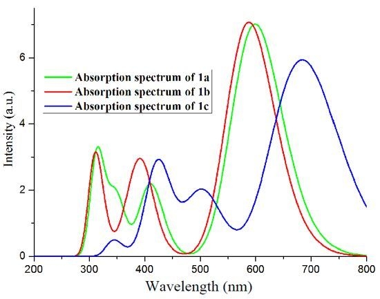
A series of diketopyrrolopyrrole-based small molecules have been designed toexplore their optical, electronic, and charge transport properties as organic solar cell(OSCs) materials. The calculation results showed that the designed molecules can lowerthe band gap and extend the absorption spectrum towards longer wavelengths.The designed molecules own the large longest wavelength of absorption spectra,the oscillator strength, and absorption region values. The optical, electronic, and chargetransport properties of the designed molecules are affected by the introduction of differentπ-bridges and end groups. We have also predicted the mobility of the designed moleculewith the lowest total energies. Our results reveal that the designed molecules are expectedto be promising candidates for OSC materials. Additionally, the designed molecules areexpected to be promising candidates for electron and/or hole transport materials. On thebasis of our results, we suggest that molecules under investigation are suitable donors for[6,6]-phenyl-C61-butyric acid methyl ester (PCBM) and its derivatives as acceptors of OSCs. Full article
(This article belongs to the Section Physical Chemistry, Theoretical and Computational Chemistry)
Show Figures

Graphical abstract