Review

Epicardial Lineages and Cardiac Repair

Department of Cell and Developmental Biology, Cardiovascular Institute and Institute for Regenerative Medicine, Perelman School of Medicine at the University of Pennsylvania, Philadelphia, PA 19104, USA
*
Authors to whom correspondence should be addressed.
J. Dev. Biol. 2013, 1(2), 141-158; https://doi.org/10.3390/jdb1020141
Submission received: 14 June 2013 / Revised: 8 August 2013 / Accepted: 9 August 2013 / Published: 26 August 2013
(This article belongs to the Special Issue Epicardial Development and Cardiovascular Disease)

Abstract

:
The death of cardiac myocytes resulting from myocardial infarction is a major cause of heart failure worldwide. Effective therapies for regenerating lost cardiac myocytes are lacking. Recently, the epicardium has been implicated as a source of inflammatory cytokines, growth factors and progenitor cells that modulate the response to myocardial injury. During embryonic development, epicardially-derived cells have the potential to differentiate into multiple cardiac lineages, including fibroblasts, vascular smooth muscle and potentially other cell types. In the healthy adult heart, epicardial cells are thought to be generally quiescent. However, injury of the adult heart results in reactivation of a developmental gene program in the epicardium, which leads to increased epicardial cell proliferation and differentiation of epicardium-derived cells (EPDCs) into various cardiac lineages. Recent work suggests that epicardial reactivation after injury is accompanied by, and contributes to, a robust inflammatory response. In this review, we describe the current status of research related to epicardial biology in cardiac development and regeneration, highlighting important recent discoveries and ongoing controversies.

1. Introduction

The development of the epicardium is conserved through the evolution of vertebrates. The epicardial layer of the heart derives from an embryonic anlage, known as the proepicardial organ (PEO), which is a transient structure that arises from the mesothelium of the septum transversum. The outer lining of the PEO is covered with epithelial cells, while mesenchymal cells compose the core [1,2,3,4]. Early heart development begins with the formation of the linear heart tube derived from lateral plate mesoderm. Prior to heart looping, the heart tube has only two layers: endocardium and myocardium. After looping has occurred, at ~E9.5 in the mouse, cells from the PEO migrate onto the surface of the heart to form a third heart layer: embryonic epicardium [1,2,3,4]. Proepicardial cells migrate either as individual cells (in mammals) or as a sheet of cells (in birds) and cover the developing myocardium [5]. Soon after the epicardium is formed, a subset of these epithelial cells undergoes mesenchymal transformation and invades the subepicardial space and underlying myocardium and differentiates into multiple cardiac cell types, while other cells remain on the surface of the heart and form the definitive epicardium [1,4,6,7,8] (Figure 1). Many secreted factors, extracellular matrix molecules, enzymes, transcription factors and signaling molecules, have been implicated in PEO formation, epicardium formation, epicardial epithelial-to-mesenchymal transformation (EMT) and epicardially-derived cell (EPDC) proliferation, survival and migration [6,9,10]. For example, α4-integrin, a cell adhesion receptor expressed on epicardial cells, binds to fibronectin and vascular cell adhesion molecule-1 (VCAM-1) and regulates PEO migration. Genetic deletion of α4-integrin or VCAM-1 results in a heart without an epicardium, due to impaired proepicardial cell migration [11,12,13]. Podoplanin deletion results in reduction of PEO size and decreased attachment to myocardium [14]. Loss of Dicer, a microRNA-processing enzyme, in the epicardium results in coronary vasculature defects, due to reduced epicardial cell proliferation, differentiation and EMT [9]. Wt1 expression in the epicardium is necessary for coronary vasculature development [15,16]. Wt1 modulates epicardial EMT by regulating Wnt and retinoic acid signaling during development [15,16]. Tbx18, a member of the T-box family of transcription factors, is expressed in the proepicardium, the epicardium, the ventricular septum, the left ventricular myocardium and the sinus horns of the mouse heart [17,18]. Although loss of Tbx18 in the epicardium does not affect epicardial development and function (perhaps due to functional redundancy by other T-box factors), epicardial expression of a transcriptional activator version of Tbx18 (Tbx18VP16) results in premature smooth muscle differentiation of epicardial cells, due to activated transforming growth factor-beta receptor (TGFßR) and Notch signaling in the embryonic epicardium [19]. Nfatc1, a transcription factor regulated by calcium and calcineurin, is expressed by a subset of cells in the PEO, epicardium and coronary vessels [20]. Genetic studies suggest that Nfatc1 is required for promoting EPDCs invasion into myocardium [20]. Tallquist and colleagues identified neurofibromin (encoded by the Nf1 gene) as a key mediator of epicardial EMT [21]. Loss of Nf1 in epicardial cells results in increased EMT, EPDC proliferation and subsequent differentiation into fibroblasts and smooth muscle cells [21].
For many decades, epicardium was considered to be a passive fibrous lining surrounding the myocardium. However, studies in the last 10 years have suggested an active role for epicardium in development, disease and regeneration. For example, epicardium is a source of multipotent progenitor cells, which give rise to important components of the developing heart (Figure 1 and Figure 2) [22,23,24,25,26]. Moreover, reciprocal paracrine signaling between the epicardium and myocardium regulates coronary vasculature development, as well as cardiomyocyte proliferation and differentiation [8,10,27,28,29] (Figure 3). Myocardial injury of the adult heart leads to reactivation of an embryonic gene program in epicardial cells. Factors released from activated epicardium modulate cardiac regeneration and function, at least in part by promoting angiogenesis (Figure 3 and Figure 4) [10,27,30,31]. Despite the critical role of the epicardium during development, physiological functions in the adult heart remain poorly understood.
Figure 1. Epicardial derivatives contribute to cardiac lineages. Epicardial cells undergo epithelial-to-mesenchymal transition (EMT), delaminate from the epicardium, invade the underlying myocardium and differentiate into various cardiac lineages, including fibroblasts, smooth muscle cells, endothelial cells and, perhaps, cardiomyocytes.
Jdb 01 00141 g001
Figure 2. Heterogeneous proepicardial precursors and their fates. Distinct proepicardial sub-compartments variably give rise to coronary endothelial cells, vascular smooth muscle cells, fibroblasts and perhaps cardiomyocytes. Arrows represent differentiation potential. Dotted arrows indicate unresolved potential.
Jdb 01 00141 g002
Figure 3. Reciprocal interactions between epicardium and myocardium during cardiac development. Retinoic acid (RA) modulates epicardial EMT and coronary vascular development by regulating Fgf2 and Wnt/β-catenin signaling. RA also activates Fgf9 expression in the epicardium, which signals via receptors Fgfr1/2 to regulate myocardial proliferation and differentiation. Signals from the myocardium, such as Thymosin β4, platelet-derived growth factors (PDGFs) and fibroblast growth factors (FGFs), regulate epicardial cell differentiation.
Jdb 01 00141 g003
Figure 4. Regenerative and inflammatory response after myocardial injury. Factors accumulate in the pericardial fluid after myocardial infarction that may affect both epicardial and myocardial function and which are implicated in cardiac regeneration and inflammation. Recent studies suggest that the balance between regenerative and inflammatory factors can affect repair and subsequent cardiac function.
Jdb 01 00141 g004

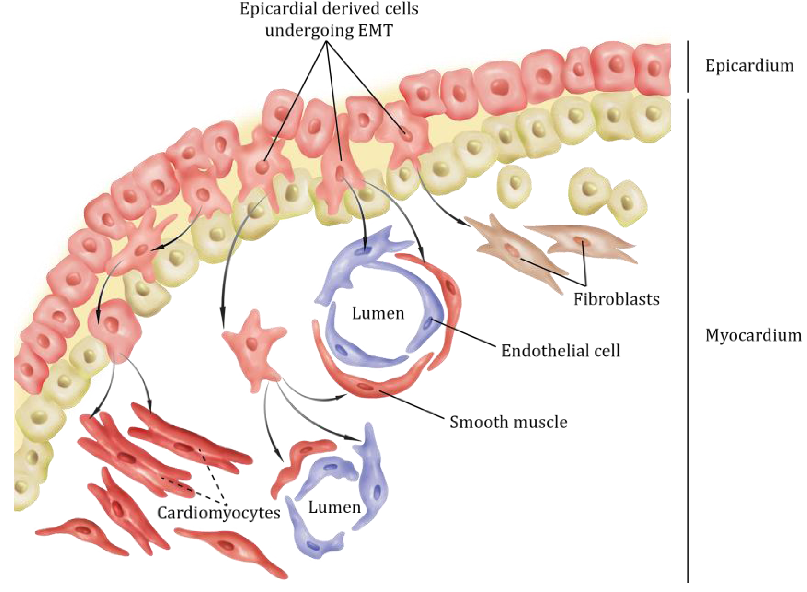
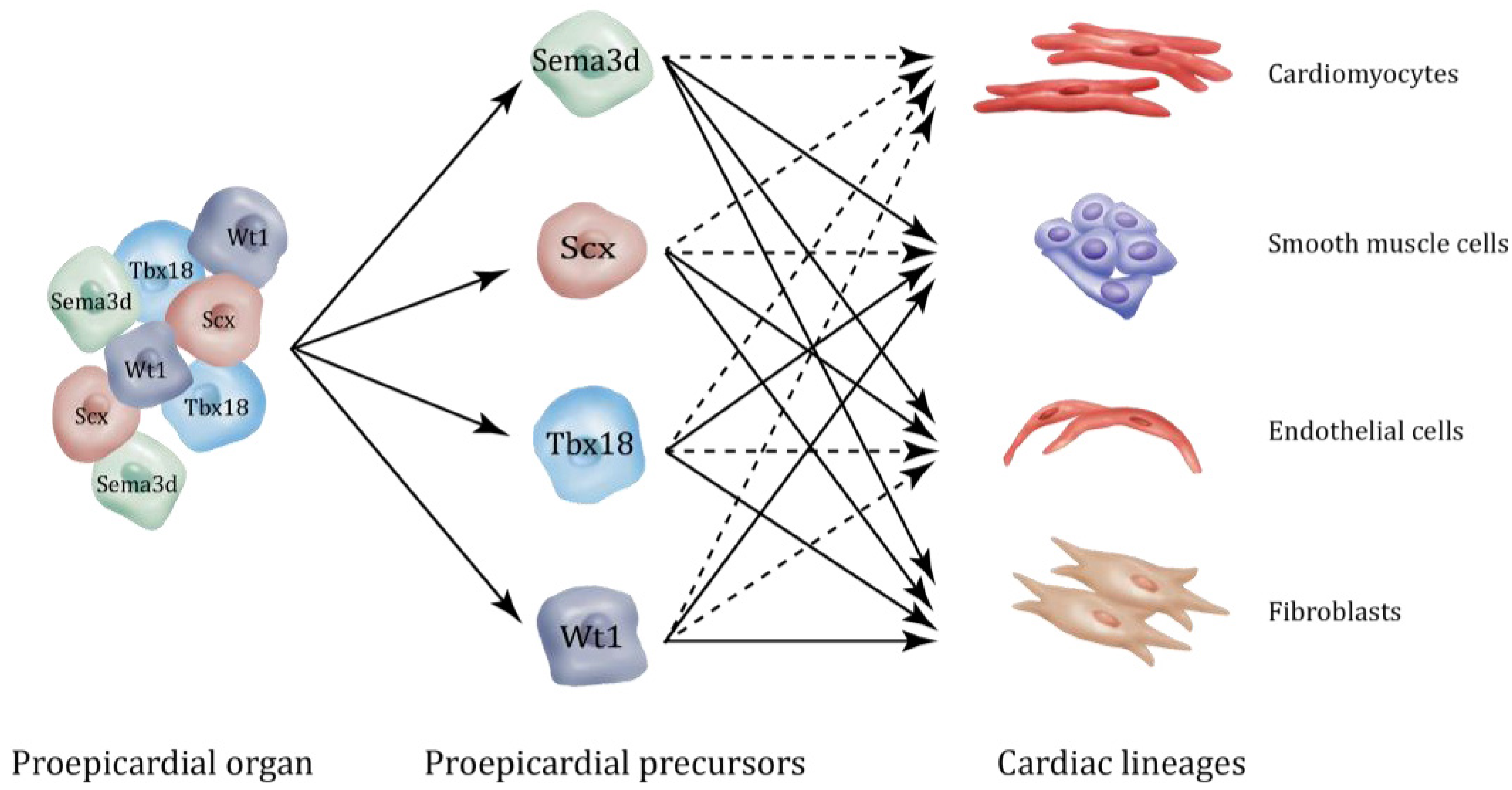
2. Epicardial Lineages

Fate mapping analysis of early cardiac multipotent precursors that express either Nkx2.5 or Isl1 suggest that these cells contribute to cardiomyocyte, smooth muscle and endothelial lineages of the mature heart [32,33]. However, the relationship between early cardiac progenitors (marked by Nkx2.5 or Isl1) and the PEO was not fully elucidated in these initial studies. Recent work by Zhou et al. demonstrates that Wilm's Tumor 1 (Wt1) is expressed in the PEO, epicardium and epicardium-derived cells (EPDCs) [24]. Wt1-expressing proepicardial progenitors are derived from Nkx2.5 and Isl1-expressing secondary heart field precursors [34]. Nkx2.5 and Wt1 are not actively co-expressed in the PEO, but are either sequentially expressed or only transiently co-expressed in a subset of PEO precursors [34]. Unlike Isl1, expression of Nkx2.5 is necessary for the formation of the PEO, since loss of Nkx2.5 resulted in abnormal PEO formation and reduced Wt1 expression [34].
The proepicardial contribution to various cardiac lineages has been disputed, due to conflicting findings in chick and mouse. The epicardial cells begin to cover the myocardium at the same time that coronary precursor cells first appear in the heart tube [1]. Retroviral labeling experiments demonstrate that single vasculogenic cells of the proepicardium differentiate into cardiac endothelial, smooth muscle cells and perivascular connective tissue cells [1]. Further studies using diI labeling and quail-chick chimeras have established the PEO as a source of both cardiovascular smooth muscle and endothelial cells in chick [35,36,37,38]. However initial fate mapping studies in mice did not identify a significant proepicardial contribution to the cardiac endothelium [19,23,24,26].
The PEO was thought to be a genetically homogeneous tissue. However, recent studies in both chick and mouse suggest that the PEO is a highly heterogeneous collection of genetically distinct sub-populations of cells with distinct functions and downstream fates (Figure 2). In a recent study, we, with our collaborators, demonstrated that semaphorin 3D (Sema3D), scleraxis (Scx), Wt1 and Tbx18 could mark distinct sub-populations of progenitor cells within the PEO. Wt1- and Tbx18-expressing proepicardial cells differentiate into smooth muscle cells and fibroblasts, but rarely, if ever, into coronary endothelium [23,24,26]. In contrast, Scx- and Sema3D-expressing progenitors within the PEO differentiate into smooth muscle cells, fibroblasts and coronary endothelium [25]. In addition, they also contribute to two other vascular endothelial tissues: the sinus venosus and the endocardium. Scx and Sema3D expression partially overlaps in the proepicardium, but is distinct from cells expressing the canonical epicardial markers, Tbx18 and Wt1 [25]. This might explain why fate mapping using mouse Wt1 and Tbx18 cre lines does not detect the proepicardial contribution tocoronary endothelium.
Further, two independent studies have recently demonstrated that during cardiac development, a subpopulation of proepicardial cells marked by either Tbx18 or Wt1 expression have the potential to differentiate into cardiomyocytes [23,24]. This could have significant clinical implications as a potential source for regenerative cardiomyocytes in adults [39]. However, the conclusions that Wt1 and Tbx18 expressing epicardial cells can give rise to cardiomyocytes have been challenged based on the suggestion that Wt1 and Tbx18 are normally expressed by some cardiomyocytes, thus confounding the interpretation of the cre-based fate-mapping results. This controversy remains unresolved [18,23,24,26].
Cardiac fibroblasts compose up to two-thirds of the cells in the heart and regulate structural, mechanical, biochemical and electrical properties during development and disease [40,41]. During embryonic development, the epicardium is considered the primary source of cardiac fibroblasts, although more work is needed to fully define the embryonic origins of all cardiac fibroblasts [42]. Despite their significant role, surprisingly little is known about the factors that determine the cardiac fibroblast lineage. PDGF receptors (PDGFRα and PDGFRβ) are expressed in the embryonic epicardium [43,44,45,46] and contribute to fibroblast lineage specification. PDGFRα deletion in the epicardium results in a deficit in cardiac fibroblast formation, whereas cardiac vascular smooth muscle cell (cVSMC) development is unperturbed [46]. Inactivation of PDGFRβ perturbs cVSMC development without affecting cardiac fibroblasts [45]. A recent study by Tallquist and colleagues identified Tcf21 (epicardin/Pod1/capsulin) as an essential regulator of cardiac fibroblast cell fate determination [47]. Epicardial fate mapping in a Tcf21 null background demonstrates that epicardial cell differentiation into cardiac fibroblasts is significantly compromised [47].
Epicardial activation and re-expression of a fetal gene program following myocardial injury contributes significantly to cardiac fibrosis and scar formation [10,22,41,48]. Recent work by Huang et al. suggests that epicardium-mediated injury induces an inflammatory and regenerative response [49]. Considering their significant role during cardiac regeneration, it is important to better understand factors that lead to fibrosis and to design strategies manipulating epicardial activation after cardiac injury to decrease fibrosis and enhance regeneration.

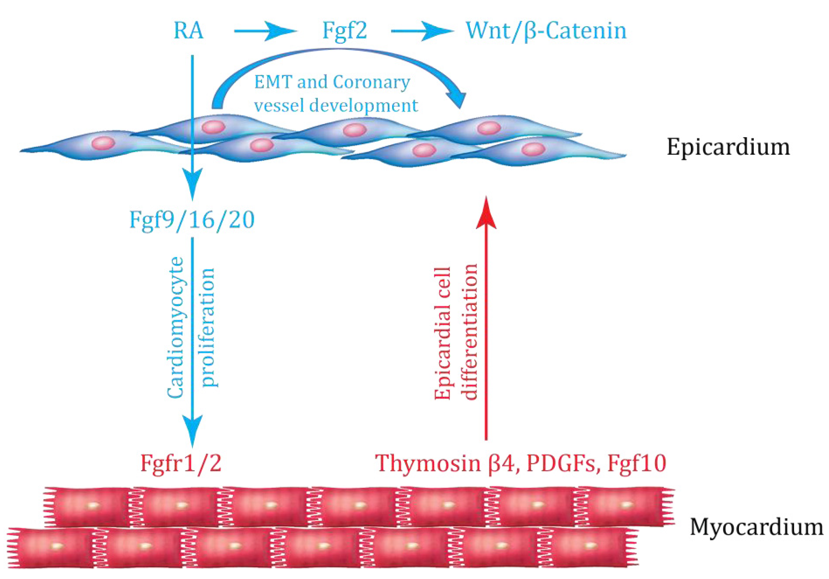
3. Interaction between Epicardium and Myocardium

The embryonic epicardium is a source of growth factors required for coronary vasculature development and for proliferation and differentiation of underlying myocardial cells [10,27,28,29]. Early evidence suggesting that epicardium plays a significant role in modulating myocardial development arose from chick experiments in which blocking or photoablation of proepicardial cells resulted in a thin myocardial compact layer within the ventricular wall, poor cardiac function and embryonic lethality [50,51,52]. This suggests that soluble factors are released from the epicardium and act in a paracrine manner to regulate myocardial proliferation and differentiation. In recent years, several of these paracrine signaling pathways have been elucidated, including retinoic acid (RA), fibroblast growth factors (FGFs) and Wnt/β-catenin (Figure 3).
A number of studies have suggested that RA is important for early development of mammalian embryos [53]. RA exerts its functions by signaling to nuclear hormone receptors, including retinoid A and retinoid X receptors (RARs and RXRs, respectively). The phenotype of mouse embryos carrying a null mutation of the gene encoding retinaldehyde dehydrogenase 2 (Raldh2), which participates in RA synthesis, suggests that RA signaling is required for cardiac looping and chamber morphogenesis [53]. Epicardial RA is required for normal myocardial growth [28,54]. Blockade of RA signaling in intact chick heart slices impairs myocardial cell proliferation [55]. However, administration of RA to heart slices cultured in the absence of the epicardium failed to rescue cardiac myocyte proliferation in these experiments, suggesting that RA does not have a direct mitogenic effect on myocardium [55]. These results suggest that epicardium may modulate myocardial growth by producing a mitogen whose expression is regulated by RA. This finding was further supported by a genetic experiment in which deletion of RXRα in ventricular cardiomyocytes did not affect the thickness of the ventricular myocardium [56]. In agreement with this model, conditional deletion of RXRα in the epicardium using Gata5-Cre resulted in abnormal cardiac development, characterized by epicardial detachment and thinning of the myocardial compartment [54].
One function of RA in the epicardium may be to induce expression of fibroblast growth factors, including Fgf2 and Fgf9, to regulate cardiomyocyte proliferation and differentiation [28]. Fgf16 and Fgf20 are also produced by epicardium, but RA does not regulate their expression [28]. Loss of Fgf9 results in ventricular hypoplasia and a significant reduction in the myocardial proliferation rate [28]. Epicardial-specific RXRα mutants show decreased Fgf2 gene expression in the epicardium [54], and Fgf2 can activate epicardial Wnt signaling through activation of Wnt9b [54]. Loss of RXRα in epicardium produces thinned myocardium and, also, persistent ventricular expression of atrial myosin light chain (mlc2a), which is normally down-regulated [54]; thus, both proliferation and differentiation appears to be affected. However, one caveat with these studies is that Gata5-Cre, used to delete RXRα in epicardium, is not entirely restricted to epicardial tissue and can display “leakiness” in myocardium, as well. Thus, further studies using alternative epicardial cre lines, such as Sema3d-Cre or Wt1-Cre, should be performed to validate this model. Recent work by Brade et al. demonstrates that hepatic RA signaling is required for induction of EPO expression in the liver [57]. Secreted EPO then travels to the epicardium and induces IGF2 expression, which is secreted by the epicardium to stimulate myocardial growth [57,58]. In adult hearts, these developmental signaling pathways are reactivated in the epicardium and modulate epicardial EMT. For example, cardiac injury activates Wnt1 expression in the epicardium and epicardial-derived fibroblasts [59].
FGFs regulate cardiac development by binding and signaling to their receptors (FGFRs). Among FGFRs, only Fgfr1 and Fgfr2 are expressed on cardiomyocytes [52,60]. Loss of Fgfr1 and Fgfr2 in the myocardium results in hypoplastic heart [28]; similar to Fgf9 nulls. Additional studies suggest that Fgf9-Fgfr1/2 signaling is also required for coronary vasculature development. Myocardial Fgf9-Fgfr1/2 signaling activates Hedgehog signaling, which is essential for vascular endothelial growth factor (VEGF)-A, VEGF-B, VEGF-C and angiopoietin-2 (Ang2) expression and, thereby, regulation of coronary vasculature development [54].
In addition to mitogenesis, FGF signaling has been implicated in regulating cardiac morphogenesis and lineage specification during embryonic development. In quail embryos, Fgfr1 is expressed in the proepicardium and epicardium-derived cells. Fgfr1 is required for epicardial EMT and coronary vasculature development [60]. Variation of Fgfr1 levels in the proepicardium affects epicardial EMT and myocardial invasion and differentiation of epicardium-derived cells [60]. Loss of Fgfr1 leads to a reduction in the ability of EPDCs to invade the underlying myocardium [60], while overexpression promotes epicardial EMT and biases the differentiation of EPDCs into endothelial cells over smooth muscle cells [60].
Signals from the myocardium can reciprocally modulate epicardial EMT (Figure 3). Early evidence to support this idea came from in vitro experiments showing that conditioned media derived from cultured cardiomyocytes can increase epicardial EMT [61,62]. Indeed, Fgf10 produced by myocardium can modulate epicardial EMT and EPDC migration [63]. Multiple other factors expressed by myocardium have been shown to regulate epicardial development and function. Thymosin β4 is an actin-binding protein expressed in the developing myocardium, but not in the epicardium [64,65]. Cardiomyocyte-specific deletion of Thymosin β4 produces impaired formation of coronary vasculature, suggesting that Thymosin β4 acts on epicardium in a paracrine manner [65]. The addition of Thymosin β4 to epicardial explants significantly increases the number of epicardial-derived endothelial and smooth muscle cells [65]. In adult hearts, systemic administration of Thymosin β4 leads to induction of coronary vessel growth, myocardial progenitor mobilization and activation of embryonic developmental programs in adult epicardium [66]. Notably, however, recent work by Banerjee et al. demonstrates that Thymosin β4 is dispensable for embryonic viability and vascular development in mice [67,68].
Platelet-derived growth factors (PDGFs) may also participate in myocardial-epicardial communication. During development, PdgfA is expressed by the myocardium, while PDGF receptors, Pdgfrα and Pdgfrβ, are expressed by epicardial cells [45,46]. Genetic deletion of both PDGF receptors in the epicardium results in abnormal epicardial EMT and EPDC formation [45,46]. Taken together, these studies suggest that a PDGF signal from the myocardium is important for epicardial development and function.

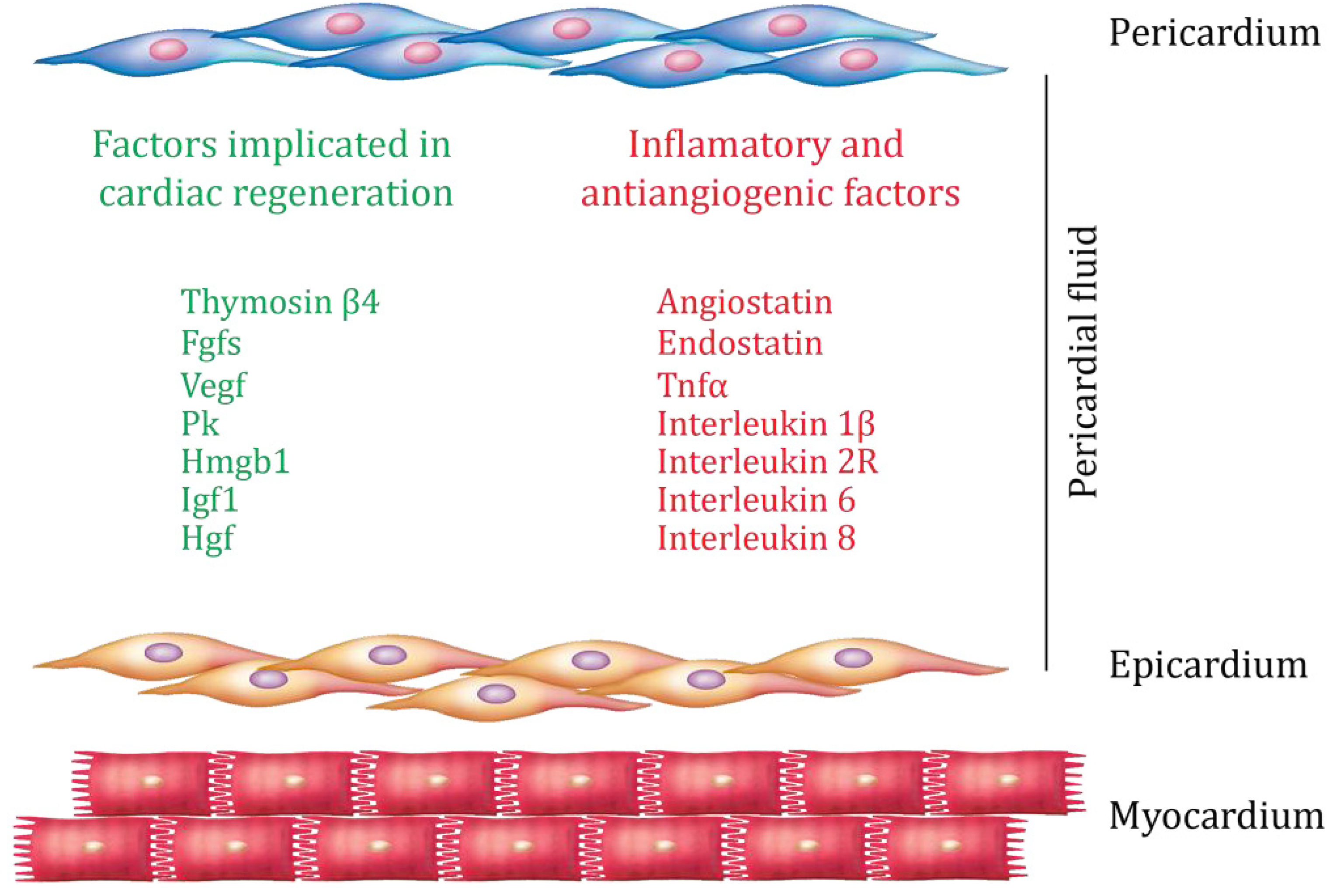
4. Epicardium and Cardiac Regeneration

The adult mammalian heart does not undergo significant regeneration following injury and, as a consequence, ischemic heart disease results in permanent myocardial damage, impaired pump function and heart failure. In lower vertebrates, however, efficient myocardial regeneration occurs after damage [69,70,71,72,73,74,75,76]. For example, the zebrafish heart undergoes minimal scarring and exhibits a robust regenerative response upon injury [71,74]. Zebrafish hearts can fully regenerate within a period of 30 to 60 days after surgical removal of as much as 20% of the ventricle [71], a cryoinjury or a genetic ablation of over 60% of the cardiomyocytes [77,78,79,80]. Cardiac regeneration in adult zebrafish is not achieved by proliferation and differentiation of cardiac stem cells, as initially suggested, but by activation, de-differentiation and proliferation of mature cardiomyocytes [75,76,81]. After de-differentiation, cardiomyocytes undergo a subsequent maturation step to completely restore cardiac function. Ultimately, newly generated cardiac myocytes are functionally integrated with the preexisting myocardium with little or no evidence of the injury, suggesting complete myocardial regeneration [75,76,81]. These studies have encouraged scientists to determine whether the adult mammalian heart is incapable of cardiac myocyte replacement or whether it retains a low-level capacity for regeneration, which could be therapeutically amplified.
The traditional view that the adult mammalian heart is incapable of cardiomyocyte renewal has been recently challenged. Studies have shown that humans and other mammals have a limited capacity for myocardial regeneration that is insufficient to restore cardiac function after injury [7,39,82,83,84,85]. Porrello et al. demonstrated that the neonatal mouse heart has a robust regeneration potential similar to the zebrafish heart [86]. A one-day-old mouse has the capacity to fully regenerate after partial surgical resection of the ventricles (~15% of the ventricle) within a period of 21 days [86]. Resected hearts show complete restoration of cardiac function after 60 days [86]. This regenerative response is characterized by cardiomyocyte proliferation with minimal hypertrophy or fibrosis. Genetic lineage tracing studies using an inducible cardiomyocyte specific cre line (αMHC-MerCreMer mice) demonstrate that the majority of newly formed cardiomyocytes within the regenerated ventricle are derived from preexisting cardiomyocytes [86]. Myocardial infarction of the neonatal heart also results in a robust regenerative response and recovery within 21 days. During this regenerative process, a possible contribution from stem or epicardial progenitor cells was not ruled out. Importantly, the regenerative capability of the neonatal heart is lost by seven days of age.
The role of the epicardium during cardiac regeneration has been recently explored [69,70,71,72,73,74,75,76]. During zebrafish heart regeneration, the epicardium is activated and re-expresses developmental genes [73,74]. Upon myocardial resection, epicardial cells rapidly expand and create a new epithelial cover for the exposed myocardium [73]. A subset of activated epicardial cells undergoes EMT and contributes to new vasculature for the regenerating cardiac muscle [73,74]. In zebrafish, cardiac injury induces expression of Fgf17b by myocardium and its receptors (Fgfr2 and Fgfr4) on EPDCs [73]. Blocking FGF signaling results in reduction of epicardial EMT and impaired neovascularization, leading to poor cardiac regeneration [73]. Although different organisms have different regenerative capacities, components of the injury response are conserved, including the activation of the epicardium and the contribution of epicardium-derived cells (EPDCs) to the repair process [27,48,49,73,74].
In mammals, the epicardium is reactivated after myocardial infarction [10,27,31,48,49,59]. Activated epicardial cells then undergo EMT and differentiate into fibroblasts and smooth muscle cells. However, little or no epicardial contribution towards cardiomyocytes or endothelial cells is observed in the adult after injury [27,48]. Activated epicardium secretes angiogenic factors, and treatment with epicardium-derived conditioned media significantly reduces infarct size and improves cardiac function [27].
Although available data suggest that differentiation of EPDCs into cardiomyocytes after myocardial damage is rare or non-existent, Riley and co-workers demonstrated that thymosin β4 priming before myocardial infarction results in a significant increase in the number of epicardium-derived cardiomyocytes [87]. However, work by Pu and coworkers demonstrated that thymosin β4 treatment after myocardial infarction did not alter the fate of epicardially-derived cells [88,89]. Differentiation of EPDCs into either cardiomyocyte or endothelial lineages was not observed [88,89]. Thus, while intriguing, the potential clinical utility and mechanism of action of thymosin ß4 remains to be fully elucidated.
Additional lines of evidence have implicated epicardium not just as a signaling center, but also as a source of cardiovascular progenitors. Harvey and colleagues identified a population of cardiac resident progenitor cells, called cardiac colony-forming units-fibroblast (cCFU-Fs) [90,91]. Genetic lineage tracing experiments showed that cCFU-Fs are derived from epicardium and have the potential to differentiate into smooth muscle cells, cardiac muscle and adipose tissue in vitro [90]. The role of cCFU-Fs during cardiac regeneration will require further examination.
Recent work by Olson and colleagues highlights the critical role of inflammation and inflammatory mediators produced by epicardium in the cardiac response to injury [49]. By analyzing conserved enhancer regions in genes expressed during epicardial activation, the authors implicated the family of transcription factor C/EBPs (CCAAT-enhancer binding proteins) as critical mediators of epicardial gene expression after injury [49]. C/EBP inhibition resulted in significantly improved contractile function and decreased myocardial fibrosis after injury [49]. Interestingly, the authors found that the inflammatory response after injury, marked by neutrophil influx to the infarcted area, was significantly reduced after C/EBP inhibition [49]. This study implicates epicardial-derived cytokines in the inflammatory response and the neutrophil infiltration that occurs after cardiac injury. Modulation of this response may have both beneficial and damaging effects and may offer an opportunity for modulation of the postulated balance between fibrosis and regeneration during the injury response.
Consistent with this finding, molecules released from adult epicardium and other cardiac tissues can be found in pericardial fluid, and these factors may affect cardiac function under normal or pathological conditions [10]. Several molecules have been identified in the pericardial fluid of normal and diseased heart, and their levels correlate with regenerative [92,93,94,95,96,97,98] and inflammatory responses [99,100,101,102] (Figure 4). Examples include FGFs [92,93], VEGF [92,94,95], hepatocyte growth factor [96], high-mobility group box 1 protein-96 and insulin-like growth factor-1 [96,98]. Growth factors present in pericardial fluid can affect proliferation and differentiation of cardiac progenitors towards myocardial and vascular lineages [103,104,105]. The pericardial fluid of ischemic heart patients also shows higher levels of inflammatory factors [99,100,101,102]. For example, high levels of angiostatin [99], endostatin [100], tumor necrosis factor-alpha [102] and interleukins [101,102] were detected in ischemic heart disease patients compared to controls. An elevated level of inflammatory factors may adversely affect the regenerative process, causing more fibrosis and less regeneration.

5. Conclusions and Future Perspectives

In summary, the epicardium has emerged as a fascinating and dynamic tissue that can contribute to a variety of cardiac lineages in the embryo and in the adult. Reciprocal signaling between epicardium and myocardium regulates cardiac growth and cell fate decisions, and paracrine factors emerging from activated epicardium after injury can modulate the repair process. A fine balance between scar formation and myocardial regeneration may provide a therapeutic window to enhance the degree to which new myocytes are produced after damage to the heart, and epicardial regulation of the inflammatory response may significantly impact this process. Rather than a quiescent and uninteresting covering for the heart, the epicardium is an active participant in homeostasis, development and disease and will be the focus of exciting investigation for years to come.

Acknowledgements

The authors would like to thank Kurt Engleka for critical reading of the manuscript. This work was supported by UO1 HL100405, the Spain Fund and the WW Smith Endowed Chair.

Conflict of Interest

The authors declare no conflict of interest.

References

  1. Mikawa, T.; Gourdie, R.G. Pericardial mesoderm generates a population of coronary smooth muscle cells migrating into the heart along with ingrowth of the epicardial organ. Dev. Biol. 1996, 174, 221–232. [Google Scholar] [CrossRef]
  2. Manner, J.; Perez-Pomares, J.M.; Macias, D.; Munoz-Chapuli, R. The origin, formation and developmental significance of the epicardium: a review. Cells Tissues Organs 2001, 169, 89–103. [Google Scholar] [CrossRef]
  3. Gittenberger-de Groot, A.C.; Vrancken Peeters, M.P.; Mentink, M.M.; Gourdie, R.G.; Poelmann, R.E. Epicardium-derived cells contribute a novel population to the myocardial wall and the atrioventricular cushions. Circ. Res. 1998, 82, 1043–1052. [Google Scholar] [CrossRef]
  4. Mikawa, T.; Fischman, D.A. Retroviral analysis of cardiac morphogenesis: discontinuous formation of coronary vessels. Proc. Natl. Acad. Sci. USA 1992, 89, 9504–9508. [Google Scholar] [CrossRef]
  5. Lie-Venema, H.; van den Akker, N.M.; Bax, N.A.; Winter, E.M.; Maas, S.; Kekarainen, T.; Hoeben, R.C.; deRuiter, M.C.; Poelmann, R.E.; Gittenberger-de Groot, A.C. Origin, fate, and function of epicardium-derived cells (EPDCs) in normal and abnormal cardiac development. Sci. World J. 2007, 7, 1777–1798. [Google Scholar] [CrossRef]
  6. von Gise, A.; Pu, W.T. Endocardial and epicardial epithelial to mesenchymal transitions in heart development and disease. Circ. Res. 2012, 110, 1628–1645. [Google Scholar] [CrossRef]
  7. Epstein, J.A.; Franklin, H. Epstein Lecture. Cardiac development and implications for heart disease. N. Engl. J. Med. 2010, 363, 1638–1647. [Google Scholar] [CrossRef]
  8. Degenhardt, K.; Singh, M.K.; Epstein, J.A. New approaches under development: Cardiovascular embryology applied to heart disease. J. Clin. Invest. 2013, 123, 71–74. [Google Scholar] [CrossRef]
  9. Singh, M.K.; Lu, M.M.; Massera, D.; Epstein, J.A. MicroRNA-processing enzyme Dicer is required in epicardium for coronary- vasculature development. J. Biol. Chem. 2011, 286, 41036–41045. [Google Scholar]
  10. Limana, F.; Capogrossi, M.C.; Germani, A. The epicardium in cardiac repair: From the stem cell view. Pharmacol. Ther. 2011, 129, 82–96. [Google Scholar] [CrossRef]
  11. Kwee, L.; Baldwin, H.S.; Shen, H.M.; Stewart, C.L.; Buck, C.; Buck, C.A.; Labow, M.A. Defective development of the embryonic and extraembryonic circulatory systems in vascular cell adhesion molecule (VCAM-1) deficient mice. Development 1995, 121, 489–503. [Google Scholar]
  12. Yang, J.T.; Rayburn, H.; Hynes, R.O. Cell adhesion events mediated by alpha 4 integrins are essential in placental and cardiac development. Development 1995, 121, 549–560. [Google Scholar]
  13. Sengbusch, J.K.; He, W.; Pinco, K.A.; Yang, J.T. Dual functions of [alpha]4[beta]1 integrin in epicardial development: Initial migration and long-term attachment. J. Cell. Biol. 2002, 157, 873–882. [Google Scholar] [CrossRef]
  14. Mahtab, E.A.; Wijffels, M.C.; Van Den Akker, N.M.; Hahurij, N.D.; Lie-Venema, H.; Wisse, L.J.; Deruiter, M.C.; Uhrin, P.; Zaujec, J.; Binder, B.R.; Schalij, M.J.; Poelmann, R.E.; Gittenberger-De Groot, A.C. Cardiac malformations and myocardial abnormalities in podoplanin knockout mouse embryos: Correlation with abnormal epicardial development. Dev. Dyn. 2008, 237, 847–857. [Google Scholar] [CrossRef]
  15. von Gise, A.; Zhou, B.; Honor, L.B.; Ma, Q.; Petryk, A.; Pu, W.T. WT1 regulates epicardial epithelial to mesenchymal transition through beta-catenin and retinoic acid signaling pathways. Dev. Biol. 2011, 356, 421–431. [Google Scholar] [CrossRef]
  16. Guadix, J.A.; Ruiz-Villalba, A.; Lettice, L.; Velecela, V.; Muñoz-Chápuli, R.; Hastie, N.D.; Pérez-Pomares, J.M.; Martínez-Estrada, O.M. Wt1 controls retinoic acid signalling in embryonic epicardium through transcriptional activation of Raldh2. Development 2011, 138, 1093–1097. [Google Scholar] [CrossRef]
  17. Kraus, F.; Haenig, B.; Kispert, A. Cloning and expression analysis of the mouse T-box gene Tbx18. Mech. Dev. 2001, 100, 83–86. [Google Scholar] [CrossRef]
  18. Christoffels, V.M.; Grieskamp, T.; Norden, J.; Mommersteeg, M.T.; Rudat, C.; Kispert, A. Tbx18 and the fate of epicardial progenitors. Nature 2009, 458, E8–E9; discussion E9–E10. [Google Scholar] [CrossRef]
  19. Greulich, F.; Farin, H.F.; Schuster-Gossler, K.; Kispert, A. Tbx18 function in epicardial development. Cardiovasc. Res. 2012, 96, 476–483. [Google Scholar] [CrossRef]
  20. Combs, M.D.; Braitsch, C.M.; Lange, A.W.; James, J.F.; Yutzey, K.E. NFATC1 promotes epicardium-derived cell invasion into myocardium. Development 2011, 138, 1747–1757. [Google Scholar] [CrossRef]
  21. Baek, S.T.; Tallquist, M.D. Nf1 limits epicardial derivative expansion by regulating epithelial to mesenchymal transition and proliferation. Development 2012, 139, 2040–2049. [Google Scholar] [CrossRef]
  22. Winter, E.M.; Gittenberger-de Groot, A.C. Epicardium-derived cells in cardiogenesis and cardiac regeneration. Cell. Mol. Life Sci. 2007, 64, 692–703. [Google Scholar] [CrossRef]
  23. Cai, C.L.; Martin, J.C.; Sun, Y.; Cui, L.; Wang, L.; Ouyang, K.; Yang, L.; Bu, L.; Liang, X.; Zhang, X.; Stallcup, W.B.; Denton, C.P.; McCulloch, A.; Chen, J.; Evans, S.M. A myocardial lineage derives from Tbx18 epicardial cells. Nature 2008, 454, 104–108. [Google Scholar] [CrossRef]
  24. Zhou, B.; Ma, Q.; Rajagopal, S.; Wu, S.M.; Domian, I.; Rivera-Feliciano, J.; Jiang, D.; von Gise, A.; Ikeda, S.; Chien, K.R.; Pu, W.T. Epicardial progenitors contribute to the cardiomyocyte lineage in the developing heart. Nature 2008, 454, 109–113. [Google Scholar] [CrossRef]
  25. Katz, T.C.; Singh, M.K.; Degenhardt, K.; Rivera-Feliciano, J.; Johnson, R.L.; Epstein, J.A.; Tabin, C.J. Distinct compartments of the proepicardial organ give rise to coronary vascular endothelial cells. Dev. Cell 2012, 22, 639–650. [Google Scholar] [CrossRef]
  26. Grieskamp, T.; Rudat, C.; Ludtke, T.H.; Norden, J.; Kispert, A. Notch signaling regulates smooth muscle differentiation of epicardium-derived cells. Circ. Res. 2011, 108, 813–823. [Google Scholar] [CrossRef]
  27. Zhou, B.; Honor, L.B.; He, H.; Ma, Q.; Oh, J.H.; Butterfield, C.; Lin, R.Z.; Melero-Martin, J.M.; Dolmatova, E.; Duffy, H.S.; Gise, Av.; Zhou, P.; Hu, Y.W.; Wang, G.; Zhang, B.; Wang, L.; Hall, J.L.; Moses, M.A.; McGowan, F.X.; Pu, W.T. Adult mouse epicardium modulates myocardial injury by secreting paracrine factors. J. Clin. Invest. 2011, 121, 1894–1904. [Google Scholar] [CrossRef]
  28. Lavine, K.J.; Lavine, K.J.; Yu, K.; White, A.C.; Zhang, X.; Smith, C.; Partanen, J.; Ornitz, D.M. Endocardial and epicardial derived FGF signals regulate myocardial proliferation and differentiation in vivo. Dev. Cell. 2005, 8, 85–95. [Google Scholar] [CrossRef]
  29. Tian, Y.; Morrisey, E.E. Importance of myocyte-nonmyocyte interactions in cardiac development and disease. Circ. Res. 2012, 110, 1023–1034. [Google Scholar] [CrossRef]
  30. Choi, W.Y.; Poss, K.D. Cardiac regeneration. Curr. Top. Dev. Biol. 2012, 100, 319–344. [Google Scholar] [CrossRef]
  31. Laflamme, M.A.; Murry, C.E. Heart regeneration. Nature 2011, 473, 326–335. [Google Scholar] [CrossRef]
  32. Laugwitz, K.L.; Moretti, A.; Caron, L.; Nakano, A.; Chien, K.R. Islet1 cardiovascular progenitors: a single source for heart lineages? Development 2008, 135, 193–205. [Google Scholar]
  33. Wu, S.M.; Fujiwara, Y.; Cibulsky, S.M.; Clapham, D.E.; Lien, C.L.; Schultheiss, T.M.; Orkin, S.H. Developmental origin of a bipotential myocardial and smooth muscle cell precursor in the mammalian heart. Cell 2006, 127, 1137–1150. [Google Scholar] [CrossRef]
  34. Zhou, B.; von Gise, A.; Ma, Q.; Rivera-Feliciano, J.; Pu, W.T. Nkx2–5- and Isl1-expressing cardiac progenitors contribute to proepicardium. Biochem. Biophys. Res. Commun. 2008, 375, 450–453. [Google Scholar] [CrossRef]
  35. Manner, J. Does the subepicardial mesenchyme contribute myocardioblasts to the myocardium of the chick embryo heart? A quail-chick chimera study tracing the fate of the epicardial primordium. Anat. Rec. 1999, 255, 212–226. [Google Scholar] [CrossRef]
  36. Gourdie, R.G.; Cheng, G.; Thompson, R.P.; Mikawa, T. Retroviral cell lineage analysis in the developing chick heart. Meth. Mol. Biol 2000, 135, 297–304. [Google Scholar]
  37. Perez-Pomares, J.M.; Carmona, R.; González-Iriarte, M.; Atencia, G.; Wessels, A.; Muñoz-Chápuli, R. Origin of coronary endothelial cells from epicardial mesothelium in avian embryos. Int. J. Dev. Biol. 2002, 46, 1005–1013. [Google Scholar]
  38. Guadix, J.A.; Carmona, R.; Munoz-Chapuli, R.; Perez-Pomares, J.M. In vivo and in vitro analysis of the vasculogenic potential of avian proepicardial and epicardial cells. Dev. Dyn. 2006, 235, 1014–1026. [Google Scholar] [CrossRef]
  39. Parmacek, M.S.; Epstein, J.A. Cardiomyocyte renewal. N. Engl. J. Med. 2009, 361, 86–88. [Google Scholar] [CrossRef]
  40. Nag, A.C. Study of non-muscle cells of the adult mammalian heart: a fine structural analysis and distribution. Cytobios 1980, 28, 41–61. [Google Scholar]
  41. Souders, C.A.; Bowers, S.L.; Baudino, T.A. Cardiac fibroblast: the renaissance cell. Circ. Res. 2009, 105, 1164–1176. [Google Scholar] [CrossRef]
  42. Zeisberg, E.M.; Kalluri, R. Origins of cardiac fibroblasts. Circ. Res. 2010, 107, 1304–1312. [Google Scholar] [CrossRef]
  43. Van den Akker, N.M.; Winkel, L.C.; Nisancioglu, M.H.; Maas, S.; Wisse, L.J.; Armulik, A.; Poelmann, R.E.; Lie-Venema, H.; Betsholtz, C.; Gittenberger-de Groot, A.C. PDGF-B signaling is important for murine cardiac development: its role in developing atrioventricular valves, coronaries, and cardiac innervation. Dev. Dyn. 2008, 237, 494–503. [Google Scholar] [CrossRef]
  44. Van Den Akker, N.M.; Lie-Venema, H.; Maas, S.; Eralp, I.; DeRuiter, M.C.; Poelmann, R.E.; Gittenberger-De Groot, A.C. Platelet-derived growth factors in the developing avian heart and maturating coronary vasculature. Dev. Dyn. 2005, 233, 1579–1588. [Google Scholar] [CrossRef]
  45. Mellgren, A.M.; Mellgren, A.M.; Smith, C.L.; Olsen, G.S.; Eskiocak, B.; Zhou, B.; Kazi, M.N.; Ruiz, F.R.; Pu, W.T.; Tallquist, M.D. Platelet-derived growth factor receptor beta signaling is required for efficient epicardial cell migration and development of two distinct coronary vascular smooth muscle cell populations. Circ. Res. 2008, 103, 1393–1401. [Google Scholar] [CrossRef]
  46. Smith, C.L.; Baek, S.T.; Sung, C.Y.; Tallquist, M.D. Epicardial-derived cell epithelial-to-mesenchymal transition and fate specification require PDGF receptor signaling. Circ. Res. 2011, 108, e15–e26. [Google Scholar] [CrossRef]
  47. Acharya, A.; Baek, S.T.; Huang, G.; Eskiocak, B.; Goetsch, S.; Sung, C.Y.; Banfi, S.; Sauer, M.F.; Olsen, G.S.; Duffield, J.S.; Olson, E.N.; Tallquist, M.D. The bHLH transcription factor Tcf21 is required for lineage-specific EMT of cardiac fibroblast progenitors. Development 2012, 139, 2139–2149. [Google Scholar] [CrossRef]
  48. van Wijk, B.; Gunst, Q.D.; Moorman, A.F.; van den Hoff, M.J. Cardiac regeneration from activated epicardium. PLoS One 2012, 7, e44692. [Google Scholar]
  49. Huang, G.N.; Thatcher, J.E.; McAnally, J.; Kong, Y.; Qi, X.; Tan, W.; DiMaio, J.M.; Amatruda, J.F.; Gerard, R.D.; Hill, J.A.; Bassel-Duby, R.; Olson, E.N. C/EBP transcription factors mediate epicardial activation during heart development and injury. Science 2012, 338, 1599–1603. [Google Scholar] [CrossRef]
  50. Manner, J. Experimental study on the formation of the epicardium in chick embryos. Anat. Embryol. (Berl.) 1993, 187, 281–289. [Google Scholar] [CrossRef]
  51. Manner, J.; Schlueter, J.; Brand, T. Experimental analyses of the function of the proepicardium using a new microsurgical procedure to induce loss-of-proepicardial-function in chick embryos. Dev. Dyn. 2005, 233, 1454–1463. [Google Scholar] [CrossRef]
  52. Pennisi, D.J.; Ballard, V.L.; Mikawa, T. Epicardium is required for the full rate of myocyte proliferation and levels of expression of myocyte mitogenic factors FGF2 and its receptor, FGFR-1, but not for transmural myocardial patterning in the embryonic chick heart. Dev. Dyn. 2003, 228, 161–172. [Google Scholar] [CrossRef]
  53. Niederreither, K.; Subbarayan, V.; Dolle, P.; Chambon, P. Embryonic retinoic acid synthesis is essential for early mouse post-implantation development. Nat. Genet. 1999, 21, 444–448. [Google Scholar] [CrossRef]
  54. Merki, E.; Zamora, M.; Raya, A.; Kawakami, Y.; Wang, J.; Zhang, X.; Burch, J.; Kubalak, S.W.; Kaliman, P.; Izpisua Belmonte, J.C.; Chien, K.R.; Ruiz-Lozano, P. Epicardial retinoid X receptor alpha is required for myocardial growth and coronary artery formation. Proc. Natl. Acad. Sci. USA 2005, 102, 18455–18460. [Google Scholar] [CrossRef]
  55. Stuckmann, I.; Evans, S.; Lassar, A.B. Erythropoietin and retinoic acid, secreted from the epicardium, are required for cardiac myocyte proliferation. Dev. Biol 2003, 255, 334–349. [Google Scholar] [CrossRef]
  56. Chen, J.; Kubalak, S.W.; Chien, K.R. Ventricular muscle-restricted targeting of the RXRalpha gene reveals a non-cell-autonomous requirement in cardiac chamber morphogenesis. Development 1998, 125, 1943–1949. [Google Scholar]
  57. Brade, T.; Kumar, S.; Cunningham, T.J.; Chatzi, C.; Zhao, X.; Cavallero, S.; Li, P.; Sucov, H.M.; Ruiz-Lozano, P.; Duester, G. Retinoic acid stimulates myocardial expansion by induction of hepatic erythropoietin which activates epicardial Igf2. Development 2011, 138, 139–148. [Google Scholar] [CrossRef]
  58. Li, P.; Cavallero, S.; Gu, Y.; Chen, T.H.; Hughes, J.; Hassan, A.B.; Brüning, J.C.; Pashmforoush, M.; Sucov, H.M. IGF signaling directs ventricular cardiomyocyte proliferation during embryonic heart development. Development 2011, 138, 1795–1805. [Google Scholar] [CrossRef]
  59. Duan, J.; Gherghe, C.; Liu, D.; Hamlett, E.; Srikantha, L.; Rodgers, L.; Regan, J.N.; Rojas, M.; Willis, M.; Leask, A.; Majesky, M.; Deb, A. Wnt1/betacatenin injury response activates the epicardium and cardiac fibroblasts to promote cardiac repair. EMBO J. 2012, 31, 429–442. [Google Scholar]
  60. Pennisi, D.J.; Mikawa, T. FGFR-1 is required by epicardium-derived cells for myocardial invasion and correct coronary vascular lineage differentiation. Dev. Biol. 2009, 328, 148–159. [Google Scholar] [CrossRef]
  61. Chen, T.; Chang, T.C.; Kang, J.O.; Choudhary, B.; Makita, T.; Tran, C.M.; Burch, J.B.; Eid, H.; Sucov, H.M. Epicardial induction of fetal cardiomyocyte proliferation via a retinoic acid-inducible trophic factor. Dev. Biol. 2002, 250, 198–207. [Google Scholar] [CrossRef]
  62. Morabito, C.J.; Dettman, R.W.; Kattan, J.; Collier, J.M.; Bristow, J. Positive and negative regulation of epicardial-mesenchymal transformation during avian heart development. Dev. Biol. 2001, 234, 204–215. [Google Scholar] [CrossRef]
  63. Vega-Hernandez, M.; Kovacs, A.; De Langhe, S.; Ornitz, D.M. FGF10/FGFR2b signaling is essential for cardiac fibroblast development and growth of the myocardium. Development 2011, 138, 3331–3340. [Google Scholar] [CrossRef]
  64. Bock-Marquette, I.; Saxena, A.; White, M.D.; Dimaio, J.M.; Srivastava, D. Thymosin beta4 activates integrin-linked kinase and promotes cardiac cell migration, survival and cardiac repair. Nature 2004, 432, 466–472. [Google Scholar] [CrossRef]
  65. Smart, N.; Risebro, C.A.; Melville, A.A.; Moses, K.; Schwartz, R.J.; Chien, K.R.; Riley, P.R. Thymosin beta4 induces adult epicardial progenitor mobilization and neovascularization. Nature 2007, 445, 177–182. [Google Scholar] [CrossRef]
  66. Bock-Marquette, I.; Shrivastava, S.; Pipes, G.C.; Thatcher, J.E.; Blystone, A.; Shelton, J.M.; Galindo, C.L.; Melegh, B.; Srivastava, D.; Olson, E.N.; DiMaio, J.M. Thymosin beta4 mediated PKC activation is essential to initiate the embryonic coronary developmental program and epicardial progenitor cell activation in adult mice in vivo. J. Mol. Cell. Cardiol. 2009, 46, 728–738. [Google Scholar] [CrossRef]
  67. Banerjee, I.; Zhang, J.; Moore-Morris, T.; Lange, S.; Shen, T.; Dalton, N.D.; Gu, Y.; Peterson, K.L.; Evans, S.M.; Chen, J. Thymosin beta 4 is dispensable for murine cardiac development and function. Circ. Res. 2012, 110, 456–464. [Google Scholar] [CrossRef]
  68. Banerjee, I.; Morris, T.M.; Evans, S.M.; Chen, J. Thymosin beta4 is not required for embryonic viability or vascular development. Circ. Res. 2013, 112, e25–e28. [Google Scholar] [CrossRef]
  69. Becker, R.O.; Chapin, S.; Sherry, R. Regeneration of the ventricular myocardium in amphibians. Nature 1974, 248, 145–147. [Google Scholar] [CrossRef]
  70. Neff, A.W.; Dent, A.E.; Armstrong, J.B. Heart development and regeneration in urodeles. Int. J. Dev. Biol. 1996, 40, 719–725. [Google Scholar]
  71. Poss, K.D.; Wilson, L.G.; Keating, M.T. Heart regeneration in zebrafish. Science 2002, 298, 2188–2190. [Google Scholar] [CrossRef]
  72. Flink, I.L. Cell cycle reentry of ventricular and atrial cardiomyocytes and cells within the epicardium following amputation of the ventricular apex in the axolotl, Amblystoma mexicanum: Confocal microscopic immunofluorescent image analysis of bromodeoxyuridine-labeled nuclei. Anat. Embryol.(Berl.) 2002, 205, 235–244. [Google Scholar] [CrossRef]
  73. Lepilina, A.; Coon, A.N.; Kikuchi, K.; Holdway, J.E.; Roberts, R.W.; Burns, C.G.; Poss, K.D. A dynamic epicardial injury response supports progenitor cell activity during zebrafish heart regeneration. Cell 2006, 127, 607–619. [Google Scholar] [CrossRef]
  74. Wills, A.A.; Holdway, J.E.; Major, R.J.; Poss, K.D. Regulated addition of new myocardial and epicardial cells fosters homeostatic cardiac growth and maintenance in adult zebrafish. Development 2008, 135, 183–192. [Google Scholar]
  75. Jopling, C.; Sleep, E.; Raya, M.; Martí, M.; Raya, A.; Izpisúa Belmonte, J.C. Zebrafish heart regeneration occurs by cardiomyocyte dedifferentiation and proliferation. Nature 2010, 464, 606–609. [Google Scholar] [CrossRef]
  76. Kikuchi, K.; Holdway, J.E.; Werdich, A.A.; Anderson, R.M.; Fang, Y.; Egnaczyk, G.F.; Evans, T.; Macrae, C.A.; Stainier, D.Y.; Poss, K.D. Primary contribution to zebrafish heart regeneration by gata4(+) cardiomyocytes. Nature 2010, 464, 601–605. [Google Scholar] [CrossRef]
  77. Chablais, F.; Veit, J.; Rainer, G.; Jazwinska, A. The zebrafish heart regenerates after cryoinjury-induced myocardial infarction. BMC Dev. Biol. 2011, 11, 21. [Google Scholar] [CrossRef]
  78. Gonzalez-Rosa, J.M.; Martin, V.; Peralta, M.; Torres, M.; Mercader, N. Extensive scar formation and regression during heart regeneration after cryoinjury in zebrafish. Development 2011, 138, 1663–1674. [Google Scholar] [CrossRef]
  79. Schnabel, K.; Wu, C.C.; Kurth, T.; Weidinger, G. Regeneration of cryoinjury induced necrotic heart lesions in zebrafish is associated with epicardial activation and cardiomyocyte proliferation. PLoS One 2011, 6, e18503. [Google Scholar] [CrossRef]
  80. Wang, J.; Panáková, D.; Kikuchi, K.; Holdway, J.E.; Gemberling, M.; Burris, J.S.; Singh, S.P.; Dickson, A.L.; Lin, Y.F.; Sabeh, M.K.; Werdich, A.A.; Yelon, D.; Macrae, C.A.; Poss, K.D. The regenerative capacity of zebrafish reverses cardiac failure caused by genetic cardiomyocyte depletion. Development 2011, 138, 3421–3430. [Google Scholar] [CrossRef]
  81. Kikuchi, K.; Gupta, V.; Wang, J.; Holdway, J.E.; Wills, A.A.; Fang, Y.; Poss, K.D. tcf21+ epicardial cells adopt non-myocardial fates during zebrafish heart development and regeneration. Development 2011, 138, 2895–2902. [Google Scholar] [CrossRef]
  82. Bergmann, O.; Bhardwaj, R.D.; Bernard, S.; Zdunek, S.; Barnabé-Heider, F.; Walsh, S.; Zupicich, J.; Alkass, K.; Buchholz, B.A.; Druid, H.; Jovinge, S.; Frisén, J. Evidence for cardiomyocyte renewal in humans. Science 2009, 324, 98–102. [Google Scholar] [CrossRef]
  83. Senyo, S.E.; Steinhauser, M.L.; Pizzimenti, C.L.; Yang, V.K.; Cai, L.; Wang, M.; Wu, T.D.; Guerquin-Kern, J.L.; Lechene, C.P.; Lee, R.T. Mammalian heart renewal by pre-existing cardiomyocytes. Nature 2013, 493, 433–436. [Google Scholar]
  84. Mummery, C.L.; Lee, R.T. Is heart regeneration on the right track? Nat. Med. 2013, 19, 412–413. [Google Scholar] [CrossRef]
  85. Hsieh, P.C.; Segers, V.F.; Davis, M.E.; MacGillivray, C.; Gannon, J.; Molkentin, J.D.; Robbins, J.; Lee, R.T. Evidence from a genetic fate-mapping study that stem cells refresh adult mammalian cardiomyocytes after injury. Nat. Med. 2007, 13, 970–974. [Google Scholar] [CrossRef]
  86. Porrello, E.R.; Mahmoud, A.I.; Simpson, E.; Hill, J.A.; Richardson, J.A.; Olson, E.N.; Sadek, H.A. Transient regenerative potential of the neonatal mouse heart. Science 2011, 331, 1078–1080. [Google Scholar] [CrossRef]
  87. Smart, N.; Bollini, S.; Dubé, K.N.; Vieira, J.M.; Zhou, B.; Davidson, S.; Yellon, D.; Riegler, J.; Price, A.N.; Lythgoe, M.F.; Pu, W.T.; Riley, P.R. De novo cardiomyocytes from within the activated adult heart after injury. Nature 2011, 474, 640–644. [Google Scholar] [CrossRef]
  88. Zhou, B.; Honor, L.B.; Ma, Q.; Oh, J.H.; Lin, R.Z.; Melero-Martin, J.M.; von Gise, A.; Zhou, P.; Hu, T.; He, L.; Wu, K.H.; Zhang, H.; Zhang, Y.; Pu, W.T. Thymosin beta 4 treatment after myocardial infarction does not reprogram epicardial cells into cardiomyocytes. J. Mol. Cell. Cardiol. 2012, 52, 43–47. [Google Scholar] [CrossRef]
  89. Kispert, A. No muscle for a damaged heart: thymosin beta 4 treatment after myocardial infarction does not induce myocardial differentiation of epicardial cells. J. Mol. Cell. Cardiol. 2012, 52, 10–12. [Google Scholar] [CrossRef]
  90. Chong, J.J.; Chandrakanthan, V.; Xaymardan, M.; Asli, N.S.; Li, J.; Ahmed, I.; Heffernan, C.; Menon, M.K.; Scarlett, C.J.; Rashidianfar, A.; Biben, C.; Zoellner, H.; Colvin, E.K.; Pimanda, J.E.; Biankin, A.V.; Zhou, B.; Pu, W.T.; Prall, O.W.; Harvey, R.P. Adult cardiac-resident MSC-like stem cells with a proepicardial origin. Cell Stem Cell 2011, 9, 527–540. [Google Scholar] [CrossRef]
  91. Singh, M.K.; Epstein, J.A. Epicardium-derived cardiac mesenchymal stem cells: expanding the outer limit of heart repair. Circ. Res. 2012, 110, 904–906. [Google Scholar] [CrossRef]
  92. Fujita, M.; Ikemoto, M.; Kishishita, M.; Otani, H.; Nohara, R.; Tanaka, T.; Tamaki, S.; Yamazato, A.; Sasayama, S. Elevated basic fibroblast growth factor in pericardial fluid of patients with unstable angina. Circulation 1996, 94, 610–613. [Google Scholar] [CrossRef]
  93. Yoneda, T.; Fujita, M.; Kihara, Y.; Hasegawa, K.; Sawamura, T.; Tanaka, T.; Inanami, M.; Nohara, R.; Sasayama, S. Pericardial fluid from patients with ischemic heart disease accelerates the growth of human vascular smooth muscle cells. Jpn. Circ. J. 2000, 64, 495–498. [Google Scholar] [CrossRef]
  94. Iwakura, A.; Fujita, M.; Hasegawa, K.; Sawamura, T.; Nohara, R.; Sasayama, S.; Komeda, M. Pericardial fluid from patients with unstable angina induces vascular endothelial cell apoptosis. J. Am. Coll. Cardiol. 2000, 35, 1785–1790. [Google Scholar] [CrossRef]
  95. Kubota, T.; Namiki, A.; Fukazawa, M.; Ishikawa, M.; Moroi, M.; Ebine, K.; Yamaguchi, T. Concentrations of hepatocyte growth factor, basic fibroblast growth factor, and vascular endothelial growth factor in pericardial fluid and plasma. Jpn. Heart J. 2004, 45, 989–998. [Google Scholar] [CrossRef]
  96. Limana, F.; Bertolami, C.; Mangoni, A.; Di Carlo, A.; Avitabile, D.; Mocini, D.; Iannelli, P.; De Mori, R.; Marchetti, C.; Pozzoli, O.; Gentili, C.; Zacheo, A.; Germani, A.; Capogrossi, M.C. Myocardial infarction induces embryonic reprogramming of epicardial c-kit(+) cells: role of the pericardial fluid. J. Mol. Cell. Cardiol. 2010, 48, 609–618. [Google Scholar] [CrossRef]
  97. Ikemoto, M.; Hasegawa, K.; Kihara, Y.; Iwakura, A.; Komeda, M.; Yamazato, A.; Fujita, M. Development of enzyme-linked immunosorbent assay for acidic fibroblast growth factor and its clinical application. Clin. Chim. Acta. 1999, 283, 171–182. [Google Scholar] [CrossRef]
  98. Abe, N.; Matsunaga, T.; Kameda, K.; Tomita, H.; Fujiwara, T.; Ishizaka, H.; Hanada, H.; Fukui, K.; Fukuda, I.; Osanai, T.; Okumura, K. Increased level of pericardial insulin-like growth factor-1 in patients with left ventricular dysfunction and advanced heart failure. J. Am. Coll. Cardiol. 2006, 48, 1387–1395. [Google Scholar] [CrossRef]
  99. Matsunaga, T.; Chilian, W.M.; March, K. Angiostatin is negatively associated with coronary collateral growth in patients with coronary artery disease. Am. J. Physiol. Heart Circ. Physiol. 2005, 288, H2042–H2046. [Google Scholar] [CrossRef]
  100. Namiki, A.; Kubota, T.; Fukazawa, M.; Ishikawa, M.; Moroi, M.; Aikawa, J.; Ebine, K.; Yamaguchi, T. Endothelin-1 concentrations in pericardial fluid are more elevated in patients with ischemic heart disease than in patients with nonischemic heart disease. Jpn. Heart J. 2003, 44, 633–644. [Google Scholar] [CrossRef]
  101. Oyama, J.; Shimokawa, H.; Morita, S.; Yasui, H.; Takeshita, A. Elevated interleukin-1beta in pericardial fluid of patients with ischemic heart disease. Coron. Artery Dis. 2001, 12, 567–571. [Google Scholar] [CrossRef]
  102. Ege, T.; Canbaz, S.; Yuksel, V.; Duran, E. Effect of pericardial fluid pro-inflammatory cytokines on hemodynamic parameters. Cytokine 2003, 23, 47–51. [Google Scholar] [CrossRef]
  103. Urbanek, K.; Rota, M.; Cascapera, S.; Bearzi, C.; Nascimbene, A.; De Angelis, A.; Hosoda, T.; Chimenti, S.; Baker, M.; Limana, F.; Nurzynska, D.; Torella, D.; Rotatori, F.; Rastaldo, R.; Musso, E.; Quaini, F.; Leri, A.; Kajstura, J.; Anversa, P. Cardiac stem cells possess growth factor-receptor systems that after activation regenerate the infarcted myocardium, improving ventricular function and long-term survival. Circ. Res. 2005, 97, 663–673. [Google Scholar] [CrossRef]
  104. Limana, F.; Germani, A.; Zacheo, A.; Kajstura, J.; Di Carlo, A.; Borsellino, G.; Leoni, O.; Palumbo, R.; Battistini, L.; Rastaldo, R.; Müller, S.; Pompilio, G.; Anversa, P.; Bianchi, M.E.; Capogrossi, M.C. Exogenous high-mobility group box 1 protein induces myocardial regeneration after infarction via enhanced cardiac C-kit+ cell proliferation and differentiation. Circ. Res. 2005, 97, e73–e83. [Google Scholar] [CrossRef]
  105. Linke, A.; Müller, P.; Nurzynska, D.; Casarsa, C.; Torella, D.; Nascimbene, A.; Castaldo, C.; Cascapera, S.; Böhm, M.; Quaini, F.; Urbanek, K.; Leri, A.; Hintze, T.H.; Kajstura, J.; Anversa, P. Stem cells in the dog heart are self-renewing, clonogenic, and multipotent and regenerate infarcted myocardium, improving cardiac function. Proc. Natl. Acad. Sci. USA 2005, 102, 8966–8971. [Google Scholar] [CrossRef]

