
Effectiveness of a Novel Compound HAIR & SCALP COMPLEX on Hair Follicle Regeneration

1
Department of Biological, Geological and Environmental Sciences, University of Catania, Via Androne 81, 95124 Catania, Italy
2
Institute for Microelectronics and Microsystems, National Research Council of Italy (CNR-IMM), 95123 Catania, Italy
*
Author to whom correspondence should be addressed.
Cosmetics 2024, 11(1), 10; https://doi.org/10.3390/cosmetics11010010
Submission received: 3 December 2023
/
Revised: 31 December 2023
/
Accepted: 12 January 2024
/
Published: 16 January 2024
/
Corrected: 21 February 2024
Abstract
:Background: People lose between 50 and 100 hairs a day and generate new ones from stem cells in hair follicles, but in those suffering from baldness, the stem cells remain inactive and are unable to regenerate new hair. Although 9% of hair follicles remain in telogen at any time, a variety of factors, including growth factors and cytokines, promote the transition from telogen to anagen and the subsequent stimulation of hair growth. Methods: We compared in vitro, on cultures of human hair follicles, the effect on hair growth and regeneration of the dermal papilla of plant-derived nanovesicles, exosomes from cord blood stem cells and bovine colostrum, a mixture of growth factors and cytokines purified from bovine colostrum, called GF20, and a new compound called HAIR & SCALP COMPLEX obtained by adding exosomes isolated from colostrum to GF20. Results: The analyses demonstrated a significant increase in the growth of the bulb and the regeneration of the dermal papilla in the samples treated with HAIR & SCALP COMPLEX compared to the other elements tested. Conclusions: In this research, we propose a possible new treatment that could help significantly slow down hair loss and encourage new hair growth: HAIR & SCALP COMPLEX.
1. Introduction
Alopecia is the general medical term for hair loss, which is highly prevalent in society, and beyond its sociological meaning, it has become a very important part of self-identity, causing a negative impact on an individual’s life [1,2]. Traditionally, alopecia is classified into two categories of hair loss disorders: scarring alopecia and non-scarring alopecia. Scarring alopecia is a rare hair condition that causes the destruction of the hair follicle, replaced by fibrous scar tissue, resulting in irreversible hair loss [3]. Non-scarring alopecia, on the other hand, is more common than scarring alopecia and affects both men and women, with a prevalence of 3% to 7%. This condition does not lead to the destruction of the hair follicle bulbs, and although the hair cycle is altered, the follicles are preserved, with the ability to regenerate, allowing hair to regrow if the condition is treated during the earlier stage of disease. Non-scarring alopecia includes male- and female-pattern hair loss, also known as androgenetic alopecia, alopecia areata, and other less-common conditions [4]. Androgenetic alopecia (AGA), particularly male-pattern baldness, is one of the most prevalent forms of hair loss with an age-related hair loss prevalence: up to 50% of white men at age 50 and 80% at age 70 will have AGA, and it is known that Caucasian populations are largely affected by it [5]. The occurrence and course of hair loss depend on the interaction of endocrine factors (testosterone and dihydrotestosterone (DHT) being the most important regulators) and genetic predisposition [6]. Abnormal levels of testosterone and DHT alter the cycle of some hair follicles, hindering the hair growth process. In men, high levels of these hormones result in frontal hairline depletion with hair loss, hair thinning, and eventually, hair loss in the upper scalp, leading to vellus transformation of terminal hair [7]. Based on the knowledge of the hair follicle cycle, which is divided into three phases, anagen (the active phase, in which the hair follicle works to produce hair fiber, determining hair length), catagen (the transitional phase characterized by hair follicle regression), and telogen (the resting phase, in which hair shaft growth does not occur), androgenetic alopecia results from an alteration of this cycle: the duration of the anagen phase decreases, while that of the telogen phase increases [8,9].
Furthermore, it is well known that the hair follicle is a complicated biological system finely regulated by the action of several growth factors involved in the correct communication between epithelial cells and mesenchymal stem cells, resulting in the proper progression of the hair growth cycle [10]. Abnormal levels of testosterone and DHT result in the inappropriate activation of pathways leading to the abnormal release of factors such as Transforming Growth Factor (TGF-β), Insulin-like Growth Factor (IGF-1), Basic Fibroblast Growth Factor (bFGF), and Epidermal Growth Factor (EGF) [11,12]. IGF-1 and bFGF are crucial factors for follicle survival and growth in the anagen phase, while their expression is inhibited during the catagen phase [13,14]. IGF-1 has been shown to influence follicular proliferation, tissue remodeling, and the hair growth cycle, as well as follicular differentiation, identifying IGF-1 signaling as an important mitogenic and morphogenetic regulator in hair follicle biology [15]. During the growth (anagen) phase, dermal papilla cells (fibroblasts) produce IGF-1, which promotes cell division in hair matrix cells and hair growth and maintenance. In the anagen phase, IGF-1 is supported by other factors such as Hepatocyte Growth Factor (HGF) and Vascular Endothelial Growth Factor (VEGF), which are activated in dermal papilla cells, affecting follicular keratinocytes and melanocytes via a paracrine mechanism [16,17]. EGF promotes the growth of the outer sheath (ORS) in the anagen phase and enhances proliferation and migration of ORS cells during the early stage of hair follicle growth [18]. TGF-β helps orchestrate the apoptosis that characterizes the catagen phase, accompanied by the removal of the hair shaft from the dermal papilla [19]. Finally, the combination of IGF-1 and EGF promotes the transition of the hair cycle from telogen to anagen and stimulates hair shaft growth. All growth factors are closely related and influence each other in regulatory and controlling ways [20].
In recent years, research in this field has been focused on developing possible treatments that could stimulate hair follicle growth in the anagenic phase. Particular attention has been given to the use of exosomes, which are known to act as cargoes for the transport of bioactive molecules, some of which are involved in regenerative mechanisms [21,22]. Exosomes are a subset of extracellular microvesicles, with an average diameter of 30–150 nanometers, involved in intercellular communication and cellular trafficking. Exosomes are critically involved in human health, including development, immunity, and tissue homeostasis [23]. They are produced by various human and animal cells, such as red blood cells, lymphocytes, and dendritic cells, as well as being concentrated in various biological fluids such as milk, urine, and blood plasma [24,25]. More recently, exosome-like vesicles have also been isolated in plants [26,27]. Exosomes are produced through the inward budding of endosomes, resulting in the formation of membrane-surrounded multivesicular bodies (MVBs), which are secreted through the fusion of MVBs with the cell membrane. Once internalized, they can fuse with the membrane of endosomes, allowing the transfer of their contents into the cytoplasm of target cells. Their functional components include proteins, lipids, amino acids, and different RNA patterns [28,29]. Due to their high biocompatibility, exosomes are utilized in many biomedical and therapeutic applications, from drug delivery in various types of cancer (pancreatic, liver, gastric) [30,31], to the uptake of pathogenic molecules involved in the etiogenesis of neurodegenerative diseases, such as Parkinson’s and Alzheimer’s [32,33], to tissue stimulation in the field of regenerative medicine [34,35]. Although the extracellular vesicle field has grown significantly in recent years, and several studies have been conducted to understand their physiology and molecular mechanism, as well as possible uses, the controversy associated with the exosome literature is fueled by isolation and characterization methods that result in impure preparation [36,37].
The purpose of the present study was to evaluate in vitro the action of some compounds on hair growth and regeneration in the dermal papilla and to compare their efficacy. We have tested the effectiveness of plant-derived nanovesicles (PDNVs), exosomes from human umbilical cord blood stem cells (CBSCs), and colostrum. Plant nanovesicles and stem cell exosomes have already been extensively studied and characterized in previous years by several authors [38,39]. Plant-derived nanovesicles have already demonstrated their regenerative function, particularly in the context of skin repair mechanisms and their action as scavengers in reducing oxidative stress [40,41]; PDNVs’ biological properties also include anti-proliferative and pro-apoptotic effects, which account for their anticancer activities [42,43,44]. Anti-cancerous and anti-inflammatory properties have also been investigated for stem cells exosomes [45,46,47,48]. Their possible role in the dynamics of hair follicle growth, however, has not been discussed. Exosomes and nanovesicles from these matrices have not yet been investigated regarding their possible effects on hair regeneration, whereas interesting results have been achieved with exosomes and factors present in colostrum, using mouse models [49,50]. Thanks to their load of bioactive molecules, exosomes play a crucial role in intercellular communication and tissue homeostasis, and their use in regenerative aesthetic medicine for both skin rejuvenation and hair growth represents a novelty. In addition, the potential regenerative action of a mixture of exosomes and 20 growth factors (GF20), both derived from bovine colostrum, called HAIR & SCALP COMPLEX (HSC50+), to counteract the loss and/or the failure to regenerate in the hair follicles that we examined. GF20 is a mixture of growth factors and cytokines isolated from colostrum, which contains 20 different biologically active factors essential for several functions.
2. Materials and Methods
2.1. Cord Blood Stem Cell (CBSC)-Derived Exosomes
Exosomes derived from human cord blood stem cells (CBSCs) were provided by our collaborators. All samples were collected from healthy mothers after obtaining consent in Tehran’s Taleghani Hospital. The morphology of exosomes was evaluated via transmission electron microscopy [51].
2.2. Plant-Derived Nanovesicles (PDNVs)
We used lyophilized nanovesicles derived from Citrus bergamia. The PDNVs, obtained according to [52], were measured through nanoparticle tracking analysis to analyze number and size.
2.3. Isolation and Characterization of Colostrum-Derived Exosomes
The vesicle size normally associated with exosomes is 30–150 nm in diameter, as reported in several scientific articles. This range depends on the sample, as well as the method used for purification and imaging, as well as evaluation of purity of exosomes derived from different matrices via ultracentrifugation, commonly used for purification [53,54]. In the present study, the colostrum sample was obtained from healthy cows in the first 6 h postcalving. Exosomes from bovine colostrum (BC) were isolated through successive ultracentrifugation at ultrahigh speeds up to 100,000× g (Sorvall WX Ultra 100, Thermo Scientific, Waltham, MA, USA). Briefly, the sample was centrifuged for 10 min at 2000× g and 4 °C. The upper layer of fat globules and the pellet of dead cells were discarded, while the supernatant was collected. The supernatant was then ultracentrifuged at 10,000× g and 4 °C for 30 min to remove cell debris, and, as in the previous step, the pellet of cell debris was discarded, and the supernatant was collected. The supernatant was subjected to 2 consecutive ultracentrifugations steps at 100,000× g for 70 min to purify exosomes. After the first of the 2 ultracentrifugations, the resultant pellet, containing exosomes and potential contaminating proteins, was collected, resuspended in a large volume of filtered phosphate-buffered saline (PBS, Sigma-Aldrich, St. Louis, MO, USA), and centrifuged for at least 70 min at the same high speed to wash the pellet enriched for exosomes.
Approaches like dynamic light scattering (DLS) and other biophysical methods provide size distribution measurements of exosomes and are being adapted for exosome data acquisition [53]. In this paper, we developed a light-scattering system for colostrum exosomes according to Zimbone et al. [55,56]. Measurements were performed with a homemade apparatus using a quartz scattering cell, confocal collecting optics, a Hamamatsu photomultiplier mounted on a rotating arm, and a BI-9100AT hardware correlator (Brookhaven Instruments Corporation, Holtsville, NY, USA), and the sample was illuminated with a 660 nm laser. The power ranged between 5 and 15 mW. Low power intensity was used to avoid convective motions due to local heating. Light scattering is a technique used to measure the size and concentration of exosomes based on the analysis of Brownian motion. The scattered light of the particles suspended in solution was determined by tracking changes in a single particle set and then converted to a hydrodynamic diameter.
2.4. Colostrum-Derived Exosome Imaging with Scanning Electron Microscopy
Electron microscopy is the key method by which the morphology, integrity, and size of the isolated exosomes could be determined simultaneously and precisely. Approaches usually used include scanning electron microscopy (SEM) [53]. Pellets containing exosomes isolated from BC were vortexed and resuspended in 2.5% EMS-quality glutaraldehyde in PBS for 15 min at 4 °C. Subsequently, 500 μL of sample was resuspended in 1.5 mL of PBS and brought to volume to be ultracentrifuged at 100,000× g for 1 h at 4 °C. In the following step, the sample was dehydrated with alcohol at 50° and 100° and ultracentrifugated at the same speed of 100,000× g for 1 h at 4 °C. After collecting, the pellet was air-dried overnight on specific carbon fiber-covered stubs and observed via SEM (Coxem EM-30 plus).
2.5. GF20 Preparation and Detection of Bioactive Factors via ELISA
The colostrum sample for GF20 preparation was processed according to the procedure described by Sacerdote et al. [57]. Bovine colostrum was collected from Holstein cows from 1 up to 6 h after parturition. It was first diluted 1:10 in deionized water and then was added with sodium chloride (NaCl, Sigma-Aldrich) to reach the concentration of 0.9%. Starting colostrum was centrifuged at 12,400× g at 20 °C to 25 °C: the pellet containing the cream phase was discarded, while the supernatant was ultrafiltered, always maintaining the same temperature to remove large proteins and pathogenetic microorganisms. The product obtained was then dialyzed with a 0.2 µm membranes (SEPRA srl, Via Como 69/A, 20811 Cesano Maderno, MB, Italy) and subsequently frozen. At the end of the preparatory process, after also removing the immunoglobulins, the mixture of biological factors was lyophilized, obtaining a sterile powder free from allergens.
The concentrations of bioactive factors of GF20 (Table 1) were determined using commercially available ELISA tests specific to human molecules (Quantikine, RD Systems Inc., Minneapolis, MN, USA); all procedures were performed according to the instructions of the manufacturer. Since the samples used came from non-human matrices, specifically from bovine ones, it was necessary to validate the ELISA tests used. We evaluated the use of the commercial immunometric methods Quantikine human IGF-1 Immunoassay (DG100B) and Quantikine human TGF-β1 Immunoassay (DB100B) (S1, S2). This validation was possible due to the degree of analogy sequential between the human-derived molecule and the bovine-derived molecule (95% for IGF-1 and 94% for TGF-β).
2.6. In Vitro Culture of Human Hair Follicle
Human follicles were micro-dissected and isolated from the occipital scalp during hair transplantation procedures of 10 healthy male individuals. All individuals signed informed consent, according to the Helsinki Declaration (2001). Intact and anagen phase VI follicles, observed and selected via stereomicroscopy, were cultured in 24-well plates (1 per well) with 500 µL Williams E medium (Gibco BRL, Rockville, MD, USA), with insulin 10 µg/mL, hydrocortisone 10 ng/mL, streptomycin 10 µg/mL, and penicillin 100 U/mL (Wuppertal, Germany), at 37 °C and 5% CO2. A total of 72 follicles were distributed in different plates to test various matrices previously dissolved in Williams E. medium. The growth parameters of the follicles were analyzed every 3 days for 18 days. Hair length was assessed using an inverted microscope (LEICA DM IRB), and the images obtained were measured using ImageJ. Three replicates were performed.
2.7. Identification and Analysis of Hair Follicle Derma Papilla Cells
After 18 days of exposure, hair follicles were stained with DAPI to highlight the nuclei in blue and allowed them to be identified. We used a fast-staining method for hair follicles that required us to place the samples directly on microscope slides, upon which a drop of DAPI (Sigma-Aldrich) was added. A coverslip glass was applied, and the hair follicles were immediately visualized under the fluorescence microscope (Nikon Eclipse Ci, Melville, NY, USA) with a magnification of 10×.
2.8. Statistical Analysis
The analyzed parameters were represented by mean ± standard deviation. Statistical and multiple comparisons of the data were performed by ANOVA, followed by Tukey’s test, considering p values less than 0.05 as significant. Significant data were represented with the symbol * (p < 0.05), while strong statistical differences were reported with the symbol ** (p < 0.01).
3. Results
3.1. Characterization of CBSC-Derived Exosomes
The isolated CBSC-derived exosomes showed a spherical morphology and a diameter between 30 and 120 nm and a concentration of 3.4 × 10⁹ particles/mL.
3.2. Characterization of Plant-Derived Nanovesicles
The plant-derived nanovesicles (PDNVs) of C. bergamia within the separated and purified suspension showed an average of 182.5 ± 1.3 nm and a concentration of 2 × 10¹⁰ mL−1.
3.3. Characterization of Colostrum-Derived Exosomes
Exosomes were obtained from bovine colostrum collected in the first 6 h. The particle size was verified via dynamic light scattering (DLS), and the morphology was analyzed via scanning electron microscopy (SEM) (Figure 1). The average diameter size was 90–120 nm, and exosomes presented a spherical morphology. The concentration mean was 4.2 × 10¹² particles/mL.
3.4. Bioactive Components of Colostrum
All bioactive factors were measured in colostrum after lyophilization. Figure 2 shows the concentration (pg/mL) of main bioactive substances isolated from GF20. The graph shows that all components found in colostrum had an elevated concentration, especially GM-CSF, bFGF, TGF-β, for which the concentrations, expressed in logarithmic scale, corresponded to 3.963 ± 0.38, 3.955 ± 0.492, and 3.5 ± 0.15, respectively. All measurements obtained are shown in Table S1.
3.5. In Vitro Culture of Human Hair Follicles
The biological model used for this test was represented by hair follicles micro-dissected from human skin fragments. The aim of the test was to verify hair follicle growth in the presence or absence of the testing substances: the hair follicles were grown in William culture medium without adding other compositions (negative control) and in the same culture medium with the addition of all exosomes tested. However, the samples exposed to the nanovesicles and exosomes of various matrices showed a significant difference in the width of the germinative growth area of the hair bulb compared with the negative control (CTRL). As shown in Figure 3, the higher growth was evidenced in the samples treated primarily with HSC50+, followed by GF20. The expansion of the germinative area was lesser in bulbs treated with colostrum-derived exosomes applied alone, followed by CBSC-derived exosomes and, finally, plant-derived nanovesicles. This evidence was also confirmed via the analysis of dermal papilla nuclei detected via DAPI. As shown in Figure 4, a progressive increase in cell number was found in samples treated with colostrum exosomes and HSC50+. The obtained images were analyzed using ImageJ software 1.53, which provided the fluorescence intensity. The graph in Figure 5 shows a significant increase in the measured parameter indirectly related to the number of cells in samples treated with colostrum exosomes (25.477 ± 0.02, * p < 0.05) and HSC50+ (43.766 ± 0.02, ** p < 0.01) compared with the CTRL (17.984 ± 0.02), plant-derived nanovesicles (19.463 ± 0.03), and CB-SC exosomes (21.244 ± 0.04).
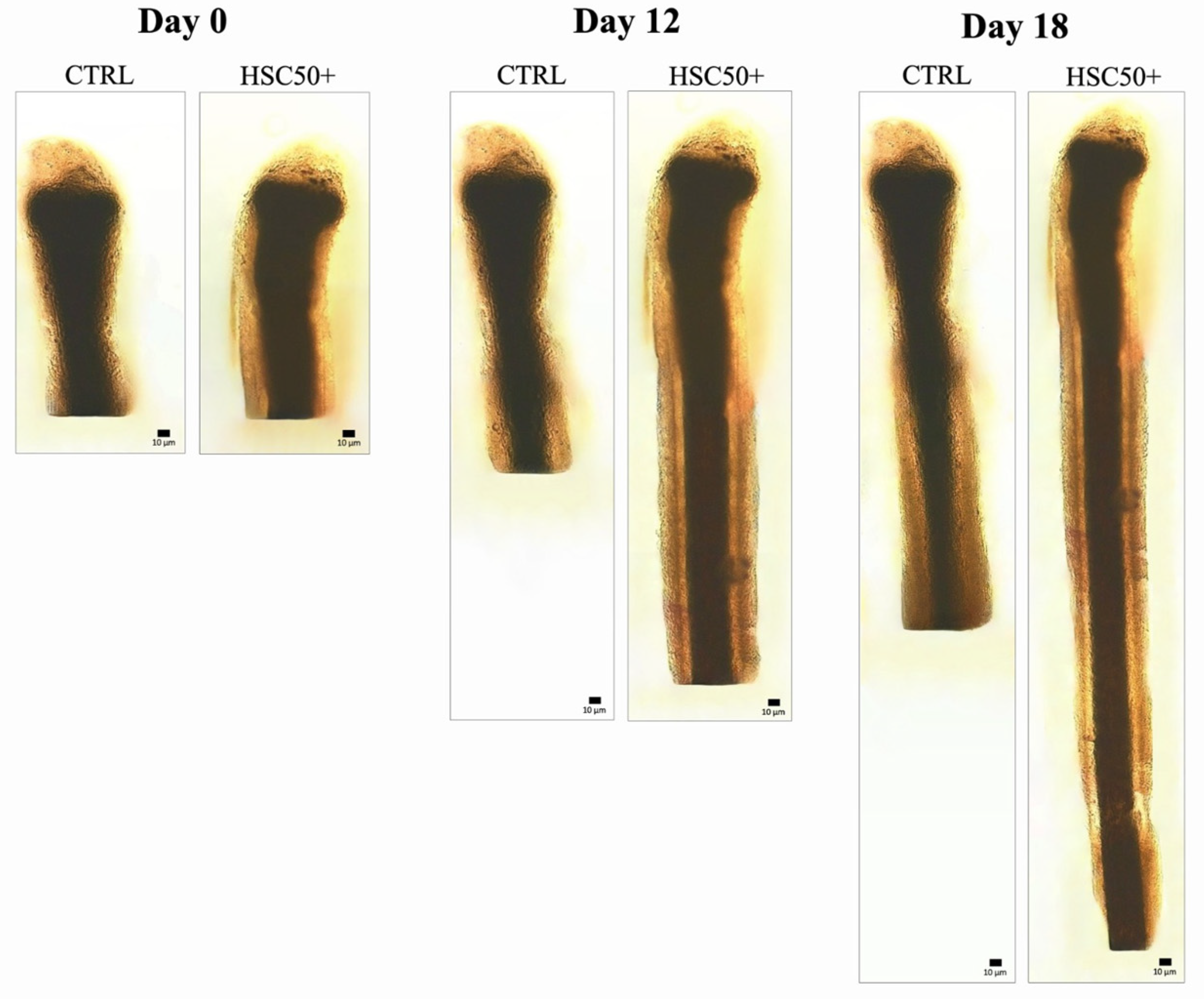
Hair follicle growth was followed for 18 days, with its length measured at regular intervals (every 3 days). The results showed the effectiveness of HSC50+ compared with the other compounds tested (Figure 6). Specifically, at the end of treatment, follicles exposed to the new HSC50+ showed a length of 2.81 ± 0.16 mm. Lower growth was shown following treatment with colostrum exosomes used alone, where the length corresponded to 2.31 ± 0.15 mm; plant-derived nanovesicles, with a length of 1.72 ± 0.14 mm; and CBSC exosomes, with a length corresponding to 1.96 ± 0.16 mm. Finally, the negative control (CTRL) showed a value of 1.83 ± 0.13 mm. It can be seen from the graph that highly significant values (** p < 0.01) were obtained following treatment with HSC50+ from day 12. Treatment with colostrum exosomes also showed a significant (* p < 0.05) increase in the measured parameter, but lower than the previously mentioned formulation. In contrast, no significant difference in follicle growth was found in the samples treated with exosomes of CBSCs and PDNVs, compared with the untreated negative control (Figure 7).
4. Discussion
The preliminary part of our study was focused on the isolation and characterization of exosomes derived from colostrum, whose size and concentration corresponded to 90–120 and 4.2 × 10¹² particles/mL, respectively. Compared with those obtained from plant nanovesicles and exosomes from stem cells, colostrum-derived exosomes showed higher concentrations (particles/mL), while the average size was lower than in C. Bergamia, but higher than CBSC exosomes. However, exosomes derived from the matrices, in comparison, exhibited a similar and regular spherical morphology. Following isolation and characterization, colostrum exosomes, known for their high biocompatibility [58,59], were combined with bioactive molecules present in GF20, a mixture isolated from colostrum and purified of potentially allergenic components, e.g., immunoglobulins and molecules not involved in tissue regeneration mechanisms [60,61,62]. Thus, GF20, as described in our study via an ELISA test, is composed of a high variety of growth factors, cytokines, and other protein molecules known to act synergistically during the hair follicle growth process. bFGF and TGF-β, among the characterized factors, showed the highest concentrations. These growth factors are crucial to providing substantial help in regenerating hair tissue, and each of them is engaged in an explicit biomolecular activity. bFGF promotes hair growth by inducing the anagen phase, stimulating the proliferation of papilla cells, leading to an increase in the size of the hair follicle [63]. TGF-β plays a crucial role in regulating hair growth: in hair follicles, it is involved in the development of the dermal papilla and hair matrix during the anagen phase, but also in the apoptotic process that characterizes the catagen phase, accompanied by the removal of the hair shaft from the dermal papilla [64].
The final goal of the present work was to evaluate the effects of a new compound called HSC50+ that combines the action of colostrum-derived exosomes with the growth factors and cytokines of GF20. The stimulatory effect on hair growth was evaluated through an in vitro test involving treatment for 18 days. At the end of the exposure, GF20 and HSC50+ resulted not only in an extension of follicle growth compared to all other compounds tested, but also in an increase in the number of dermal papilla cells. The positive results observed could be related partly to the action of the exosomes and partly to that of the free factors. The exosomes, in fact, act as carriers of the same free factors present in GF20. It is known that exosomes, through encapsulation into their structure, protect bioactive molecules from enzymatic and nonenzymatic degradation systems that could partially neutralize the action of free factors [65]. Exosomes also play an important role in cell communication because they are recognized by target cells fusing with their membranes and releasing their contents (endocytosis) [66]. This caused more factors to recognize specific receptors, triggering the enzymatic cascades that ultimately led to the stimulation of hair growth. Although the current clinical evidence supporting the use of exosome treatment is limited, there is a growing body of evidence suggesting its therapeutic potential. Our study has allowed us to identify a natural product with high potential in the treatment of hair regeneration. The effectiveness of HSC50+ is certainly due to the high concentration of growth factors and cytokines which, by stimulating the anagen phase of the hair cycle and reducing the telogen phase of the hair cycle, help to restart the entire hair cycle [67]. Moreover, HSC50+ can be stored with long-term stability and can be produced on a large scale and at a low cost.
5. Conclusions
Based on the results obtained, nanovesicles from plants and exosomes from stem cells had no significant effect on the stimulation of hair growth, unlike exosomes from colostrum. In contrast, these exosomes combined with the bioactive molecules with regenerative potential of GF20, in HSC50+, resulted in a significant increase in bulb growth and the regeneration of the dermal papilla. This evidence could represent the key to implementing new treatments to solve the problem of hair loss, with a consequent positive impact on the quality of life and psychological sphere of subjects suffering from alopecia or other conditions characterized by limited follicular growth.
Supplementary Materials
The following supporting information can be downloaded at: https://www.mdpi.com/2079-9284/11/1/10/s1, Table S1: Concentration of 20 bioactive molecules of GF20 characterized by ELISA test.
Author Contributions
Conceptualization, M.V.B.; methodology, M.V.B.; software, G.F., M.C. and M.Z.; validation, G.F. and M.C.; formal analysis, G.F., M.C. and M.Z.; investigation, G.F. and M.C.; resources, G.F. and M.C.; data curation, G.F. and M.C.; writing—original draft preparation, M.V.B.; writing—review and editing, G.F. and M.C.; visualization, G.F. and M.C.; supervision, M.V.B.; funding acquisition, M.V.B. All authors have read and agreed to the published version of the manuscript.
Funding
This research received no external funding.
Institutional Review Board Statement
This study was performed in line with the principles of the Declaration of Helsinki and does not require approval from the Ethics Committee of University of Catania.
Informed Consent Statement
Informed consent has been obtained from the volunteers to publish this paper.
Data Availability Statement
Original data are available on request.
Acknowledgments
G.F. thanks the Ph.D. program FSE Notice 1/2021. Authors acknowledge the PON project Bio-nanotech Research and Innovation Tower (BRIT), financed by the Italian Ministry for Education, University and Research (MIUR) (Grant no. PONa3_00136). The research team would like to express its sincere gratitude to Dermoaroma for the generous provision of the HSC50+ and related consumables that facilitated the successful completion of this research project.
Conflicts of Interest
The authors declare that they have no conflict of interest regarding the contents of this article.
References
- Thadanipon, K.; Suchonwanit, P. Measuring Patient Quality of Life Following Treatment for Alopecia. Patient Prefer. Adherence 2021, 15, 1601–1610. [Google Scholar] [CrossRef] [PubMed]
- Dhami, L. Psychology of Hair Loss Patients and Importance of Counseling. Indian J. Plast. Surg. 2021, 54, 411–415. [Google Scholar] [CrossRef] [PubMed]
- Lin, J.; Saknite, I.; Valdebran, M.; Balu, M.; Lentsch, G.; Williams, J.N.; Koenig, K.; Tromberg, B.J.; Atanaskova Mesinkovska, N. Feature characterization of scarring and non-scarring types of alopecia by multiphoton microscopy. Lasers Surg. Med. 2019, 51, 95–103. [Google Scholar] [CrossRef] [PubMed]
- Vidal, C.I. Overview of Alopecia: A Dermatopathologist’s Perspective. Mo. Med. 2015, 112, 308–312. [Google Scholar]
- Piraccini, B.M.; Alessandrini, A. Androgenetic alopecia. G. Ital. Dermatol. Venereol. 2014, 149, 15–24. [Google Scholar] [PubMed]
- Lolli, F.; Pallotti, F.; Rossi, A.; Fortuna, M.C.; Caro, G.; Lenzi, A.; Sansone, A.; Lombardo, F. Androgenetic alopecia: A review. Endocrine 2017, 57, 9–17. [Google Scholar] [CrossRef]
- Grymowicz, M.; Rudnicka, E.; Podfigurna, A.; Napierala, P.; Smolarczyk, R.; Smolarczyk, K.; Meczekalski, B. Hormonal Effects on Hair Follicles. Int. J. Mol. Sci. 2020, 21, 5342. [Google Scholar] [CrossRef] [PubMed]
- Nestor, M.S.; Ablon, G.; Gade, A.; Han, H.; Fischer, D.L. Treatment options for androgenetic alopecia: Efficacy, side effects, compliance, financial considerations, and ethics. J. Cosmet. Dermatol. 2021, 20, 3759–3781. [Google Scholar] [CrossRef]
- Natarelli, N.; Gahoonia, N.; Sivamani, R.K. Integrative and Mechanistic Approach to the Hair Growth Cycle and Hair Loss. J. Clin. Med. 2023, 12, 893. [Google Scholar] [CrossRef]
- Lin, X.; Zhu, L.; He, J. Morphogenesis, Growth Cycle and Molecular Regulation of Hair Follicles. Front. Cell Dev. Biol. 2022, 10, 899095. [Google Scholar] [CrossRef]
- Hibino, T.; Nishiyama, T. Role of TGF-beta2 in the human hair cycle. J. Dermatol. Sci. 2004, 35, 9–18. [Google Scholar] [CrossRef] [PubMed]
- Shin, S.H.; Koh, Y.G.; Lee, W.G.; Seok, J.; Park, K.Y. The use of epidermal growth factor in dermatological practice. Int. Wound J. 2023, 20, 2414–2423. [Google Scholar] [CrossRef] [PubMed]
- Ahn, S.Y.; Pi, L.Q.; Hwang, S.T.; Lee, W.S. Effect of IGF-I on Hair Growth Is Related to the Anti-Apoptotic Effect of IGF-I and Up-Regulation of PDGF-A and PDGF-B. Ann. Dermatol. 2012, 24, 26–31. [Google Scholar] [CrossRef] [PubMed]
- Woo, J.; Suh, W.; Sung, J.H. Hair Growth Regulation by Fibroblast Growth Factor 12 (FGF12). Int. J. Mol. Sci. 2022, 23, 9467. [Google Scholar] [CrossRef] [PubMed]
- Trüeb, R.M. Further Clinical Evidence for the Effect of IGF-1 on Hair Growth and Alopecia. Skin. Appendage Disord. 2018, 4, 90–95. [Google Scholar] [CrossRef] [PubMed]
- El-Refai, A.M.; Elhabak, D.M.; Khashaba, R.A. More is Not Always Better in Hair Growth Factors. Epidermal Growth Factor: Hair Growth Factor Involved in Alopecia Areata Pathogenesis. Int. J. Trichology 2020, 12, 182–187. [Google Scholar]
- Yoon, S.Y.; Kim, K.T.; Jo, S.J.; Cho, A.R.; Jeon, S.I.; Choi, H.D.; Kim, K.H.; Park, G.S.; Pack, J.K.; Kwon, O.S.; et al. Induction of hair growth by insulin-like growth factor-1 in 1,763 MHz radiofrequency-irradiated hair follicle cells. PLoS ONE 2011, 6, e28474. [Google Scholar] [CrossRef]
- Zhang, H.; Nan, W.; Wang, S.; Zhang, T.; Si, H.; Yang, F.; Li, G. Epidermal Growth Factor Promotes Proliferation and Migration of Follicular Outer Root Sheath Cells via Wnt/β-Catenin Signaling. Cell Physiol. Biochem. 2016, 39, 360–370. [Google Scholar] [CrossRef]
- Foitzik, K.; Lindner, G.; Mueller-Roever, S.; Maurer, M.; Botchkareva, N.; Botchkarev, V.; Handjiski, B.; Metz, M.; Hibino, T.; Soma, T.; et al. Control of murine hair follicle regression (catagen) by TGF-beta1 in vivo. FASEB J. 2000, 14, 752–760. [Google Scholar] [CrossRef]
- Zhao, B.; Li, J.; Chen, Q.; Yang, N.; Bao, Z.; Hu, S.; Chen, Y.; Wu, X. A Treatment Combination of IGF and EGF Promotes Hair Growth in the Angora Rabbit. Genes 2020, 12, 24. [Google Scholar] [CrossRef]
- Xie, M.; Wu, D.; Li, G.; Yang, J.; Zhang, Y.S. Exosomes targeted towards applications in regenerative medicine. Nano Sel. 2021, 2, 880–908. [Google Scholar] [CrossRef]
- Thakur, A.; Shah, D.; Rai, D.; Parra, D.C.; Pathikonda, S.; Kurilova, S.; Cili, A. Therapeutic Values of Exosomes in Cosmetics, Skin Care, Tissue Regeneration, and Dermatological Diseases. Cosmetics 2023, 10, 65. [Google Scholar] [CrossRef]
- Kalluri, R.; LeBleu, V.S. The biology, function, and biomedical applications of exosomes. Science 2020, 367, eaau6977. [Google Scholar] [CrossRef] [PubMed]
- Isola, A.L.; Chen, S. Exosomes: The Messengers of Health and Disease. Curr. Neuropharmacol. 2017, 15, 157–165. [Google Scholar] [CrossRef]
- Akuma, P.; Okagu, O.D.; Udenigwe, C.C. Naturally Occurring Exosome Vesicles as Potential Delivery Vehicle for Bioactive Compounds. Front. Sustain. Food Syst. 2019, 3, 23. [Google Scholar] [CrossRef]
- Mu, N.; Li, J.; Zeng, L.; You, J.; Li, R.; Qin, A.; Liu, X.; Yan, F.; Zhou, Z. Plant-Derived Exosome-Like Nanovesicles: Current Progress and Prospects. Int. J. Nanomed. 2023, 18, 4987–5009. [Google Scholar] [CrossRef]
- Sarasati, A.; Syahruddin, M.H.; Nuryanti, A.; Ana, I.D.; Barlian, A.; Wijaya, C.H.; Ratnadewi, D.; Wungu, T.D.K.; Takemori, H. Plant-Derived Exosome-like Nanoparticles for Biomedical Applications and Regenerative Therapy. Biomedicines 2023, 11, 1053. [Google Scholar] [CrossRef]
- Stoorvogel, W.; Kleijmeer, M.J.; Geuze, H.J.; Raposo, G. The biogenesis and functions of exosomes. Traffic 2002, 3, 321–330. [Google Scholar] [CrossRef] [PubMed]
- Doyle, L.M.; Wang, M.Z. Overview of Extracellular Vesicles, Their Origin, Composition, Purpose, and Methods for Exosome Isolation and Analysis. Cells 2019, 8, 727. [Google Scholar] [CrossRef]
- Zhou, Y.; Zhang, Y.; Gong, H.; Luo, S.; Cui, Y. The Role of Exosomes and Their Applications in Cancer. Int. J. Mol. Sci. 2021, 22, 12204. [Google Scholar] [CrossRef]
- Xie, F.; Huang, Y.; Zhan, Y.; Bao, L. Exosomes as drug delivery system in gastrointestinal cancer. Front. Oncol. 2023, 12, 1101823. [Google Scholar] [CrossRef]
- Lee, J.Y.; Kim, H.S. Extracellular Vesicles in Neurodegenerative Diseases: A Double-Edged Sword. Tissue Eng. Regen. Med. 2017, 14, 667–678. [Google Scholar] [CrossRef] [PubMed]
- Rademacher, D.J. Potential for Therapeutic-Loaded Exosomes to Ameliorate the Pathogenic Effects of α-Synuclein in Parkinson’s Disease. Biomedicines 2023, 11, 1187. [Google Scholar] [CrossRef]
- De Jong, O.G.; Van Balkom, B.W.; Schiffelers, R.M.; Bouten, C.V.; Verhaar, M.C. Extracellular vesicles: Potential roles in regenerative medicine. Front. Immunol. 2014, 5, 608. [Google Scholar] [CrossRef] [PubMed]
- Muthu, S.; Bapat, A.; Jain, R.; Jeyaraman, N.; Jeyaraman, M. Exosomal therapy-a new frontier in regenerative medicine. Stem Cell Investig. 2021, 8, 7. [Google Scholar] [CrossRef]
- Mathivanan, S.; Ji, H.; Simpson, R.J. Exosomes: Extracellular organelles important in intercellular communication. J. Proteom. 2010, 73, 1907–1920. [Google Scholar] [CrossRef] [PubMed]
- Wu, Y.; Deng, W.; Klinke, D.J., 2nd. Exosomes: Improved methods to characterize their morphology, RNA content, and surface protein biomarkers. Analyst 2015, 140, 6631–6642. [Google Scholar] [CrossRef]
- Yaghoubi, Y.; Movassaghpour, A.; Zamani, M.; Talebi, M.; Mehdizadeh, A.; Yousefi, M. Human umbilical cord mesenchymal stem cells derived-exosomes in diseases treatment. Life Sci. 2019, 233, 116733. [Google Scholar] [CrossRef]
- Shkryl, Y.; Tsydeneshieva, Z.; Degtyarenko, A.; Yugay, Y.; Balabanova, L.; Rusapetova, T.; Bulgakov, V. Plant Exosomal Vesicles: Perspective Information Nanocarriers in Biomedicine. Appl. Sci. 2022, 12, 8262. [Google Scholar] [CrossRef]
- Lian, M.Q.; Chng, W.H.; Liang, J.; Yeo, H.Q.; Lee, C.K.; Belaid, M.; Tollemeto, M.; Wacker, M.G.; Czarny, B.; Pastorin, G. Plant-derived extracellular vesicles: Recent advancements and current challenges on their use for biomedical applications. J. Extracell. Vesicles 2022, 11, e12283. [Google Scholar] [CrossRef]
- Kim, M.; Jang, H.; Kim, W.; Kim, D.; Park, J.H. Therapeutic Applications of Plant-Derived Extracellular Vesicles as Antioxidants for Oxidative Stress-Related Diseases. Antioxidants 2023, 12, 1286. [Google Scholar] [CrossRef]
- Di Giulio, S.; Carata, E.; Mariano, S.; Panzarini, E. Plant Extracellular Vesicles: Investigating Their Utilization as Beneficial Nutrients in Diet. Appl. Sci. 2023, 13, 6656. [Google Scholar] [CrossRef]
- Di Gioia, S.; Hossain, M.N.; Conese, M. Biological properties and therapeutic effects of plant-derived nanovesicles. Open Med. 2020, 15, 1096–1122. [Google Scholar] [CrossRef] [PubMed]
- Liu, Y.; Wu, S.; Koo, Y.; Yang, A.; Dai, Y.; Khant, H.; Osman, S.R.; Chowdhury, M.; Wei, H.; Li, Y.; et al. Characterization of and isolation methods for plant leaf nanovesicles and small extracellular vesicles. Nanomedicine 2020, 29, 102271. [Google Scholar] [CrossRef]
- Naeem, P.; Baumgartner, A.; Ghaderi, N.; Sefat, F.; Alhawamdeh, M.; Heidari, S.; Shahzad, F.; Swaminathan, K.; Akhbari, P.; Isreb, M.; et al. Anticarcinogenic impact of extracellular vesicles (exosomes) from cord blood stem cells in malignant melanoma: A potential biological treatment. J. Cell Mol. Med. 2023, 27, 222–231. [Google Scholar] [CrossRef] [PubMed]
- Cardoso, R.M.S.; Rodrigues, S.C.; Gomes, C.F.; Duarte, F.V.; Romao, M.; Leal, E.C.; Freire, P.C.; Neves, R.; Simões-Correia, J. Development of an optimized and scalable method for isolation of umbilical cord blood-derived small extracellular vesicles for future clinical use. Stem Cells Transl. Med. 2021, 10, 910–921. [Google Scholar] [CrossRef] [PubMed]
- Harrell, C.R.; Jovicic, N.; Djonov, V.; Arsenijevic, N.; Volarevic, V. Mesenchymal Stem Cell-Derived Exosomes and Other Extracellular Vesicles as New Remedies in the Therapy of Inflammatory Diseases. Cells 2019, 8, 1605. [Google Scholar] [CrossRef]
- Hu, Y.; Rao, S.S.; Wang, Z.X.; Cao, J.; Tan, Y.J.; Luo, J.; Li, H.M.; Zhang, W.S.; Chen, C.Y.; Xie, H. Exosomes from human umbilical cord blood accelerate cutaneous wound healing through miR-21-3p-mediated promotion of angiogenesis and fibroblast function. Theranostics 2018, 8, 169–184. [Google Scholar] [CrossRef]
- Gupta, A.K.; Wang, T.; Rapaport, J.A. Systematic review of exosome treatment in hair restoration: Preliminary evidence, safety, and future directions. J. Cosmet. Dermatol. 2023, 22, 2424–2433. [Google Scholar] [CrossRef]
- Kim, H.; Jang, Y.; Kim, E.H.; Jang, H.; Cho, H.; Han, G.; Song, H.K.; Kim, S.H.; Yang, Y. Potential of Colostrum-Derived Exosomes for Promoting Hair Regeneration Through the Transition From Telogen to Anagen Phase. Front. Cell Dev. Biol. 2022, 10, 815205. [Google Scholar] [CrossRef]
- Li, X.; Li, X.; Zhang, B.; He, B. The Role of Cancer Stem Cell-Derived Exosomes in Cancer Progression. Stem Cells Int. 2022, 4, 9133658. [Google Scholar] [CrossRef]
- Logozzi, M.; Di Raimo, R.; Mizzoni, D.; Fais, S. Nanovesicles from Organic Agriculture-Derived Fruits and Vegetables: Characterization and Functional Antioxidant Content. Int. J. Mol. Sci. 2021, 22, 8170. [Google Scholar] [CrossRef] [PubMed]
- Szatanek, R.; Baj-Krzyworzeka, M.; Zimoch, J.; Lekka, M.; Siedlar, M.; Baran, J. The Methods of Choice for Extracellular Vesicles (EVs) Characterization. Int. J. Mol. Sci. 2017, 18, 1153. [Google Scholar] [CrossRef]
- Théry, C.; Amigorena, S.; Raposo, G.; Clayton, A. Isolation and characterization of exosomes from cell culture supernatants and biological fluids. Curr. Protoc. Cell Biol. 2006, 3, 3–22. [Google Scholar] [CrossRef] [PubMed]
- Zimbone, M.; Musumeci, P.; Baeri, P.; Messina, E.; Boninelli, S.; Compagnini, G.; Calcagno, L. Rotational dynamics of gold nanoparticle chains in water solution. J. Nanopart Res. 2012, 14, 1308. [Google Scholar] [CrossRef]
- Zimbone, M.; Baeri, P.; Calcagno, L.; Musumeci, P.; Contino, A.; Barcellona, M.L.; Bonaventura, G. Dynamic light scattering on bioconjugated laser generated gold nanoparticles. PLoS ONE 2014, 9, e89048. [Google Scholar] [CrossRef] [PubMed]
- Sacerdote, P.; Mussano, F.; Franchi, S.; Panerai, A.E.; Bussolati, G.; Carossa, S.; Bartorelli, A.; Bussolati, B. Biological components in a standardized derivative of bovine colostrum. J. Dairy. Sci. 2013, 96, 1745–1754. [Google Scholar] [CrossRef]
- Somiya, M.; Yoshioka, Y.; Ochiya, T. Biocompatibility of highly purified bovine milk-derived extracellular vesicles. J. Extracell. Vesicles 2018, 7, 1440132. [Google Scholar] [CrossRef]
- Zhong, J.; Xia, B.; Shan, S.; Zheng, A.; Zhang, S.; Chen, J.; Liang, X.J. High-quality milk exosomes as oral drug delivery system. Biomaterials 2021, 277, 121126. [Google Scholar] [CrossRef]
- Wheeler, T.T.; Hodgkinson, A.J.; Prosser, C.G.; Davis, S.R. Immune components of colostrum and milk—A historical perspective. J. Mammary Gland. Biol. Neoplasia 2007, 12, 237–247. [Google Scholar] [CrossRef]
- El-Agamy, E.I. The challenge of cow milk protein allergy. Small Rumin. Res. 2007, 68, 64–72. [Google Scholar] [CrossRef]
- Ulfman, L.H.; Leusen, J.H.W.; Savelkoulm, H.F.J.; Warnerm, J.O.; van Neervenm, R.J.J. Effects of Bovine Immunoglobulins on Immune Function, Allergy, and Infection. Front. Nutr. 2018, 5, 52. [Google Scholar] [CrossRef] [PubMed]
- Lin, W.H.; Xiang, L.J.; Shi, H.X.; Zhang, J.; Jiang, L.P.; Cai, P.T.; Lin, Z.L.; Lin, B.B.; Huang, Y.; Zhang, H.L.; et al. Fibroblast growth factors stimulate hair growth through β-catenin and Shh expression in C57BL/6 mice. Biomed. Res. Int. 2015, 2015, 730139. [Google Scholar]
- Rishikaysh, P.; Dev, K.; Diaz, D.; Qureshi, W.M.; Filip, S.; Mokry, J. Signaling involved in hair follicle morphogenesis and development. Int. J. Mol. Sci. 2014, 15, 1647–1670. [Google Scholar] [CrossRef]
- Feng, X.; Chen, X.; Zheng, X.; Zhu, H.; Qi, Q.; Liu, S.; Zhang, H.; Che, J. Latest Trend of Milk Derived Exosomes: Cargos, Functions, and Applications. Front. Nutr. 2021, 8, 747294. [Google Scholar] [CrossRef]
- Rashidi, M.; Bijari, S.; Khazaei, A.H.; Shojaei-Ghahrizjani, F.; Rezakhani, L. The role of milk-derived exosomes in the treat ment of diseases. Front. Genet. 2022, 13, 1009338. [Google Scholar] [CrossRef]
- Hoffmann, R.; Eicheler, W.; Huth, A.; Wenzel, E.; Happle, R. Cytokines and growth factors influence hair growth in vitro. Possible implications for the pathogenesis and treatment of alopecia areata. Arch. Dermatol. Res. 1996, 288, 153–156. [Google Scholar] [CrossRef]
Figure 1.
Colostrum exosome characterization. (a) Analysis of size and concentration via DLS; (b) observation of exosome morphology via SEM.
Figure 1.
Colostrum exosome characterization. (a) Analysis of size and concentration via DLS; (b) observation of exosome morphology via SEM.

Figure 2.
Concentrations of the main bioactive factors present in GF20, expressed on a logarithmic scale.
Figure 2.
Concentrations of the main bioactive factors present in GF20, expressed on a logarithmic scale.
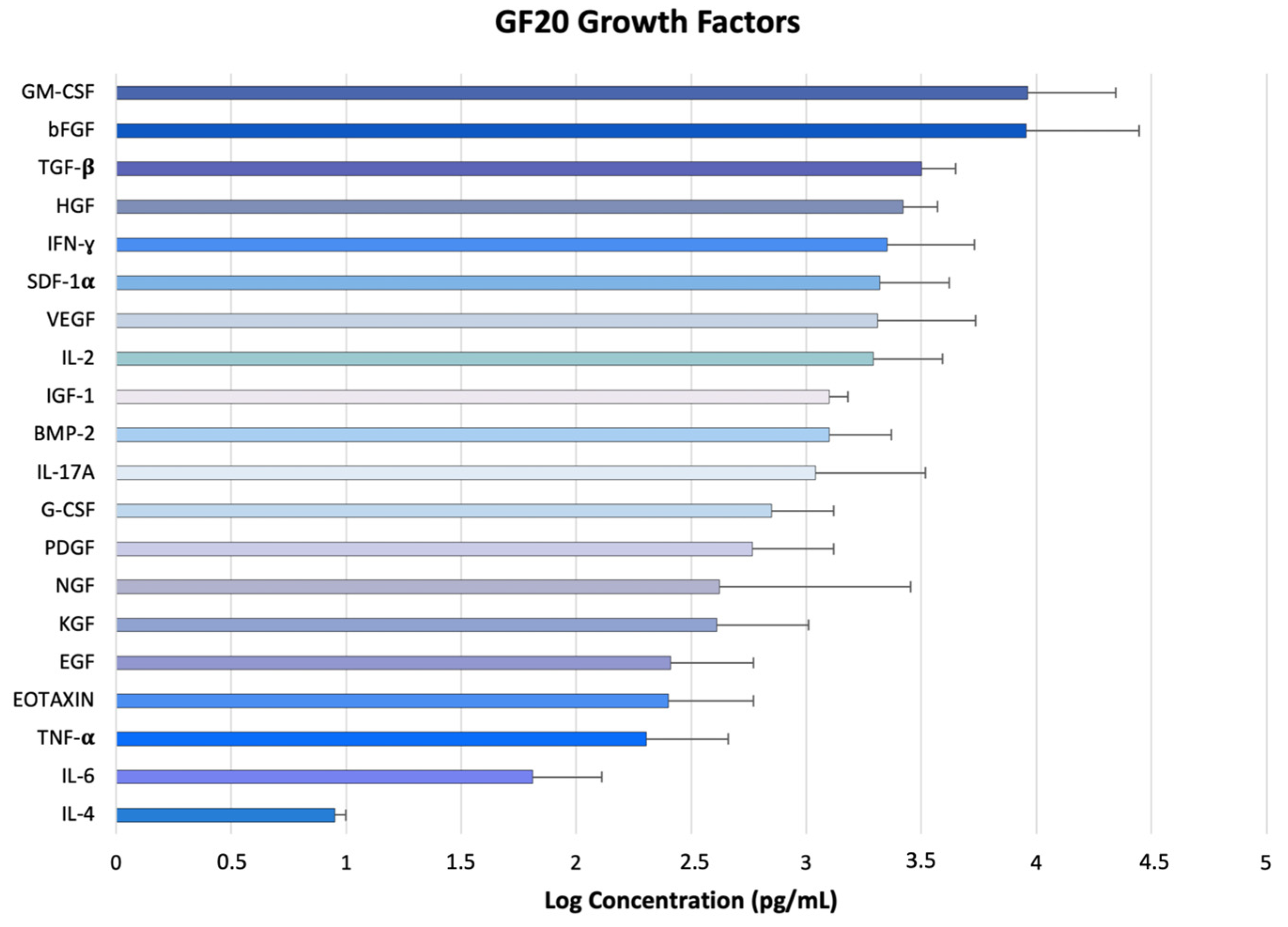
Figure 3.
In vitro culture of human hair follicle. (a) CTRL; (b) samples exposed to plant-derived nanovesicles; (c) samples exposed to CBSC exosomes; (d) samples exposed to colostrum exosomes; (e) samples exposed to GF20; (f) samples exposed to HSC50+. The red arrow indicates the expanding germinative area of the hair bulb.
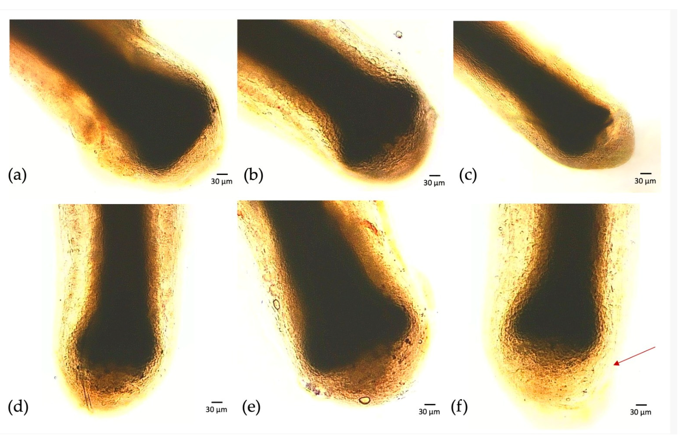
Figure 4.
Hair follicles stained with DAPI and the identification of papilla dermis cells. White dotted lines indicate the dermal papilla (a) CTRL; (b) samples exposed to plant-derived nanovesicles; (c) samples exposed to CBSC exosomes; (d) samples exposed to colostrum exosomes; (e) samples exposed to GF20; (f) samples exposed to HSC50+.
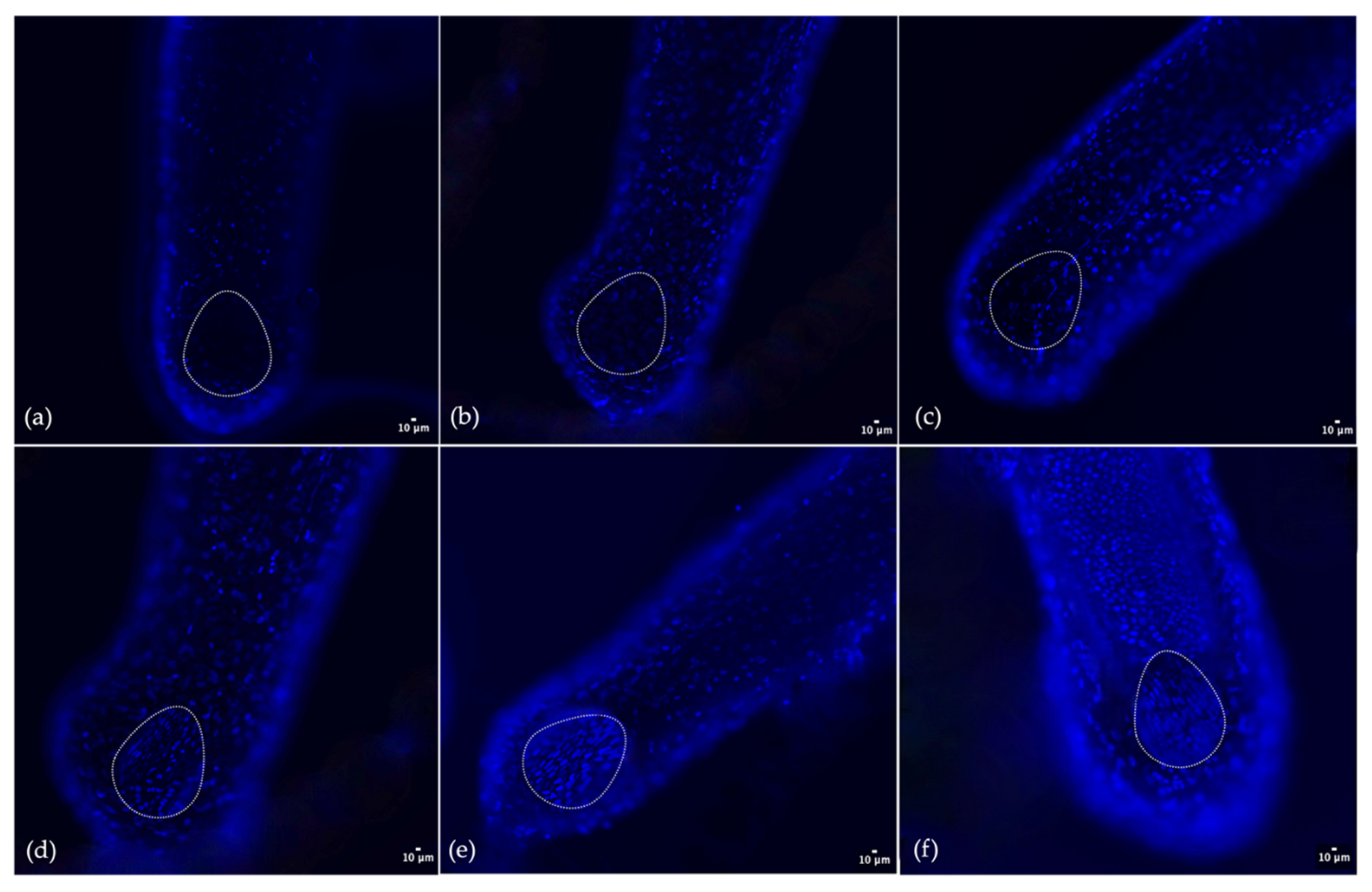
Figure 5.
Analysis of the fluorescence intensity of the hair bulb via ImageJ. Statistical differences are indicated with the symbol * (p < 0.05), while more significant data are reported with the symbol ** (p < 0.01).
Figure 5.
Analysis of the fluorescence intensity of the hair bulb via ImageJ. Statistical differences are indicated with the symbol * (p < 0.05), while more significant data are reported with the symbol ** (p < 0.01).

Figure 6.
Comparison of hair length variation between samples exposed to exosomes from different matrices. Statistical differences are indicated with the symbol * (p < 0.05), while more significant data are reported with the symbol ** (p < 0.01).
Figure 6.
Comparison of hair length variation between samples exposed to exosomes from different matrices. Statistical differences are indicated with the symbol * (p < 0.05), while more significant data are reported with the symbol ** (p < 0.01).
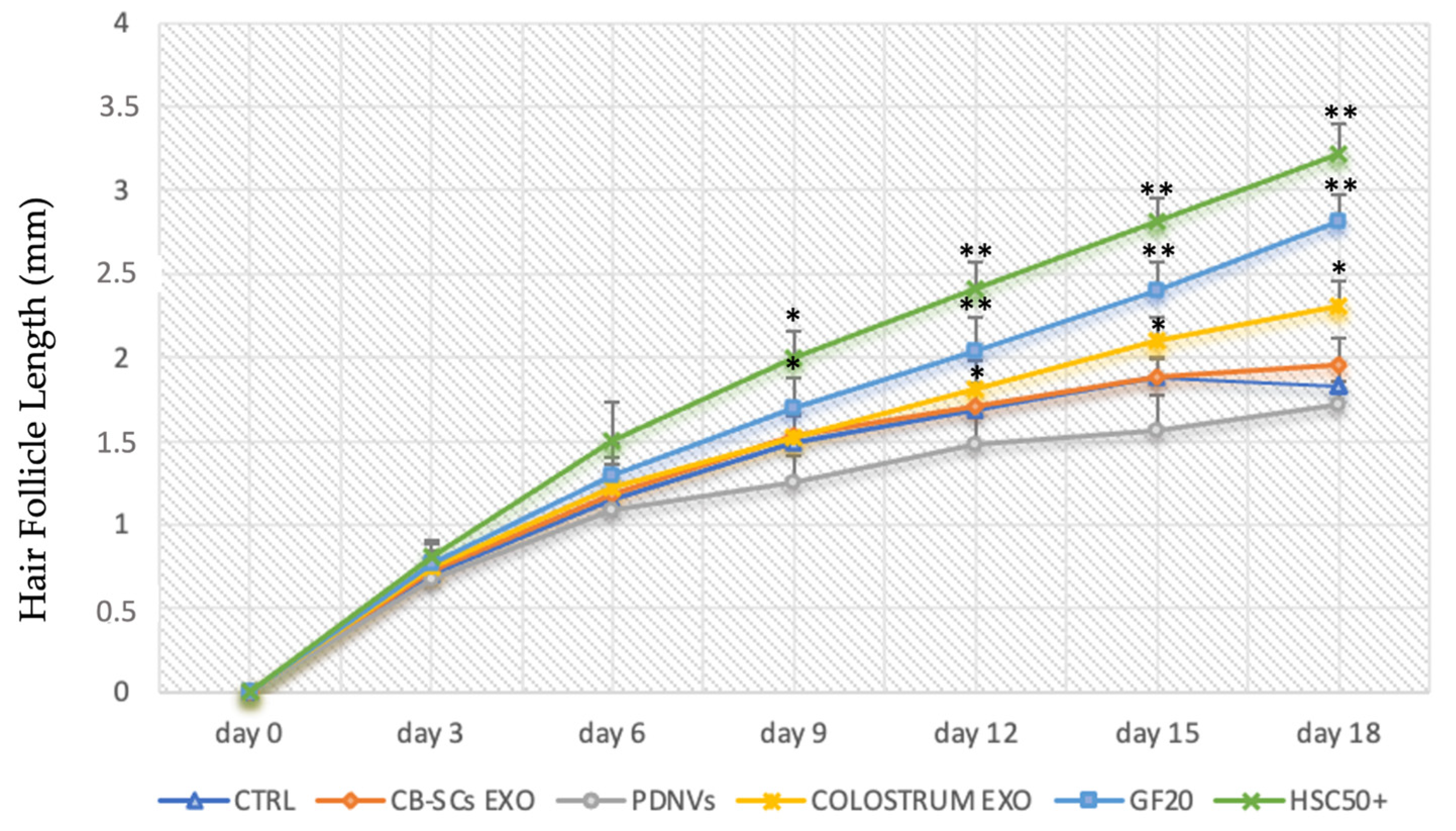
Figure 7.
Observation of the hair bulb length in the CTRL and samples exposed to HSC50+ at day 0, 2, and 18.
Figure 7.
Observation of the hair bulb length in the CTRL and samples exposed to HSC50+ at day 0, 2, and 18.

Table 1.
Growth factors and cytokines detected via ELISA kit in GF20.
| Growth Factors and Cytokines Detected via ELISA |
|---|
| Transforming Growth Factors β (TGF-β) |
| Insulin-Like Growth Factor 1 (IGF-1) |
| Basic Fibroblast Growth Factor (bFGF) |
| Vascular Endothelial Growth Factor (VEGF) |
| Epidermal Growth Factor (EGF) |
| Platelet-Derived Growth Factor (PDGF) |
| Keratinocyte Growth Factor (KGF) |
| Hepatocyte Growth Factor (HGF) |
| Granulocyte Macrophage-Colony Stimulating Factor (GM-CSF) |
| Granulocyte-Colony Stimulating Factor (G-CSF) |
| EOTAXIN-CCL11 |
| Tumor Necrosis Factor α (TNF-α) |
| Nerve Growth Factor (NGF) |
| Gamma Interferon (INF-γ) |
| Bone Morphogenetic Protein 2 (BMP-2) |
| Stromal-Cell-Derived Factor 1α (SDF1-α) |
| Interleukin-2 (IL-2) |
| Interleukin-4 (IL-4) |
| Interleukin-6 (IL-6) |
| Interleukin-17A (IL-17A) |
Disclaimer/Publisher’s Note: The statements, opinions and data contained in all publications are solely those of the individual author(s) and contributor(s) and not of MDPI and/or the editor(s). MDPI and/or the editor(s) disclaim responsibility for any injury to people or property resulting from any ideas, methods, instructions or products referred to in the content. |
© 2024 by the authors. Licensee MDPI, Basel, Switzerland. This article is an open access article distributed under the terms and conditions of the Creative Commons Attribution (CC BY) license (https://creativecommons.org/licenses/by/4.0/).
Share and Cite
MDPI and ACS Style
Ferruggia, G.; Contino, M.; Zimbone, M.; Brundo, M.V. Effectiveness of a Novel Compound HAIR & SCALP COMPLEX on Hair Follicle Regeneration. Cosmetics 2024, 11, 10. https://doi.org/10.3390/cosmetics11010010
AMA Style
Ferruggia G, Contino M, Zimbone M, Brundo MV. Effectiveness of a Novel Compound HAIR & SCALP COMPLEX on Hair Follicle Regeneration. Cosmetics. 2024; 11(1):10. https://doi.org/10.3390/cosmetics11010010
Chicago/Turabian StyleFerruggia, Greta, Martina Contino, Massimo Zimbone, and Maria Violetta Brundo. 2024. "Effectiveness of a Novel Compound HAIR & SCALP COMPLEX on Hair Follicle Regeneration" Cosmetics 11, no. 1: 10. https://doi.org/10.3390/cosmetics11010010
Note that from the first issue of 2016, this journal uses article numbers instead of page numbers. See further details here.