Next Issue
Volume 14, April
Previous Issue
Volume 14, February


Biosensors, Volume 14, Issue 3 (March 2024) – 42 articles

Cover Story (view full-size image): 
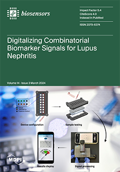
We are introducing a groundbreaking smartphone-based analyzing and reporting system (SBARS) for lupus nephritis (LN) biomarker detection. Traditional healthcare systems often face challenges in efficiency and patient coverage. The SBARS utilizes a portable microscopic reader and smartphone app, eliminating the need for expensive large equipment. Through innovative image processing techniques, it analyzes the intensity of biomarker signals with impressive accuracy. Our system demonstrated comparable performance to commercial scanners in detecting potential LN biomarkers and it allows for discriminating LN patients with active renal disease from healthy controls with remarkable precision. The SBARS may help revolutionize healthcare with accessible, cost-effective, and accurate biomarker detection. View this paper
  • Issues are regarded as officially published after their release is announced to the table of contents alert mailing list.
  • You may sign up for e-mail alerts to receive table of contents of newly released issues.
  • PDF is the official format for papers published in both, html and pdf forms. To view the papers in pdf format, click on the "PDF Full-text" link, and use the free Adobe Reader to open them.
Order results
Result details
Section
Select all
Export citation of selected articles as:
14 pages, 4465 KiB  
Article
Sensitivity of Electrocardiogram on Electrode-Pair Locations for Wearable Devices: Computational Analysis of Amplitude and Waveform Distortion
by Kiyoto Sanjo, Kazuki Hebiguchi, Cheng Tang, Essam A. Rashed, Sachiko Kodera, Hiroyoshi Togo and Akimasa Hirata
Biosensors 2024, 14(3), 153; https://doi.org/10.3390/bios14030153 - 21 Mar 2024
Viewed by 760
Abstract
An electrocardiogram (ECG) is used to observe the electrical activity of the heart via electrodes on the body surface. Recently, an ECG with fewer electrodes, such as a bipolar ECG in which two electrodes are attached to the chest, has been employed as [...] Read more.
An electrocardiogram (ECG) is used to observe the electrical activity of the heart via electrodes on the body surface. Recently, an ECG with fewer electrodes, such as a bipolar ECG in which two electrodes are attached to the chest, has been employed as wearable devices. However, the effect of different geometrical factors and electrode-pair locations on the amplitude and waveform of ECG signals remains unclear. In this study, we computationally evaluated the effects of body morphology, heart size and orientation, and electrode misalignment on ECG signals for 48 scenarios using 35 bipolar electrode pairs (1680 waveforms) with a dynamic time warping (DTW) algorithm. It was observed that the physique of the human body model predominantly affected the amplitude and waveform of the ECG signals. A multivariate analysis indicated that the heart–electrode distance and the solid angle of the heart from the electrode characterized the amplitude and waveform of the ECG signals, respectively. Furthermore, the electrode locations for less individual variability and less waveform distortion were close to the location of electrodes V2 and V3 in the standard 12-lead. These findings will facilitate the placement of ECG electrodes and interpretation of the measured ECG signals for wearable devices. Full article
Show Figures

Figure 1

15 pages, 2258 KiB  
Article
Customizable Nichrome Wire Heaters for Molecular Diagnostic Applications
by Juhee Lim, Won Han, Le Tran Huy Thang, Yong Wook Lee and Joong Ho Shin
Biosensors 2024, 14(3), 152; https://doi.org/10.3390/bios14030152 - 20 Mar 2024
Viewed by 811
Abstract
Accurate sample heating is vital for nucleic acid extraction and amplification, requiring a sophisticated thermal cycling process in nucleic acid detection. Traditional molecular detection systems with heating capability are bulky, expensive, and primarily designed for lab settings. Consequently, their use is limited where [...] Read more.
Accurate sample heating is vital for nucleic acid extraction and amplification, requiring a sophisticated thermal cycling process in nucleic acid detection. Traditional molecular detection systems with heating capability are bulky, expensive, and primarily designed for lab settings. Consequently, their use is limited where lab systems are unavailable. This study introduces a technique for performing the heating process required in molecular diagnostics applicable for point-of-care testing (POCT), by presenting a method for crafting customized heaters using freely patterned nichrome (NiCr) wire. This technique, fabricating heaters by arranging protrusions on a carbon black-polydimethylsiloxane (PDMS) cast and patterning NiCr wire, utilizes cost-effective materials and is not constrained by shape, thereby enabling customized fabrication in both two-dimensional (2D) and three-dimensional (3D). To illustrate its versatility and practicality, a 2D heater with three temperature zones was developed for a portable device capable of automatic thermocycling for polymerase chain reaction (PCR) to detect Escherichia coli (E. coli) O157:H7 pathogen DNA. Furthermore, the detection of the same pathogen was demonstrated using a customized 3D heater surrounding a microtube for loop-mediated isothermal amplification (LAMP). Successful DNA amplification using the proposed heater suggests that the heating technique introduced in this study can be effectively applied to POCT. Full article
(This article belongs to the Special Issue Biosensors Aiming for Practical Uses)
Show Figures

Figure 1

18 pages, 3412 KiB  
Article
Development of Highly Sensitive Fluorescent Sensors for Separation-Free Detection and Quantitation Systems of Pepsin Enzyme Applying a Structure-Guided Approach
by Aya M. Mostafa, Stephen J. Barton, Stephen P. Wren and James Barker
Biosensors 2024, 14(3), 151; https://doi.org/10.3390/bios14030151 - 20 Mar 2024
Viewed by 819
Abstract
Two fluorescent molecularly imprinted polymers (MIPs) were developed for pepsin enzyme utilising fluorescein and rhodamine b. The main difference between both dyes is the presence of two (diethylamino) groups in the structure of rhodamine b. Consequently, we wanted to investigate the effect of [...] Read more.
Two fluorescent molecularly imprinted polymers (MIPs) were developed for pepsin enzyme utilising fluorescein and rhodamine b. The main difference between both dyes is the presence of two (diethylamino) groups in the structure of rhodamine b. Consequently, we wanted to investigate the effect of these functional groups on the selectivity and sensitivity of the resulting MIPs. Therefore, two silica-based MIPs for pepsin enzyme were developed using 3-aminopropyltriethoxysilane as a functional monomer and tetraethyl orthosilicate as a crosslinker to achieve a one-pot synthesis. Results of our study revealed that rhodamine b dyed MIPs (RMIPs) showed stronger binding, indicated by a higher binding capacity value of 256 mg g−1 compared to 217 mg g−1 for fluorescein dyed MIPs (FMIPs). Moreover, RMIPs showed superior sensitivity in the detection and quantitation of pepsin with a linear range from 0.28 to 42.85 µmol L−1 and a limit of detection (LOD) as low as 0.11 µmol L−1. In contrast, FMIPs covered a narrower range from 0.71 to 35.71 µmol L−1, and the LOD value reached 0.34 µmol L−1, which is three times less sensitive than RMIPs. Finally, the developed FMIPs and RMIPs were applied to a separation-free quantification system for pepsin in saliva samples without interference from any cross-reactors. Full article
(This article belongs to the Section Biosensors and Healthcare)
Show Figures

Figure 1

23 pages, 7835 KiB  
Review
Hydrogel-Gated FETs in Neuromorphic Computing to Mimic Biological Signal: A Review
by Sankar Prasad Bag, Suyoung Lee, Jaeyoon Song and Jinsink Kim
Biosensors 2024, 14(3), 150; https://doi.org/10.3390/bios14030150 - 19 Mar 2024
Viewed by 945
Abstract
Hydrogel-gated synaptic transistors offer unique advantages, including biocompatibility, tunable electrical properties, being biodegradable, and having an ability to mimic biological synaptic plasticity. For processing massive data with ultralow power consumption due to high parallelism and human brain-like processing abilities, synaptic transistors have been [...] Read more.
Hydrogel-gated synaptic transistors offer unique advantages, including biocompatibility, tunable electrical properties, being biodegradable, and having an ability to mimic biological synaptic plasticity. For processing massive data with ultralow power consumption due to high parallelism and human brain-like processing abilities, synaptic transistors have been widely considered for replacing von Neumann architecture-based traditional computers due to the parting of memory and control units. The crucial components mimic the complex biological signal, synaptic, and sensing systems. Hydrogel, as a gate dielectric, is the key factor for ionotropic devices owing to the excellent stability, ultra-high linearity, and extremely low operating voltage of the biodegradable and biocompatible polymers. Moreover, hydrogel exhibits ionotronic functions through a hybrid circuit of mobile ions and mobile electrons that can easily interface between machines and humans. To determine the high-efficiency neuromorphic chips, the development of synaptic devices based on organic field effect transistors (OFETs) with ultra-low power dissipation and very large-scale integration, including bio-friendly devices, is needed. This review highlights the latest advancements in neuromorphic computing by exploring synaptic transistor developments. Here, we focus on hydrogel-based ionic-gated three-terminal (3T) synaptic devices, their essential components, and their working principle, and summarize the essential neurodegenerative applications published recently. In addition, because hydrogel-gated FETs are the crucial members of neuromorphic devices in terms of cutting-edge synaptic progress and performances, the review will also summarize the biodegradable and biocompatible polymers with which such devices can be implemented. It is expected that neuromorphic devices might provide potential solutions for the future generation of interactive sensation, memory, and computation to facilitate the development of multimodal, large-scale, ultralow-power intelligent systems. Full article
(This article belongs to the Section Biosensor and Bioelectronic Devices)
Show Figures

Figure 1

22 pages, 9533 KiB  
Article
Microwave Imaging System Based on Signal Analysis in a Planar Environment for Detection of Abdominal Aortic Aneurysms
by Andrea Martínez-Lozano, Roberto Gutierrez, Carlos G. Juan, Carolina Blanco-Angulo, Héctor García-Martínez, Germán Torregrosa, José María Sabater-Navarro and Ernesto Ávila-Navarro
Biosensors 2024, 14(3), 149; https://doi.org/10.3390/bios14030149 - 18 Mar 2024
Viewed by 871
Abstract
A proof-of-concept of a microwave imaging system for the fast detection of abdominal aortic aneurysms is shown. This experimental technology seeks to overcome the factors hampering the fast screening for these aneurysms with the usual equipment, such as high cost, long-time operation or [...] Read more.
A proof-of-concept of a microwave imaging system for the fast detection of abdominal aortic aneurysms is shown. This experimental technology seeks to overcome the factors hampering the fast screening for these aneurysms with the usual equipment, such as high cost, long-time operation or hazardous exposure to chemical substances. The hardware system is composed of 16 twin antennas mastered by a microcontroller through a switching network, which connects the antennas to the measurement instrument for sequential measurement. The software system is run by a computer, mastering the whole system, automatizing the measurement process and running the signal processing and medical image generation algorithms. Two image generation algorithms are tested: Delay-and-Sum (DAS) and Improved Delay-and-Sum (IDAS). Own-modified versions of these algorithms adapted to the requirements of our system are proposed. The system is carefully calibrated and fine-tuned with known objects placed at known distances. An experimental proof-of-concept is shown with a human torso phantom, including an aorta phantom and an aneurysm phantom placed in different positions. The results show good imaging capabilities with the potential for detecting and locating possible abdominal aortic aneurysms and reporting acceptable errors. Full article
(This article belongs to the Special Issue Development of Novel Biosensors for Point-of-Care Detection)
Show Figures

Figure 1

24 pages, 1503 KiB  
Review
Continuous and Non-Invasive Lactate Monitoring Techniques in Critical Care Patients
by Jose-Luis Lafuente, Samuel González, Clara Aibar, Desirée Rivera, Eva Avilés and Juan-Jose Beunza
Biosensors 2024, 14(3), 148; https://doi.org/10.3390/bios14030148 - 18 Mar 2024
Viewed by 999
Abstract
Lactate, once merely regarded as an indicator of tissue hypoxia and muscular fatigue, has now gained prominence as a pivotal biomarker across various medical disciplines. Recent research has unveiled its critical role as a high-value prognostic marker in critical care medicine. The current [...] Read more.
Lactate, once merely regarded as an indicator of tissue hypoxia and muscular fatigue, has now gained prominence as a pivotal biomarker across various medical disciplines. Recent research has unveiled its critical role as a high-value prognostic marker in critical care medicine. The current practice of lactate detection involves periodic blood sampling. This approach is invasive and confined to measurements at six-hour intervals, leading to resource expenditure, time consumption, and patient discomfort. This review addresses non-invasive sensors that enable continuous monitoring of lactate in critical care patients. After the introduction, it discusses the iontophoresis system, followed by a description of the structural materials that are universally employed to create an interface between the integumentary system and the sensor. Subsequently, each method is detailed according to its physical principle, outlining its advantages, limitations, and pertinent aspects. The study concludes with a discussion and conclusions, aiming at the design of an intelligent sensor (Internet of Medical Things or IoMT) to facilitate continuous lactate monitoring and enhance the clinical decision-making support system in critical care medicine. Full article
(This article belongs to the Section Biosensors and Healthcare)
Show Figures

Figure 1

15 pages, 4265 KiB  
Article
Advancing Point-of-Care Diagnosis: Digitalizing Combinatorial Biomarker Signals for Lupus Nephritis
by Jiechang Guo, Aygun Teymur, Chenling Tang, Ramesh Saxena and Tianfu Wu
Biosensors 2024, 14(3), 147; https://doi.org/10.3390/bios14030147 - 18 Mar 2024
Viewed by 892
Abstract
To improve the efficiency and patient coverage of the current healthcare system, user-friendly novel homecare devices are urgently needed. In this work, we developed a smartphone-based analyzing and reporting system (SBARS) for biomarker detection in lupus nephritis (LN). This system offers a cost-effective [...] Read more.
To improve the efficiency and patient coverage of the current healthcare system, user-friendly novel homecare devices are urgently needed. In this work, we developed a smartphone-based analyzing and reporting system (SBARS) for biomarker detection in lupus nephritis (LN). This system offers a cost-effective alternative to traditional, expensive large equipment in signal detection and quantification. This innovative approach involves using a portable and affordable microscopic reader to capture biomarker signals. Through smartphone-based image processing techniques, the intensity of each biomarker signal is analyzed. This system exhibited comparable performance to a commercial Genepix scanner in the detection of two potential novel biomarkers of LN, VISG4 and TNFRSF1b. Importantly, this smartphone-based analyzing and reporting system allows for discriminating LN patients with active renal disease from healthy controls with the area-under-the-curve (AUC) value = 0.9 for TNFRSF1b and 1.0 for VSIG4, respectively, indicating high predictive accuracy. Full article
(This article belongs to the Special Issue Biochips and Biosensors for Health-Care and Diagnostics)
Show Figures

Figure 1

37 pages, 5678 KiB  
Review
Aptamers and Nanobodies as New Bioprobes for SARS-CoV-2 Diagnostic and Therapeutic System Applications
by Ki Sung Park, Tae-In Park, Jae Eon Lee, Seo-Yeong Hwang, Anna Choi and Seung Pil Pack
Biosensors 2024, 14(3), 146; https://doi.org/10.3390/bios14030146 - 15 Mar 2024
Viewed by 1053
Abstract
The global challenges posed by the severe acute respiratory syndrome coronavirus 2 (SARS-CoV-2) pandemic have underscored the critical importance of innovative and efficient control systems for addressing future pandemics. The most effective way to control the pandemic is to rapidly suppress the spread [...] Read more.
The global challenges posed by the severe acute respiratory syndrome coronavirus 2 (SARS-CoV-2) pandemic have underscored the critical importance of innovative and efficient control systems for addressing future pandemics. The most effective way to control the pandemic is to rapidly suppress the spread of the virus through early detection using a rapid, accurate, and easy-to-use diagnostic platform. In biosensors that use bioprobes, the binding affinity of molecular recognition elements (MREs) is the primary factor determining the dynamic range of the sensing platform. Furthermore, the sensitivity relies mainly on bioprobe quality with sufficient functionality. This comprehensive review investigates aptamers and nanobodies recently developed as advanced MREs for SARS-CoV-2 diagnostic and therapeutic applications. These bioprobes might be integrated into organic bioelectronic materials and devices, with promising enhanced sensitivity and specificity. This review offers valuable insights into advancing biosensing technologies for infectious disease diagnosis and treatment using aptamers and nanobodies as new bioprobes. Full article
Show Figures

Figure 1

21 pages, 1474 KiB  
Review
Detection of Parasites in the Field: The Ever-Innovating CRISPR/Cas12a
by Xin Li, Zhisheng Dang, Wenqiang Tang, Haoji Zhang, Jianwei Shao, Rui Jiang, Xu Zhang and Fuqiang Huang
Biosensors 2024, 14(3), 145; https://doi.org/10.3390/bios14030145 - 14 Mar 2024
Viewed by 1218
Abstract
The rapid and accurate identification of parasites is crucial for prompt therapeutic intervention in parasitosis and effective epidemiological surveillance. For accurate and effective clinical diagnosis, it is imperative to develop a nucleic-acid-based diagnostic tool that combines the sensitivity and specificity of nucleic acid [...] Read more.
The rapid and accurate identification of parasites is crucial for prompt therapeutic intervention in parasitosis and effective epidemiological surveillance. For accurate and effective clinical diagnosis, it is imperative to develop a nucleic-acid-based diagnostic tool that combines the sensitivity and specificity of nucleic acid amplification tests (NAATs) with the speed, cost-effectiveness, and convenience of isothermal amplification methods. A new nucleic acid detection method, utilizing the clustered regularly interspaced short palindromic repeats (CRISPR)-associated (Cas) nuclease, holds promise in point-of-care testing (POCT). CRISPR/Cas12a is presently employed for the detection of Plasmodium falciparum, Toxoplasma gondii, Schistosoma haematobium, and other parasites in blood, urine, or feces. Compared to traditional assays, the CRISPR assay has demonstrated notable advantages, including comparable sensitivity and specificity, simple observation of reaction results, easy and stable transportation conditions, and low equipment dependence. However, a common issue arises as both amplification and cis-cleavage compete in one-pot assays, leading to an extended reaction time. The use of suboptimal crRNA, light-activated crRNA, and spatial separation can potentially weaken or entirely eliminate the competition between amplification and cis-cleavage. This could lead to enhanced sensitivity and reduced reaction times in one-pot assays. Nevertheless, higher costs and complex pre-test genome extraction have hindered the popularization of CRISPR/Cas12a in POCT. Full article
Show Figures

Figure 1

13 pages, 6824 KiB  
Article
Ultrasensitive 3D Stacked Silicon Nanosheet Field-Effect Transistor Biosensor with Overcoming Debye Shielding Effect for Detection of DNA
by Yinglu Li, Shuhua Wei, Enyi Xiong, Jiawei Hu, Xufang Zhang, Yanrong Wang, Jing Zhang, Jiang Yan, Zhaohao Zhang, Huaxiang Yin and Qingzhu Zhang
Biosensors 2024, 14(3), 144; https://doi.org/10.3390/bios14030144 - 14 Mar 2024
Viewed by 857
Abstract
Silicon nanowire field effect (SiNW-FET) biosensors have been successfully used in the detection of nucleic acids, proteins and other molecules owing to their advantages of ultra-high sensitivity, high specificity, and label-free and immediate response. However, the presence of the Debye shielding effect in [...] Read more.
Silicon nanowire field effect (SiNW-FET) biosensors have been successfully used in the detection of nucleic acids, proteins and other molecules owing to their advantages of ultra-high sensitivity, high specificity, and label-free and immediate response. However, the presence of the Debye shielding effect in semiconductor devices severely reduces their detection sensitivity. In this paper, a three-dimensional stacked silicon nanosheet FET (3D-SiNS-FET) biosensor was studied for the high-sensitivity detection of nucleic acids. Based on the mainstream Gate-All-Around (GAA) fenestration process, a three-dimensional stacked structure with an 8 nm cavity spacing was designed and prepared, allowing modification of probe molecules within the stacked cavities. Furthermore, the advantage of the three-dimensional space can realize the upper and lower complementary detection, which can overcome the Debye shielding effect and realize high-sensitivity Point of Care Testing (POCT) at high ionic strength. The experimental results show that the minimum detection limit for 12-base DNA (4 nM) at 1 × PBS is less than 10 zM, and at a high concentration of 1 µM DNA, the sensitivity of the 3D-SiNS-FET is approximately 10 times higher than that of the planar devices. This indicates that our device provides distinct advantages for detection, showing promise for future biosensor applications in clinical settings. Full article
Show Figures

Figure 1

42 pages, 4165 KiB  
Review
Extremozyme-Based Biosensors for Environmental Pollution Monitoring: Recent Developments
by Cristina Purcarea, Robert Ruginescu, Roberta Maria Banciu and Alina Vasilescu
Biosensors 2024, 14(3), 143; https://doi.org/10.3390/bios14030143 - 14 Mar 2024
Viewed by 986
Abstract
Extremozymes combine high specificity and sensitivity with the ability to withstand extreme operational conditions. This work presents an overview of extremozymes that show potential for environmental monitoring devices and outlines the latest advances in biosensors utilizing these unique molecules. The characteristics of various [...] Read more.
Extremozymes combine high specificity and sensitivity with the ability to withstand extreme operational conditions. This work presents an overview of extremozymes that show potential for environmental monitoring devices and outlines the latest advances in biosensors utilizing these unique molecules. The characteristics of various extremozymes described so far are presented, underlining their stability and operational conditions that make them attractive for biosensing. The biosensor design is discussed based on the detection of photosynthesis-inhibiting herbicides as a case study. Several biosensors for the detection of pesticides, heavy metals, and phenols are presented in more detail to highlight interesting substrate specificity, applications or immobilization methods. Compared to mesophilic enzymes, the integration of extremozymes in biosensors faces additional challenges related to lower availability and high production costs. The use of extremozymes in biosensing does not parallel their success in industrial applications. In recent years, the “collection” of recognition elements was enriched by extremozymes with interesting selectivity and by thermostable chimeras. The perspectives for biosensor development are exciting, considering also the progress in genetic editing for the oriented immobilization of enzymes, efficient folding, and better electron transport. Stability, production costs and immobilization at sensing interfaces must be improved to encourage wider applications of extremozymes in biosensors. Full article
(This article belongs to the Special Issue Women in Biosensors (Volume II))
Show Figures

Figure 1

12 pages, 2355 KiB  
Article
Systematic Assessment of Human CCR7 Signalling Using NanoBRET Biosensors Points towards the Importance of the Cellular Context
by Nathan Vanalken, Katrijn Boon, Martyna Szpakowska, Andy Chevigné, Dominique Schols and Tom Van Loy
Biosensors 2024, 14(3), 142; https://doi.org/10.3390/bios14030142 - 14 Mar 2024
Viewed by 1307
Abstract
The human CC chemokine receptor 7 (CCR7) is activated by two natural ligands, CC chemokine ligand 19 (CCL19) and 21 (CCL21). The CCL19-CCL21-CCR7 axis has been extensively studied in vitro, but there is still debate over whether CCL21 is an overall weaker agonist [...] Read more.
The human CC chemokine receptor 7 (CCR7) is activated by two natural ligands, CC chemokine ligand 19 (CCL19) and 21 (CCL21). The CCL19-CCL21-CCR7 axis has been extensively studied in vitro, but there is still debate over whether CCL21 is an overall weaker agonist or if the axis displays biased signalling. In this study, we performed a systematic analysis at the transducer level using NanoBRET-based methodologies in three commonly used cellular backgrounds to evaluate pathway and ligand preferences, as well as ligand bias and the influence of the cellular system thereon. We found that both CCL19 and CCL21 activated all cognate G proteins and some non-cognate couplings in a cell-type-dependent manner. Both ligands recruited β-arrestin1 and 2, but the potency was strongly dependent on the cellular system. Overall, CCL19 and CCL21 showed largely conserved pathway preferences, but small differences were detected. However, these differences only consolidated in a weak ligand bias. Together, these data suggest that CCL19 and CCL21 share mostly overlapping, weakly biased, transducer profiles, which can be influenced by the cellular context. Full article
(This article belongs to the Section Nano- and Micro-Technologies in Biosensors)
Show Figures

Figure 1

14 pages, 2596 KiB  
Article
Enhanced BSA Detection Precision: Leveraging High-Performance Dual-Gate Ion-Sensitive Field-Effect-Transistor Scheme and Surface-Treated Sensing Membranes
by Yeong-Ung Kim and Won-Ju Cho
Biosensors 2024, 14(3), 141; https://doi.org/10.3390/bios14030141 - 13 Mar 2024
Viewed by 957
Abstract
Bovine serum albumin (BSA) is commonly incorporated in vaccines to improve stability. However, owing to potential allergic reactions in humans, the World Health Organization (WHO) mandates strict adherence to a BSA limit (≤50 ng/vaccine). BSA detection with conventional techniques is time-consuming and requires [...] Read more.
Bovine serum albumin (BSA) is commonly incorporated in vaccines to improve stability. However, owing to potential allergic reactions in humans, the World Health Organization (WHO) mandates strict adherence to a BSA limit (≤50 ng/vaccine). BSA detection with conventional techniques is time-consuming and requires specialized equipment. Efficient alternatives such as the ion-sensitive field-effect transistor (ISFET), despite rapid detection, affordability, and portability, do not detect BSA at low concentrations because of inherent sensitivity limitations. This study proposes a silicon-on-insulator (SOI) substrate-based dual-gate (DG) ISFET platform to overcome these limitations. The capacitive coupling DG structure significantly enhances sensitivity without requiring external circuits, owing to its inherent amplification effect. The extended-gate (EG) structure separates the transducer unit for electrical signal processing from the sensing unit for biological detection, preventing chemical damage to the transducer, accommodating a variety of biological analytes, and affording easy replaceability. Vapor-phase surface treatment with (3-Aminopropyl) triethoxysilane (APTES) and the incorporation of a SnO2 sensing membrane ensure high BSA detection efficiency and sensitivity (144.19 mV/log [BSA]). This DG-FET-based biosensor possesses a simple structure and detects BSA at low concentrations rapidly. Envisioned as an effective on-site diagnostic tool for various analytes including BSA, this platform addresses prior limitations in biosensing and shows promise for practical applications. Full article
(This article belongs to the Special Issue Transistor‐Based Biosensors: From Theory to Real Devices)
Show Figures

Figure 1

12 pages, 7459 KiB  
Article
Peptide-Decorated Microneedles for the Detection of Microplastics
by Suyeon Ahn, Namju Kim, Yonghyun Choi, Jiwon Kim, Hyeryun Hwang, Cholong Kim, Hee-Young Lee, Seungyoun Kim, Jin Su Kim, Hyun Ho Lee and Jonghoon Choi
Biosensors 2024, 14(3), 140; https://doi.org/10.3390/bios14030140 - 12 Mar 2024
Viewed by 1225
Abstract
The escalating utilization of plastics in daily life has resulted in pervasive environmental pollution and consequent health hazards. The challenge of detecting and capturing microplastics, which are imperceptible to the naked eye, is exacerbated by their diminutive size, hydrophobic surface properties, and capacity [...] Read more.
The escalating utilization of plastics in daily life has resulted in pervasive environmental pollution and consequent health hazards. The challenge of detecting and capturing microplastics, which are imperceptible to the naked eye, is exacerbated by their diminutive size, hydrophobic surface properties, and capacity to absorb organic compounds. This study focuses on the application of peptides, constituted of specific amino acid sequences, and microneedles for the rapid and selective identification of microplastics. Peptides, due to their smaller size and greater environmental stability compared with antibodies, emerge as a potent solution to overcome the limitations inherent in existing detection methodologies. To immobilize peptides onto microneedles, this study employed microneedles embedded with gold nanorods, augmenting them with sulfhydryl (SH) groups at the peptides’ termini. The sensor developed through this methodology exhibited efficient peptide binding to the microneedle tips, thereby facilitating the capture of microplastics. Raman spectroscopy was employed for the detection of microplastics, with the results demonstrating successful attachment to the microneedles. This novel approach not only facilitates localized analysis but also presents a viable strategy for the detection of microplastics across diverse environmental settings. Full article
(This article belongs to the Special Issue Novel Biosensors for Food Safety and Environmental Monitoring)
Show Figures

Figure 1

12 pages, 4601 KiB  
Article
A Point-of-Care Nucleic Acid Quantification Method by Counting Light Spots Formed by LAMP Amplicons on a Paper Membrane
by Yanju Chen, Yuanyuan Zhu, Cheng Peng, Xiaofu Wang, Jian Wu, Huan Chen and Junfeng Xu
Biosensors 2024, 14(3), 139; https://doi.org/10.3390/bios14030139 - 10 Mar 2024
Viewed by 984
Abstract
Nucleic acid quantification, allowing us to accurately know the copy number of target nucleic acids, is significant for diagnosis, food safety, agricultural production, and environmental protection. However, current digital quantification methods require expensive instruments or complicated microfluidic chips, making it difficult to popularize [...] Read more.
Nucleic acid quantification, allowing us to accurately know the copy number of target nucleic acids, is significant for diagnosis, food safety, agricultural production, and environmental protection. However, current digital quantification methods require expensive instruments or complicated microfluidic chips, making it difficult to popularize in the point-of-care detection. Paper is an inexpensive and readily available material. In this study, we propose a simple and cost-effective paper membrane-based digital loop-mediated isothermal amplification (LAMP) method for nucleic acid quantification. In the presence of DNA fluorescence dyes, the high background signals will cover up the amplicons-formed bright spots. To reduce the background fluorescence signals, a quencher-fluorophore duplex was introduced in LAMP primers to replace non-specific fluorescence dyes. After that, the amplicons-formed spots on the paper membrane can be observed; thus, the target DNA can be quantified by counting the spots. Take Vibrio parahaemolyticus DNA detection as an instance, a good linear relationship is obtained between the light spots and the copy numbers of DNA. The paper membrane-based digital LAMP detection can detect 100 copies target DNA per reaction within 30 min. Overall, the proposed nucleic acid quantification method has the advantages of a simple workflow, short sample-in and answer-out time, low cost, and high signal-to-noise, which is promising for application in resourced limited areas. Full article
(This article belongs to the Special Issue Biosensors for Monitoring Pathogenic Agents (Volume II))
Show Figures

Figure 1

14 pages, 1102 KiB  
Article
Towards a Self-Powered Amperometric Glucose Biosensor Based on a Single-Enzyme Biofuel Cell
by Asta Kausaite-Minkstimiene, Algimantas Kaminskas, Galina Gayda and Almira Ramanaviciene
Biosensors 2024, 14(3), 138; https://doi.org/10.3390/bios14030138 - 08 Mar 2024
Viewed by 1113
Abstract
This paper describes the study of an amperometric glucose biosensor based on an enzymatic biofuel cell consisting of a bioanode and a biocathode modified with the same enzyme—glucose oxidase (GOx). A graphite rod electrode (GRE) was electrochemically modified with a layer of Prussian [...] Read more.
This paper describes the study of an amperometric glucose biosensor based on an enzymatic biofuel cell consisting of a bioanode and a biocathode modified with the same enzyme—glucose oxidase (GOx). A graphite rod electrode (GRE) was electrochemically modified with a layer of Prussian blue (PB) nanoparticles embedded in a poly(pyrrole-2-carboxylic acid) (PPCA) shell, and an additional layer of PPCA and was used as the cathode. A GRE modified with a nanocomposite composed of poly(1,10-phenanthroline-5,6-dione) (PPD) and gold nanoparticles (AuNPs) entrapped in a PPCA shell was used as an anode. Both electrodes were modified with GOx by covalently bonding the enzyme to the carboxyl groups of PPCA. The developed biosensor exhibited a wide linear range of 0.15–124.00 mM with an R2 of 0.9998 and a sensitivity of 0.16 μA/mM. The limit of detection (LOD) and quantification (LOQ) were found to be 0.07 and 0.23 mM, respectively. The biosensor demonstrated exceptional selectivity to glucose and operational stability throughout 35 days, as well as good reproducibility, repeatability, and anti-interference ability towards common interfering substances. The studies on human serum demonstrate the ability of the newly designed biosensor to determine glucose in complex real samples at clinically relevant concentrations. Full article
(This article belongs to the Special Issue Recent Advances in Glucose Biosensors)
Show Figures

Figure 1

31 pages, 6856 KiB  
Review
Biomedical Applications of CNT-Based Fibers
by Yun Ho Jeong, Mina Kwon, Sangsoo Shin, Jaegeun Lee and Ki Su Kim
Biosensors 2024, 14(3), 137; https://doi.org/10.3390/bios14030137 - 07 Mar 2024
Viewed by 1260
Abstract
Carbon nanotubes (CNTs) have been regarded as emerging materials in various applications. However, the range of biomedical applications is limited due to the aggregation and potential toxicity of powder-type CNTs. To overcome these issues, techniques to assemble them into various macroscopic structures, such [...] Read more.
Carbon nanotubes (CNTs) have been regarded as emerging materials in various applications. However, the range of biomedical applications is limited due to the aggregation and potential toxicity of powder-type CNTs. To overcome these issues, techniques to assemble them into various macroscopic structures, such as one-dimensional fibers, two-dimensional films, and three-dimensional aerogels, have been developed. Among them, carbon nanotube fiber (CNTF) is a one-dimensional aggregate of CNTs, which can be used to solve the potential toxicity problem of individual CNTs. Furthermore, since it has unique properties due to the one-dimensional nature of CNTs, CNTF has beneficial potential for biomedical applications. This review summarizes the biomedical applications using CNTF, such as the detection of biomolecules or signals for biosensors, strain sensors for wearable healthcare devices, and tissue engineering for regenerating human tissues. In addition, by considering the challenges and perspectives of CNTF for biomedical applications, the feasibility of CNTF in biomedical applications is discussed. Full article
(This article belongs to the Special Issue Nano/Micro Biosensors for Biomedical Applications (Volume II))
Show Figures

Figure 1

17 pages, 3959 KiB  
Article
A Careful Insight into DDI-Type Receptor Layers on the Way to Improvement of Click-Biology-Based Immunosensors
by Sylwia Karoń, Marcin Drozd and Elżbieta Malinowska
Biosensors 2024, 14(3), 136; https://doi.org/10.3390/bios14030136 - 06 Mar 2024
Viewed by 941
Abstract
Protein-based microarrays are important tools for high-throughput medical diagnostics, offering versatile platforms for multiplex immunodetection. However, challenges arise in protein microarrays due to the heterogeneous nature of proteins and, thus, differences in their immobilization conditions. This article advocates DNA-directed immobilization (DDI) as a [...] Read more.
Protein-based microarrays are important tools for high-throughput medical diagnostics, offering versatile platforms for multiplex immunodetection. However, challenges arise in protein microarrays due to the heterogeneous nature of proteins and, thus, differences in their immobilization conditions. This article advocates DNA-directed immobilization (DDI) as a solution, emphasizing its rapid and cost-effective fabrication of biosensing platforms. Thiolated single-stranded DNA and its analogues, such as ZNA® and PNA probes, were used to immobilize model proteins (anti-CRP antibodies and SARS-CoV nucleoprotein). The study explores factors influencing DDI-based immunosensor performance, including the purity of protein-DNA conjugates and the stability of their duplexes with DNA and analogues. It also provides insight into backfilling agent type and probe surface density. The research reveals that single-component monolayers lack protection against protein adsorption, while mixing the probes with long-chain ligands may hinder DNA-protein conjugate anchoring. Conventional DNA probes offer slightly higher surface density, while ZNA® probes exhibit better binding efficiency. Despite no enhanced stability in different ionic strength media, the cost-effectiveness of DNA probes led to their preference. The findings contribute to advancing microarray technology, paving the way for new generations of DDI-based multiplex platforms for rapid and robust diagnostics. Full article
(This article belongs to the Special Issue Optical Sensing Technology for Point-of-Care Diagnostics)
Show Figures

Figure 1

13 pages, 3445 KiB  
Article
Simple, Visual, Point-of-Care SARS-CoV-2 Detection Incorporating Recombinase Polymerase Amplification and Target DNA–Protein Crosslinking Enhanced Chemiluminescence
by Hui Chen, Zhiyuan Zhuang, Naihan Xu, Ying Feng, Kaixin Fang, Chunyan Tan and Ying Tan
Biosensors 2024, 14(3), 135; https://doi.org/10.3390/bios14030135 - 06 Mar 2024
Viewed by 1181
Abstract
The ongoing COVID-19 pandemic, driven by persistent SARS-CoV-2 transmission, threatens human health worldwide, underscoring the urgent need for an efficient, low-cost, rapid SARS-CoV-2 detection method. Herein, we developed a point-of-care SARS-CoV-2 detection method incorporating recombinase polymerase amplification (RPA) and DNA–protein crosslinking chemiluminescence (DPCL) [...] Read more.
The ongoing COVID-19 pandemic, driven by persistent SARS-CoV-2 transmission, threatens human health worldwide, underscoring the urgent need for an efficient, low-cost, rapid SARS-CoV-2 detection method. Herein, we developed a point-of-care SARS-CoV-2 detection method incorporating recombinase polymerase amplification (RPA) and DNA–protein crosslinking chemiluminescence (DPCL) (RPADPCL). RPADPCL involves the crosslinking of biotinylated double-stranded RPA DNA products with horseradish peroxidase (HRP)-labeled streptavidin (SA-HRP). Modified products are captured using SA-labeled magnetic beads, and then analyzed using a chemiluminescence detector and smartphone after the addition of a chemiluminescent substrate. Under optimal conditions, the RPADPCL limit of detection (LOD) was observed to be 6 copies (within the linear detection range of 1–300 copies) for a plasmid containing the SARS-CoV-2 N gene and 15 copies (within the linear range of 10–500 copies) for in vitro transcribed (IVT) SARS-CoV-2 RNA. The proposed method is convenient, specific, visually intuitive, easy to use, and does not require external excitation. The effective RPADPCL detection of SARS-CoV-2 in complex matrix systems was verified by testing simulated clinical samples containing 10% human saliva or a virus transfer medium (VTM) spiked with a plasmid containing a SARS-CoV-2 N gene sequence or SARS-CoV-2 IVT RNA. Consequently, this method has great potential for detecting targets in clinical samples. Full article
(This article belongs to the Special Issue Noble Metal Nanoparticle-Based Nanoplatforms for Biosensors)
Show Figures

Figure 1

13 pages, 2488 KiB  
Article
Reagentless Glucose Biosensor Based on Combination of Platinum Nanostructures and Polypyrrole Layer
by Natalija German, Anton Popov and Almira Ramanaviciene
Biosensors 2024, 14(3), 134; https://doi.org/10.3390/bios14030134 - 04 Mar 2024
Viewed by 1007
Abstract
Two types of low-cost reagentless electrochemical glucose biosensors based on graphite rod (GR) electrodes were developed. The electrodes modified with electrochemically synthesized platinum nanostructures (PtNS), 1,10-phenanthroline-5,6-dione (PD), glucose oxidase (GOx) without and with a polypyrrole (Ppy) layer—(i) GR/PtNS/PD/GOx and (ii) GR/PtNS/PD/GOx/Ppy, respectively, were [...] Read more.
Two types of low-cost reagentless electrochemical glucose biosensors based on graphite rod (GR) electrodes were developed. The electrodes modified with electrochemically synthesized platinum nanostructures (PtNS), 1,10-phenanthroline-5,6-dione (PD), glucose oxidase (GOx) without and with a polypyrrole (Ppy) layer—(i) GR/PtNS/PD/GOx and (ii) GR/PtNS/PD/GOx/Ppy, respectively, were prepared and tested. Glucose biosensors based on GR/PtNS/PD/GOx and GR/PtNS/PD/GOx/Ppy electrodes were characterized by the sensitivity of 10.1 and 5.31 μA/(mM cm2), linear range (LR) up to 16.5 and 39.0 mM, limit of detection (LOD) of 0.198 and 0.561 mM, good reproducibility, and storage stability. The developed glucose biosensors based on GR/PtNS/PD/GOx/Ppy electrodes showed exceptional resistance to interfering compounds and proved to be highly efficient for the determination of glucose levels in blood serum. Full article
Show Figures

Figure 1

14 pages, 2952 KiB  
Article
Highly Sensitive and Wide-Range Detection of Thiabendazole via Surface-Enhanced Raman Scattering Using Bimetallic Nanoparticle-Functionalized Nanopillars
by Hyunjun Park, Gayoung Kim, Woochang Kim, Eugene Park, Joohyung Park and Jinsung Park
Biosensors 2024, 14(3), 133; https://doi.org/10.3390/bios14030133 - 04 Mar 2024
Viewed by 1039
Abstract
Thiabendazole (TBZ) is a benzimidazole; owing to its potent antimicrobial properties, TBZ is extensively employed in agriculture as a fungicide and pesticide. However, TBZ poses environmental risks, and excessive exposure to TBZ through various leakage pathways can cause adverse effects in humans. Therefore, [...] Read more.
Thiabendazole (TBZ) is a benzimidazole; owing to its potent antimicrobial properties, TBZ is extensively employed in agriculture as a fungicide and pesticide. However, TBZ poses environmental risks, and excessive exposure to TBZ through various leakage pathways can cause adverse effects in humans. Therefore, a method must be developed for early and sensitive detection of TBZ over a range of concentrations, considering both human and environmental perspectives. In this study, we used silver nanopillar structures (SNPis) and Au@Ag bimetallic nanoparticles (BNPs) to fabricate a BNP@SNPi substrate. This substrate exhibited a broad reaction surface with significantly enhanced surface-enhanced Raman scattering hotspots, demonstrating excellent Raman performance, along with high reproducibility, sensitivity, and selectivity for TBZ detection. Ultimately, the BNP@SNPi substrate successfully detected TBZ across a wide concentration range in samples of tap water, drinking water, juice, and human serum, with respective limits of detection of 146.5, 245.5, 195.6, and 219.4 pM. This study highlights BNP@SNPi as a promising sensor platform for TBZ detection in diverse environments and contributes to environmental monitoring and bioanalytical studies. Full article
(This article belongs to the Special Issue Advanced SERS Biosensors for Detection and Analysis)
Show Figures

Figure 1

13 pages, 4018 KiB  
Article
A Self-Calibrated Single Wavelength Biosensor for Measuring Oxygen Saturation
by Michal Katan, Ori Pearl, Alon Tzroya, Hamootal Duadi and Dror Fixler
Biosensors 2024, 14(3), 132; https://doi.org/10.3390/bios14030132 - 04 Mar 2024
Viewed by 1186
Abstract
Traditional methods for measuring blood oxygen use multiple wavelengths, which produce an intrinsic error due to ratiometric measurements. These methods assume that the absorption changes with the wavelength, but in fact the scattering changes as well and cannot be neglected. We found that [...] Read more.
Traditional methods for measuring blood oxygen use multiple wavelengths, which produce an intrinsic error due to ratiometric measurements. These methods assume that the absorption changes with the wavelength, but in fact the scattering changes as well and cannot be neglected. We found that if one measures in a specific angle around a cylindrical tissue, called the iso-pathlength (IPL) point, the reemitted light intensity is unaffected by the tissue’s scattering. Therefore, the absorption can be isolated from the scattering, which allows the extraction of the subject’s oxygen saturation. In this work, we designed an optical biosensor for reading the light intensity reemitted from the tissue, using a single light source and multiple photodetectors (PDs), with one of them in the IPL point’s location. Using this bio-device, we developed a methodology to extract the arterial oxygen saturation using a single wavelength light source. We proved this method is not dependent on the light source and is applicable to different measurement locations on the body, with an error of 0.5%. Moreover, we tested thirty-eight males and females with the biosensor under normal conditions. Finally, we show the results of measuring subjects in a hypoxic chamber that simulates extreme conditions with low oxygen. Full article
(This article belongs to the Special Issue Advance in Wearable Biosensors for Healthcare Monitoring)
Show Figures

Graphical abstract

18 pages, 2256 KiB  
Article
Real-Time Classification of Anxiety in Virtual Reality Therapy Using Biosensors and a Convolutional Neural Network
by Deniz Mevlevioğlu, Sabin Tabirca and David Murphy
Biosensors 2024, 14(3), 131; https://doi.org/10.3390/bios14030131 - 03 Mar 2024
Viewed by 1290
Abstract
Virtual Reality Exposure Therapy is a method of cognitive behavioural therapy that aids in the treatment of anxiety disorders by making therapy practical and cost-efficient. It also allows for the seamless tailoring of the therapy by using objective, continuous feedback. This feedback can [...] Read more.
Virtual Reality Exposure Therapy is a method of cognitive behavioural therapy that aids in the treatment of anxiety disorders by making therapy practical and cost-efficient. It also allows for the seamless tailoring of the therapy by using objective, continuous feedback. This feedback can be obtained using biosensors to collect physiological information such as heart rate, electrodermal activity and frontal brain activity. As part of developing our objective feedback framework, we developed a Virtual Reality adaptation of the well-established emotional Stroop Colour–Word Task. We used this adaptation to differentiate three distinct levels of anxiety: no anxiety, mild anxiety and severe anxiety. We tested our environment on twenty-nine participants between the ages of eighteen and sixty-five. After analysing and validating this environment, we used it to create a dataset for further machine-learning classification of the assigned anxiety levels. To apply this information in real-time, all of our information was processed within Virtual Reality. Our Convolutional Neural Network was able to differentiate the anxiety levels with a 75% accuracy using leave-one-out cross-validation. This shows that our system can accurately differentiate between different anxiety levels. Full article
(This article belongs to the Special Issue Biosensors Aiming for Practical Uses)
Show Figures

Figure 1

42 pages, 6600 KiB  
Review
Plasmonic Fluorescence Sensors in Diagnosis of Infectious Diseases
by Juiena Hasan and Sangho Bok
Biosensors 2024, 14(3), 130; https://doi.org/10.3390/bios14030130 - 02 Mar 2024
Viewed by 1389
Abstract
The increasing demand for rapid, cost-effective, and reliable diagnostic tools in personalized and point-of-care medicine is driving scientists to enhance existing technology platforms and develop new methods for detecting and measuring clinically significant biomarkers. Humanity is confronted with growing risks from emerging and [...] Read more.
The increasing demand for rapid, cost-effective, and reliable diagnostic tools in personalized and point-of-care medicine is driving scientists to enhance existing technology platforms and develop new methods for detecting and measuring clinically significant biomarkers. Humanity is confronted with growing risks from emerging and recurring infectious diseases, including the influenza virus, dengue virus (DENV), human immunodeficiency virus (HIV), Ebola virus, tuberculosis, cholera, and, most notably, SARS coronavirus-2 (SARS-CoV-2; COVID-19), among others. Timely diagnosis of infections and effective disease control have always been of paramount importance. Plasmonic-based biosensing holds the potential to address the threat posed by infectious diseases by enabling prompt disease monitoring. In recent years, numerous plasmonic platforms have risen to the challenge of offering on-site strategies to complement traditional diagnostic methods like polymerase chain reaction (PCR) and enzyme-linked immunosorbent assays (ELISA). Disease detection can be accomplished through the utilization of diverse plasmonic phenomena, such as propagating surface plasmon resonance (SPR), localized SPR (LSPR), surface-enhanced Raman scattering (SERS), surface-enhanced fluorescence (SEF), surface-enhanced infrared absorption spectroscopy, and plasmonic fluorescence sensors. This review focuses on diagnostic methods employing plasmonic fluorescence sensors, highlighting their pivotal role in swift disease detection with remarkable sensitivity. It underscores the necessity for continued research to expand the scope and capabilities of plasmonic fluorescence sensors in the field of diagnostics. Full article
(This article belongs to the Special Issue Plasmonic Based Biosensors)
Show Figures

Figure 1

13 pages, 2401 KiB  
Article
One-Step Detection of Vancomycin in Whole Blood Using the Lateral Flow Immunoassay
by Yugyung Jung, Seonjong Kim, Min-Gon Kim, Young-Eun Lee, Myung-Geun Shin and Sung Yang
Biosensors 2024, 14(3), 129; https://doi.org/10.3390/bios14030129 - 29 Feb 2024
Viewed by 1255
Abstract
Vancomycin (VAN) is an effective antibiotic against Gram-positive bacteria and the first-line therapy to prevent and treat methicillin-resistant Staphylococcus aureus (MRSA) and severe infections. However, low concentrations of VAN can result in resistant strains. High doses of VAN can cause nephrotoxicity and ototoxicity; [...] Read more.
Vancomycin (VAN) is an effective antibiotic against Gram-positive bacteria and the first-line therapy to prevent and treat methicillin-resistant Staphylococcus aureus (MRSA) and severe infections. However, low concentrations of VAN can result in resistant strains. High doses of VAN can cause nephrotoxicity and ototoxicity; thus, VAN is a representative drug for which drug monitoring is recommended. Several methods have been proposed to detect VAN. Among them, lateral flow immunoassays (LFIAs) have advantages, such as simple and user-friendly operation, low sample volume requirement, and cost effectiveness. In this study, we developed an LFIA capable of rapid on-site detection such that the VAN concentration in plasma could be monitored within 20 min by a one-step detection process using whole blood without plasma separation. VAN can be detected in whole blood over a wide range of concentrations (20−10,000 ng/mL), and the LFIA reported here has a detection limit of 18 ng/mL. The applicability of the developed LFIA compared to the results of measuring VAN with a commercial enzyme-linked immunosorbent assay kit showed a satisfactory correlation (Spearman’s rho, ρ = 0.891). Therefore, the developed LFIA enables rapid and wide-range VAN detection in whole blood and can aid in drug monitoring to evaluate patients’ responses to treatment. Full article
(This article belongs to the Section Biosensor and Bioelectronic Devices)
Show Figures

Figure 1

13 pages, 3576 KiB  
Article
Low-Cost Arduino Reverse Transcriptase Loop-Mediated Isothermal Amplification (RT-LAMP) for Sensitive Nucleic Acid Detection
by Bruno Dias Camargo, Mateus Cassaboni Stracke, Heloisa Bruna Soligo Sanchuki, Viviane Klassen de Oliveira, Hellen Cristina Ancelmo, Dayanne Mozaner Bordin, Fabricio Klerynton Marchini, Emilson Ribeiro Viana and Lucas Blanes
Biosensors 2024, 14(3), 128; https://doi.org/10.3390/bios14030128 - 29 Feb 2024
Viewed by 1159
Abstract
This work presents a low-cost transcription loop-mediated isothermal amplification (RT-LAMP) instrument for nucleic acid detection, employing an Arduino Nano microcontroller. The cooling system includes customized printed circuit boards (PCBs) that serve as electrical resistors and incorporate fans. An aluminum block is designed to [...] Read more.
This work presents a low-cost transcription loop-mediated isothermal amplification (RT-LAMP) instrument for nucleic acid detection, employing an Arduino Nano microcontroller. The cooling system includes customized printed circuit boards (PCBs) that serve as electrical resistors and incorporate fans. An aluminum block is designed to accommodate eight vials. The system also includes two PCB heaters—one for sample heating and the other for vial lid heating to prevent condensation. The color detection system comprises a TCS3200 color 8-sensor array coupled to one side of the aluminum heater body and a white 8-LED array coupled to the other side, controlled by two Multiplexer/Demultiplexer devices. LED light passes through the sample, reaching the color sensor and conveying color information crucial for detection. The top board is maintained at 110 ± 2 °C, while the bottom board is held at 65 ± 0.5 °C throughout the RT-LAMP assay. Validation tests successfully demonstrated the efficacy of the colorimetric RT-LAMP reactions using SARS-CoV-2 RNA amplification as a sample viability test, achieving 100% sensitivity and 97.3% specificity with 66 clinical samples. Our instrument offers a cost-effective (USD 100) solution with automated result interpretation and superior sensitivity compared to visual inspection. While the prototype was tested with SARS-CoV-2 RNA samples, its versatility extends to detecting other pathogens using alternative primers, showcasing its potential for broader applications in biosensing. Full article
(This article belongs to the Special Issue Biosensors Based on Isothermal Nucleic Acid Amplification Strategies)
Show Figures

Figure 1

11 pages, 2346 KiB  
Article
Integrating a Fundus Camera with High-Frequency Ultrasound for Precise Ocular Lesion Assessment
by Alfa Rossi, Yushun Zeng, Mojtaba Rahimi, Taeyoon Son, Michael J. Heiferman, Chen Gong, Xin Sun, Mohammad Soleimani, Ali R. Djalilian, Mark S. Humayun, Qifa Zhou and Xincheng Yao
Biosensors 2024, 14(3), 127; https://doi.org/10.3390/bios14030127 - 29 Feb 2024
Viewed by 1005
Abstract
Ultrasound A-scan is an important tool for quantitative assessment of ocular lesions. However, its usability is limited by the difficulty of accurately localizing the ultrasound probe to a lesion of interest. In this study, a transparent LiNbO3 single crystal ultrasound transducer was [...] Read more.
Ultrasound A-scan is an important tool for quantitative assessment of ocular lesions. However, its usability is limited by the difficulty of accurately localizing the ultrasound probe to a lesion of interest. In this study, a transparent LiNbO3 single crystal ultrasound transducer was fabricated, and integrated with a widefield fundus camera to guide the ultrasound local position. The electrical impedance, phase spectrum, pulse-echo performance, and optical transmission spectrum of the ultrasound transducer were validated. The novel fundus camera-guided ultrasound probe was tested for in vivo measurement of rat eyes. Anterior and posterior segments of the rat eye could be unambiguously differentiated with the fundus photography-guided ultrasound measurement. A model eye was also used to verify the imaging performance of the prototype device in the human eye. The prototype shows the potential of being used in the clinic to accurately measure the thickness and echogenicity of ocular lesions in vivo. Full article
Show Figures

Figure 1

22 pages, 2299 KiB  
Review
Detection of Cannabinoids in Oral Fluid Specimens as the Preferred Biological Matrix for a Point-of-Care Biosensor Diagnostic Device
by Călin Trif, Dorin Harpaz, Evgeni Eltzov, Yardnapar Parcharoen, Chiravoot Pechyen and Robert S. Marks
Biosensors 2024, 14(3), 126; https://doi.org/10.3390/bios14030126 - 27 Feb 2024
Viewed by 1373
Abstract
An increasing number of countries have started to decriminalize or legalize the consumption of cannabis for recreational and medical purposes. The active ingredients in cannabis, termed cannabinoids, affect multiple functions in the human body, including coordination, motor skills, memory, response time to external [...] Read more.
An increasing number of countries have started to decriminalize or legalize the consumption of cannabis for recreational and medical purposes. The active ingredients in cannabis, termed cannabinoids, affect multiple functions in the human body, including coordination, motor skills, memory, response time to external stimuli, and even judgment. Cannabinoids are a unique class of terpeno-phenolic compounds, with 120 molecules discovered so far. There are certain situations when people under the influence of cannabis may be a risk to themselves or the public safety. Over the past two decades, there has been a growing research interest in detecting cannabinoids from various biological matrices. There is a need to develop a rapid, accurate, and reliable method of detecting cannabinoids in oral fluid as it can reveal the recent intake in comparison with urine specimens, which only show a history of consumption. Significant improvements are continuously made in the analytical formats of various technologies, mainly concerning improving their sensitivity, miniaturization, and making them more user-friendly. Additionally, sample collection and pretreatment have been extensively studied, and specific devices for collecting oral fluid specimens have been perfected to allow rapid and effective sample collection. This review presents the recent findings regarding the use of oral fluid specimens as the preferred biological matrix for cannabinoid detection in a point-of-care biosensor diagnostic device. A critical review is presented, discussing the findings from a collection of review and research articles, as well as publicly available data from companies that manufacture oral fluid screening devices. Firstly, the various conventional methods used to detect cannabinoids in biological matrices are presented. Secondly, the detection of cannabinoids using point-of-care biosensors is discussed, emphasizing oral fluid specimens. This review presents the current pressing technological challenges and highlights the gaps where new technological solutions can be implemented. Full article
(This article belongs to the Special Issue Feature Review Papers for Biosensors)
Show Figures

Figure 1

32 pages, 6840 KiB  
Review
Advancements in Brain Research: The In Vivo/In Vitro Electrochemical Detection of Neurochemicals
by Xiaoxuan Xu, Yimei Zuo, Shu Chen, Amir Hatami and Hui Gu
Biosensors 2024, 14(3), 125; https://doi.org/10.3390/bios14030125 - 26 Feb 2024
Viewed by 1335
Abstract
Neurochemicals, crucial for nervous system function, influence vital bodily processes and their fluctuations are linked to neurodegenerative diseases and mental health conditions. Monitoring these compounds is pivotal, yet the intricate nature of the central nervous system poses challenges. Researchers have devised methods, notably [...] Read more.
Neurochemicals, crucial for nervous system function, influence vital bodily processes and their fluctuations are linked to neurodegenerative diseases and mental health conditions. Monitoring these compounds is pivotal, yet the intricate nature of the central nervous system poses challenges. Researchers have devised methods, notably electrochemical sensing with micro-nanoscale electrodes, offering high-resolution monitoring despite low concentrations and rapid changes. Implantable sensors enable precise detection in brain tissues with minimal damage, while microdialysis-coupled platforms allow in vivo sampling and subsequent in vitro analysis, addressing the selectivity issues seen in other methods. While lacking temporal resolution, techniques like HPLC and CE complement electrochemical sensing’s selectivity, particularly for structurally similar neurochemicals. This review covers essential neurochemicals and explores miniaturized electrochemical sensors for brain analysis, emphasizing microdialysis integration. It discusses the pros and cons of these techniques, forecasting electrochemical sensing’s future in neuroscience research. Overall, this comprehensive review outlines the evolution, strengths, and potential applications of electrochemical sensing in the study of neurochemicals, offering insights into future advancements in the field. Full article
Show Figures

Figure 1

11 pages, 4037 KiB  
Communication
Sensing Characteristics of SARS-CoV-2 Spike Protein Using Aptamer-Functionalized Si-Based Electrolyte-Gated Field-Effect Transistor (EGT)
by Seonghwan Shin, Sangwon Kim, Wonyeong Choi, Jeonghyeon Do, Jongmin Son, Kihyun Kim, Sungkey Jang and Jeong-Soo Lee
Biosensors 2024, 14(3), 124; https://doi.org/10.3390/bios14030124 - 26 Feb 2024
Viewed by 1104
Abstract
The sensing responses of SARS-CoV-2 spike protein using top-down-fabricated Si-based electrolyte-gated transistors (EGTs) have been investigated. An aptamer was employed as a receptor for the SARS-CoV-2 spike protein. The EGT demonstrated excellent intrinsic characteristics and higher sensitivity in the subthreshold regime compared to [...] Read more.
The sensing responses of SARS-CoV-2 spike protein using top-down-fabricated Si-based electrolyte-gated transistors (EGTs) have been investigated. An aptamer was employed as a receptor for the SARS-CoV-2 spike protein. The EGT demonstrated excellent intrinsic characteristics and higher sensitivity in the subthreshold regime compared to the linear regime. The limit of detection (LOD) was achieved as low as 0.94 pg/mL and 20 pg/mL for the current and voltage sensitivity, respectively. To analyze the sensing responses of EGT in detecting the aptamer–SARS-CoV-2 spike protein conjugate, a lumped-capacitive model with the presence of an effective dipole potential and an effective capacitance of the functionalized layer component was employed. The aptamer-functionalized EGT showed high sensitivity even in 10 mM phosphate-buffered saline (PBS) solution. These results suggest that Si-based EGTs are a highly promising method for detecting SARS-CoV-2 spike proteins. Full article
(This article belongs to the Special Issue Biosensors Based on Transistors)
Show Figures

Figure 1

Previous Issue
Next Issue
Back to TopTop