Review

Osteopontin—A Master Regulator of Epithelial-Mesenchymal Transition

1
Loyola University Medical Center, Department of Surgery, 2160 S First Ave, Maywood, IL 60153, USA
2
Burn and Shock Trauma Institute, Loyola University Chicago, 2160 S First Ave, Maywood, IL 60153, USA
3
Division of Gastroenterology and Hepatology, Medical University of South Carolina, Charleston, South Carolina, USA
4
The Oncology Institute, Cardinal Bernardin Cancer Center, Loyola University Medical Center, Maywood, IL 60153, USA
*
Author to whom correspondence should be addressed.
J. Clin. Med. 2016, 5(4), 39; https://doi.org/10.3390/jcm5040039
Submission received: 21 December 2015 / Revised: 8 February 2016 / Accepted: 14 March 2016 / Published: 23 March 2016
(This article belongs to the Special Issue Epithelial-Mesenchymal Transition)

Abstract

:
Osteopontin (OPN) plays an important functional role in both physiologic and pathologic states. OPN is implicated in the progression of fibrosis, cancer, and metastatic disease in several organ systems. The epithelial-mesenchymal transition (EMT), first described in embryology, is increasingly being recognized as a significant contributor to fibrotic phenotypes and tumor progression. Several well-established transcription factors regulate EMT and are conserved across tissue types and organ systems, including TWIST, zinc finger E-box-binding homeobox (ZEB), and SNAIL-family members. Recent literature points to an important relationship between OPN and EMT, implicating OPN as a key regulatory component of EMT programs. In this review, OPN’s interplay with traditional EMT activators, both directly and indirectly, will be discussed. Also, OPN’s ability to restructure the tissue and tumor microenvironment to indirectly modify EMT will be reviewed. Together, these diverse pathways demonstrate that OPN is able to modulate EMT and provide new targets for directing therapeutics.

1. Introduction

Osteopontin (OPN) was first described by Senger et al. in 1979 as a phosphoprotein secreted into growth medium by transformed epithelial cells [1]. OPN plays an important functional role in many biologic processes and systems [2]. Although named for its role in non-collagenous bone matrix formation, the glyco-phosphoprotein is expressed by numerous cell types including osteoclasts, osteoblasts, epithelial cells, lens cells, pericytes, fibroblasts, hepatocytes, tubular cells, and vascular smooth muscle cells [3].
A wide variety of cell types express OPN and, as a result, it plays several physiologic roles including the regulation of inflammation and immunity. OPN plays a role in both acute and chronic inflammation by modulating effector cell types including macrophages and T-cells. For example, macrophages act as both a target and source for OPN: several cytokines are capable of inducing OPN expression in macrophages and OPN is an effective chemoattractant [4]. OPN is also highly expressed in active T cells and is capable of promoting cell mediated immune responses [5]. Non-specifically, OPN can regulate immune suppression, angiogenesis in the setting of stress response, cell adhesion and chemotaxis, and cellular motility [6]. These latter functions are important for the pathologic influences of OPN.
OPN is highly implicated in fibrosis, tumorigenesis, and cancer metastasis. This is largely a function of the diverse cellular repositories for OPN across tissue types. OPN is mostly highly expressed in bone (osteoclasts, osteoblasts), breast (epithelial cells), kidney (epithelial cells, tubular cells), skin, nerves, and the liver. It is also present in plasma, urine, milk, and bile [6]. Studies show that OPN is critical for the progression of liver, renal, pulmonary, and cardiac fibrosis [7,8,9]. Further, OPN is also important for tumor initiation and invasion in liver, gastric, colorectal, and lung cancer [10,11]. In both fibrosis and cancer, the mechanisms through which OPN exerts its effect are incompletely understood. The link between OPN and the epithelial-mesenchymal transition (EMT), however, appears to be a significant molecular explanation for how OPN influences both of these processes.
EMT was first noted by work from Elizabeth Hay in 1980s in the setting of embryogenesis [12]. EMT is defined by epithelial cells taking on a mesenchymal phenotype characterized by loss of apical-basal polarity, increased cellular motility, and reorganization of their cytoskeleton [13]. Broadly, there are 3 major types of EMT: type 1 referring to embryogenesis; type 2 referring to wound healing; type 3 referring to cancer and metastases [14].
In each type of EMT, several well-established transcriptional regulators, including ZEB, SNAIL, and TWIST, can repress E-cadherin and activate key mesenchymal genes including N-cadherin (cell-cell adhesion), vimentin (an intermediate filament protein that regulates cell motility), and fibronectin 1 (cell growth and migration) [15]. Beyond these, additional regulatory factors including transcription factors, miRNAs, and the microenvironment all play integral roles in transitioning cells from an epithelial to mesenchymal phenotype [16].
Increasingly, OPN is being recognized as a key regulator of EMT through each of these diverse molecular pathways—particularly in types 2 and 3 EMT (Figure 1). This review will focus on the current progress of understanding the emerging role of OPN as an integral component of initiating EMT in multiple pathologic settings. In particular, the clinical implications, where applicable, of OPN’s regulation of EMT will be highlighted.

2. OPN Signaling and Transcriptional Regulation of EMT

OPN plays an important regulatory role in the expression of many well-known activators of the epithelial-mesenchymal transition. It is important to note that activation of EMT does not always translate to cancer progression and cells within a tumor can be at different stages of EMT [13,17]. Nevertheless, the interplay between OPN and several common EMT pathways (TWIST, ZEB, SNAIL) is critical to activation and progression in several settings. These relationships will be reviewed in this section and are presented in Table 1.

2.1. Overview of Twist and EMT

Twist-related protein 1 (Twist1) and Twist-related protein 2 (Twist2) are helix-loop-helix transcription factors encoded by the TWIST1 and TWIST2 genes, respectively. Twist1 was discovered in studies examining Drosophila melanogaster embryology and participates in type 1 EMT in several species [18]. Overexpression of Twist1 can lead to solid tumor formation and metastatic disease in cancer. Twist2 is also important in embryologic development with some overlap in function with Twist; however, does have a distinct expression profile. In cancer, this manifests as differences in role depending on tumor type [19].
Both Twist1 and Twist2 can promote tumor progression through strong induction of EMT programs. Twist1 was first implicated in cancer as an important factor in the initiation of tumors by suppressing apoptosis through the ARF/MDM2/p53 pathway [20]. More recent work, using conditional knockdown models at various phases of tumor growth, demonstrates that Twist1 is critical for tumor initiation in skin tumorigenesis. At tumor initiation, Twist1 appears to exert its regulatory function either directly or indirectly through p53, not by activating EMT [21].
In tumor growth and metastasis, however, the interaction between Twist1 and EMT is paramount. Twist1 overexpression is associated with increased invasiveness and metastatic potential in several cancers including human glioma, neuroepithelial malignancies, hepatocellcular carcinoma, breast, esophageal, and others [22,23,24,25,26]. Mechanistically, Twist1 appears to confer these properties on tumor cell populations through EMT. In human breast cancer cell lines, the absence of Twist causes previously metastatic breast cell lines to lose the ability to form lung metastases. Twist appears to regulate the metastatic phenotype by inducing EMT, evidenced by loss of cell-cell contact and acquired fibroblastic morphology with ectopic Twist expression. At the molecular level, Twist expression leads to repression of E-cadherin transcription and the consequent initiation of EMT in these cells [25]. Similarly, in hepatocellular carcinoma (HCC), overexpression of Twist is correlated with decreased E-cadherin expression based on data from patient-derived tissue arrays. Also, highly metastatic HCC cell lines have higher Twist expression profiles than low metastatic cell lines. Ectopic Twist expression in low metastatic cell lines increases their invasion and metastatic potential through decreased E-cadherin expression and subsequent EMT [24]. In glioblastoma, Twist modulates the E- to N-cadherin switch using CXCL12-induced signaling. Treatment with CXCL12 results in significant upregulation of Twist protein levels and promotion of EMT. In mice models, the activation of EMT creates an aggressive and invasive tumor phenotype [27].
Another mechanism for Twist to impact tumor behavior is by activating EMT to enrich cancer stem cell populations. Both Twist1 and Twist2 are overexpressed in breast cancer cells exhibiting a stem-like phenotype (CD44high/CD24low). More directly, ectopic expression of Twist1 leads to dedifferentiation into cancer stem cells through downregulation of CD24 following EMT. Twist2 also appears to have a functional role in activating EMT to regulate stemness in ovarian cancer cells [28,29]. As a result, Twist1 and Twist2 can generate a population of self-renewing cancer stem cells through EMT.

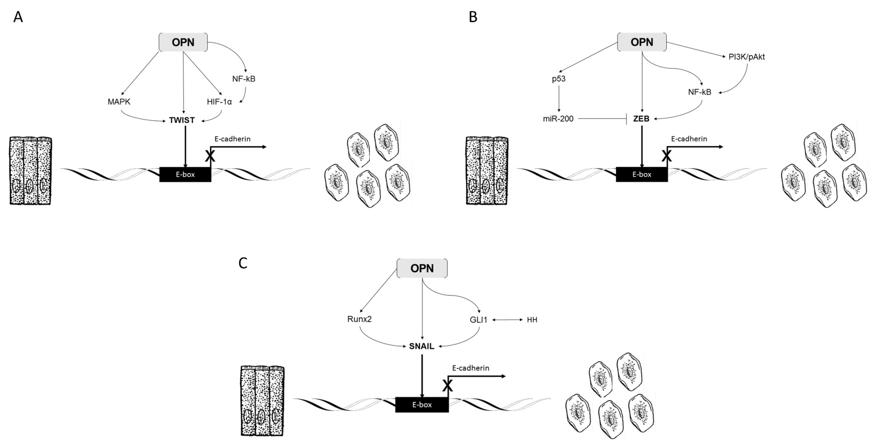
2.2. OPN, Twist, and EMT

It is clear that Twist represses E-cadherin transcription to activate EMT. However, understanding the upstream and downstream factors that impact the Twist/E-cadherin system may provide important therapeutic insight. OPN and Twist upregulation were first linked in the setting of osteogenesis. Using an osteoblast-like cell line, differential expression of Twist was shown to modify osteoblastic differentiation and, at increasing levels of Twist, OPN mRNA levels also increased [30]. This physiologic interplay between OPN and Twist extends to pathologic conditions, including cancer and fibrosis.
In breast cancer cell lines, OPN leads to an increase in expression of several EMT-related transcription factors including Twist, Snail, and Slug. OPN overexpression results in serine phosphorylation of Twist and binding to the B lymphoma Mo-MLV insertion region 1 homolog (Bmi-1) promoter The OPN/Twist/Bmi-1 pathway ultimately activates EMT programs in these cell lines [31]. The phosphorylation of Twist by OPN appears to occur through a MAPK pathway [32]. Another connection between OPN and Twist in breast cancer is through BMP-2. Compared to infiltrating breast carcinomas without microcalcifications, infiltrating breast carcinoma with microcalcifications upregulate BMP-2 and OPN, allowing them to acquire a mesenchymal characteristics and osteoblast-like phenotype [33]. This may occur through Twist modification by BMP-2 leading to activation of EMT [34]. Similarly, OPN is able to mediate EMT in HCC models through its regulation of Twist. OPN overexpression activates a PI3K-AKT-Twist pathway leading to EMT and HCC metastases [35].
OPN also functions through hypoxia-inducible factor-1α (HIF-1α) upregulation to induce EMT through Twist activation. HIF-1α is stabilized by intratumoral hypoxia and can cause tumor progression. HIF-1α can regulate the expression of Twist by binding directly to a segment in Twist’s proximal promoter. Binding to the hypoxia-response element by HIF-1α results in upregulation of Twist activity and promotes EMT [36]. In ovarian cancer, OPN expression increases the intratumoral concentration of HIF-1α through PI3K-AKT mediated signaling. A similar upregulation of HIF-1α is also seen in gastric cancer. In both ovarian and gastric cancer, overexpression of OPN leads to increased metastasis and invasiveness of tumors in in vivo models [37,38].
OPN also plays a role in maintaining the stemness of HCC through increasing the expression of HIF-1α. Unlike in ovarian and gastric cancer, induction of HIF1-α occurs through integrin αvβ3 binding and activation of NF-κB [39]. To date, no studies have shown OPN-mediated increases in HIF-1α result in Twist-dependent EMT and formation of cancer stem cells. However, based on existing literature, it appears this is a candidate signaling cascade linking OPN, EMT, and cancer cell stemness.
Recently, OPN has been demonstrated to be both a biomarker and key mechanistic contributor to metastatic disease in colorectal cancer. In postoperative patient plasma, increased OPN levels are associated with the development of future metastases. The role of OPN in this process appears to be mediated by its interaction with Twist leading to enhanced cell migration, increased invasion, and decreased cell-cell adhesion in ectopically high OPN secreting cell lines [40].
These studies together show that OPN plays an important role in the activation of Twist-dependent gene expression. OPN is positioned as an upstream and critical regulator of EMT (Figure 2A).

2.3. Overview of ZEB and EMT

Zinc finger E-box-binding homeobox (ZEB) is a family of zinc finger transcription factors that plays a major role in both normal development and disease. There have been two homologous ZEB proteins found in vertebrates, ZEB1 and ZEB2. All ZEB proteins have two zinc finger clusters that bind to ZEB boxes of the regulatory regions of target genes [41]. Mutations in ZEB genes have been shown to cause severe syndromic malformations, highlighting their importance in early embryonic developmental processes including those involving EMT. As a result, recent studies have found that ZEB1 and ZEB2 play critical roles in repressing E-cadherin leading to the activation of EMT programs [42].
ZEB1 and ZEB2 are known to be mediated by signaling cascades triggered by TGFβ, NF-κB, MAPK/ERK, and HIF-1α, amongst others, in activated tumors resulting in EMT [43]. In well-differentiated areas of carcinomas, ZEB1 is expressed at low levels, but high ZEB1 expression has been found to be inversely correlated to the expression of E-cadherin in dedifferentiated, fibroblastic-like cells at the periphery of invading tumors [44]. Specifically, high ZEB1 expressing cells have been found in invading endometrial, colorectal, lung, breast, prostate, gallbladder, hepatocellular, and pancreatic carcinomas [45].
In addition, stromal cells in colorectal, breast, lung, and bladder carcinomas have been found to be ZEB1-positive, suggesting that paracrine ZEB1 signaling could be responsible for E-cadherin down-regulation in parts of the tumor. Interestingly, ZEB2 has been shown to be highly expressed in E-cadherin-positive epithelial cells in the esophagus, stomach, colon, rectum, hepatocytes, and renal tubule, and subsequently down-regulated during transition to carcinomas [46,47,48]. This provides further evidence that both ZEB1 and ZEB2 can exert transcriptional regulation of EMT.

2.4. OPN, ZEB, and EMT

OPN-related signaling cascades interact with ZEB family members leading to modulation of EMT, both directly and indirectly. OPN is a potent activator of NF-κB and therefore can regulate NF-κB/ZEB-dependent EMT. In liver fibrosis, OPN is a key regulator of the deposition of type 1 collagen. This occurs through OPN binding to a cell surface integrin (αvβ3) and activating the PI3K/pAkt/NF-κB-signaling pathway. Activation of NF-κB, in this setting, is a direct downstream effect of OPN [49]. In the breast cancer cell line MCF10A, activation of NF-κB leads to the adoption of an EMT-like phenotype. This is related to an association between activation of NF-κB and increased expression of both ZEB1 and ZEB2 [50]. The connection between NF-κB activation and ZEB1 extends beyond mammary tumors [51]. In Helicobacter pylori infection, gastric epithelial cells undergo EMT in response to chronic exposure. Infection with H. pylori results in recruitment and activation of NF-kB leading to transactivation of ZEB1, explaining the mechanism through which EMT is induced by the bacteria [52].
OPN can also regulate ZEB-related EMT through non-NF-κB pathways. One of the most commonly cited regulatory elements of ZEB1 and ZEB2 are microRNAs (miRNA) from the miR-200 family. Members of this family suppress tumor invasiveness and metastasis by inhibiting ZEB1 and ZEB2 initiated EMT. Interestingly, p53 appears to exert anti-tumor and anti-invasive properties in part through upregulation of miR-200 family members [53]. OPN and p53 interact in many settings, suggesting a role for OPN in this miR-200/ZEB feedback loop. A summary of these interactions is shown in Figure 2B.

2.5. Overview of Snail and EMT

Snail was first discovered in 1984 and found to be essential for the development of the mesoderm and neural crest. Absence of Snail during embryonic development is lethal. The Snail family members are characterized as zinc-finger transcription factors that bind to a CAGGTG motif, known as the E-box. Much like TWIST and ZEB, Snail has also been shown to be a strong repressor of E-cadherin and a key transcription factor promoting EMT [54]. In particular, Snail is present in invasive human carcinoma cell lines and tumors where E-cadherin expression has been lost, suggesting that the same molecules are used to trigger EMT during embryonic development and tumor progression.
In addition to down-regulating E-cadherin, Snail has been associated with the down-regulation of other epithelial factors including Claudins, Occludins, and Muc1 [55,56]. Snail is also responsible for up-regulating genes associated with mesenchymal and invasive traits, such as fibronectin and MMP-9. The Snail family is the most widely studied of the major EMT program activators. As a result, numerous signaling pathways are implicated in the induction of Snail1 and Snail2 expression, including TGF-β, integrin-link kinase (ILK), phosphatidylinositol 3-kinase (PI3-K), MAPK/ERK, glycogen synthase kinase 3-β (GSK-3β), Raf kinase inhibitor protein (RKIP), NF-κβ, and Tat-interacting protein (TIP30) [57,58]. Environmental signals, such as hypoxia and stress, are also known inducers of Snail1 and Snail2. The various molecular pathways that converge on Snail1 and Snail2 are well-described in several other reviews [41,58,59].

2.6. OPN, Snail, and EMT

OPN can modify Snail1 and Snail2 signaling to modulate EMT in several settings. Blockade of OPN using an OPN-specific aptamer results in decreased expression of Snail in both a breast and hepatocellular carcinoma model [31,60]. Ectopic OPN expression can induce cell migration through overexpression of Snail and subsequent E-cadherin repression [40].
Several specific signal transduction pathways appear to regulate the interplay between OPN and Snail. Sonic hedgehog or hedgehog (HH) is critical in development and is mediated by the GLI family of transcription factors, including GLI1. HH-GLI1 signaling is implicated in progression and metastases of several malignancies [61,62,63]. One mechanism is Snail1 induction by GLI1, with resultant promotion of EMT as demonstrated in skin cancer and brain tumor models [64,65]. The link between OPN and HH pathways is through GLI1. OPN and GLI1 are co-regulated, with evidence that each have promoter binding sites for each other [66]. Clinically important relationships between OPN and GLI1 exist in nonalcoholic steatohepatitis (NASH) and melanoma, both of which demonstrate mechanisms OPN is able to modulate the EMT in the setting of fibrosis and cancer [67,68].
Another pathway regulated by OPN that converges on Snail is through Runx2. In patient-derived breast cancer clinical specimens, patients with combined Runx2/Snail positivity have shortened disease-free survival when compared to those with either Runx2-only or Snail-only positivity. This is potentially attributable to Runx2/Snail positive tumors having increased metastatic potential mediated by EMT [69]. OPN interacts directly with Runx2 signaling in breast cell lines, linking OPN to Snail-dependent EMT [70,71]. Further investigation of shared OPN and Snail pathways can be a suitable strategy for understanding and combating cancer metastasis.
Recent studies have elucidated another mechanism by which OPN induces EMT in hepatitis C virus (HCV)-induced liver disease. Elevated serum and liver OPN levels in patients with chronic hepatitis C have been correlated with advanced liver fibrosis and a reduced response to anti-viral therapy [72]. HCV induces OPN expression and, in turn, upregulates AP-1 and Sp1 transcription factors [73]. AP-1 is a downstream target of MAPK and can induce Snail-dependent EMT [74].
The pathways leveraged by OPN to control EMT through Snail transcriptional regulation are shown in Figure 2C.

3. Section 2: OPN Modifies the Microenvironment to Regulate EMT

A major regulatory component of EMT comes from the complex interaction between the cell and its surrounding environment comprised of other cells, stromal elements, infiltrating immune mediators, cytokines, microRNA, exosomes, and other factors. In a healing wound, this is broadly termed the tissue microenvironment whereas in malignancy, it is the tumor microenvironment. In each setting, the microenvironment can influence the progression of physiologic and pathologic processes.
OPN is a known mediator of the composition of the microenvironment, including playing a functional role in cell-microenvironment interplay. There is mounting evidence that OPN is able to modify the tissue and tumor microenvironment to support EMT. In the following section, the role OPN plays in influencing the structure of the microenvironment will be discussed; more specifically, how OPN regulates EMT using the microenvironment.

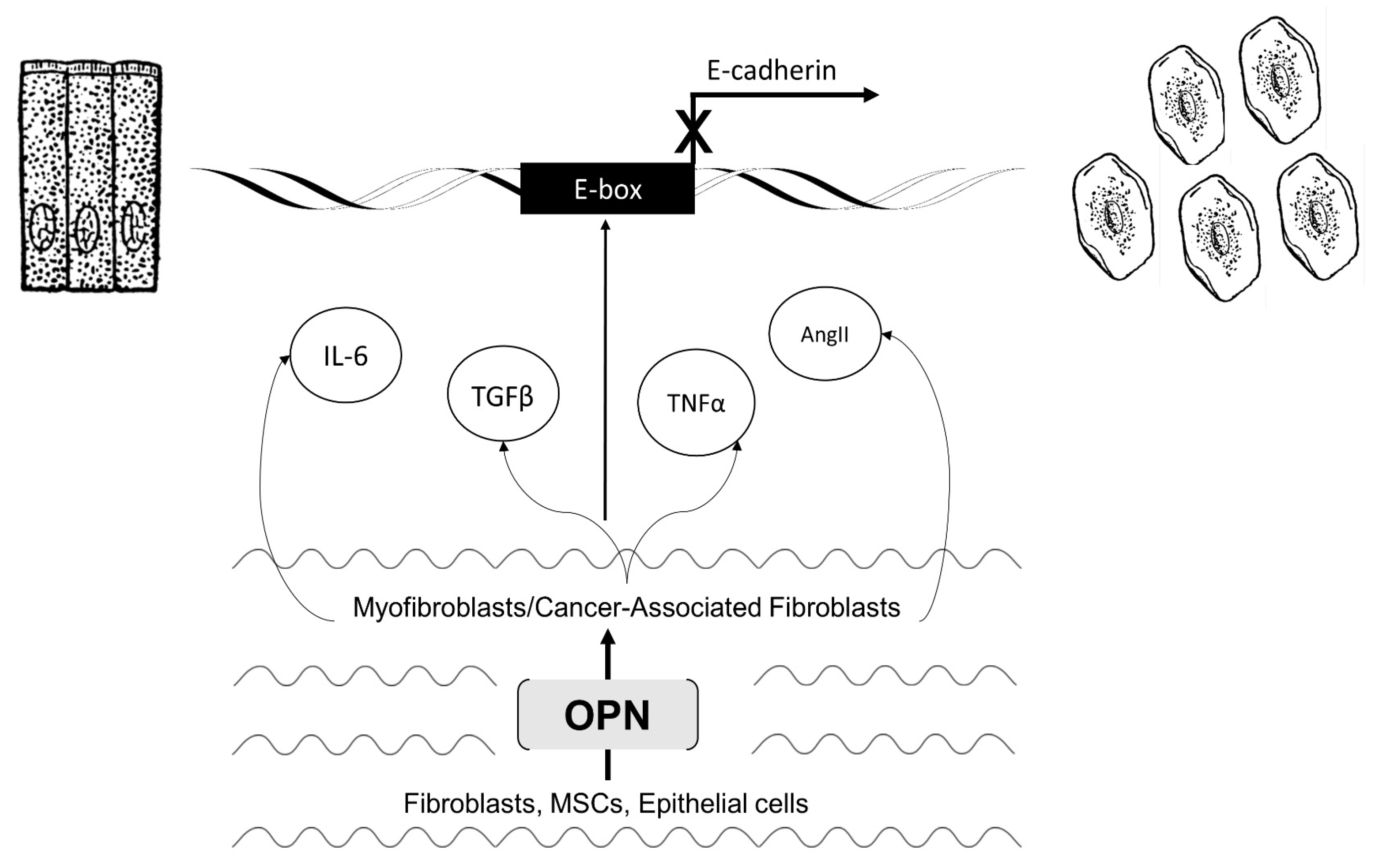
3.1. Myofibroblast Activation and Fibrosis

Fibrosis is the abnormal wound healing response characterized by the excessive deposition of extracellular matrix (ECM) proteins such as collagen and fibronectin [75]. It is seen in organs such as the lung, liver, heart, and kidney, and as the fibrosis progresses, it can cause organ failure [8,76,77]. Fibrosis is often the final outcome of chronic inflammatory diseases, and develops from the simultaneous activation of multiple profibrotic pathways [75]. In normal tissue repair and healing response, fibroblasts within the injured tissue are transformed into myofibroblasts. In turn, myofibroblasts deposit ECM to aid in closing the wound and undergo apoptosis after the wound has healed [76]. However, in chronic injury and/or abnormal tissue repair, myofibroblasts persist and resist apoptosis.
OPN is critically involved in the fibroblast to myofibroblast transformation. Research has shown that OPN expression is required for TGFβ mediated myofibroblast differentiation and subsequent cardiac fibrosis [78]. In lung fibrosis and asthma, OPN is necessary for airway remodeling in response to chronic bronchial injury. This is, in part, due to OPN activating fibroblasts into myofibroblasts that orchestrate the repair process [79]. OPN expression is capable of inducing pulmonary fibrosis by initiating the migration, adhesion, and proliferation of fibroblasts through cytokine signaling and macrophage activation [80]. An abundant source of OPN in pulmonary fibrosis comes from epithelial cells which have undergone EMT—an alternative source of myofibroblasts in the microenvironment. In idiopathic pulmonary fibrosis, OPN induces fibrosis by increasing the expression of tissue inhibitor of metalloproteinase (TIMP) and type 1 collagen in fibroblasts and increasing the expression of the OPN activating protein matrix metallopretease 7 (MMP7) in alveolar epithelial cells [81].
Another area where OPN’s role in fibrosis is well-studied is the liver. The most common causes of liver fibrosis are chronic heptatitis B and C virus infection, alcohol abuse, primary biliary cirrhosis, primary sclerosing cholangitis, and NASH [82,83,84]. Hepatic stellate cells (HSCs) are the liver analogue to myofibroblasts in other regions of injury. In chronic liver disease (CLD), a greater response and proliferation is required and leads to an abnormal fibrogenic response—a process that is driven by HSCs. In models of chronic inflammation and hepatic fibrosis, OPN is upregulated in activated HSCs and further induces HSC activation [49,85]. Patients with alcoholic liver fibrosis have high levels of OPN in liver adipose tissue, and serum elucidating the clinical relevance of OPN in fibrosis. Interestingly, it has also been shown that OPN expression upregulates the expression of type 1 collagen and type 2 transforming growth factor-β receptor mRNA, suggesting that it not only promotes fibrosis but makes HSCs more sensitive to signaling molecules that promote fibrosis and EMT.

3.2. Activated Myofibroblasts Regulate EMT

Overall, OPN is able to activate fibroblasts to take on a myofibroblast (or activated HSC) phenotype. Myofibroblasts generated directly by OPN, or through other mechanisms, produce several key cytokines and secrete a myriad of signaling molecules including angiotensin II (Ang II), TNF-α, IL-6, sphingosine-1-phosphate (S1P), and TGFβ (Figure 2D) [86]. Each of these are proinflammatory factors with the ability to induce EMT programs in several different cell lines which result in fibrotic phenotypes.
Ang II is able to activate Rho subfamily GTPases including RhoA, Rac1, and Cdc42 leading to downstream activation of key pathways that mediate fibrosis in cardiac myocytes [87]. One specific downstream consequence of Rho GTPase activation is cytoskeletal remodeling in epithelial cells—a hallmark of EMT [88]. Furthermore, inhibition of Ang II signaling, using the Angiotensin II type 1 blocker candesartan, can significantly reduce fibrosis by downregulating EMT [89].
Another cytokine increasingly recognized as a regulator of EMT is TNF-α. TNF-α, while usually associated with macrophages, is another secreted factor that can come from myofibroblasts and contributes to the fibrotic phenotype. In an in vitro model of kidney repair in response to injury, TNF-α induces renal epithelial cells to undergo EMT. In HK-2 cells exposed to TNF-α, Complement 3 (C3) is activated and E-cadherin expression repressed. Without TNF-α stimulation, HK-2 cells are unable to undergo C3-mediated EMT [90]. The role of TNF-α in renal fibrosis EMT may be related to the intermediate signaling molecule layilin, a c-type lectin-homologous protein that binds to cell surface cytoskeletal elements and is expressed in many organs. In a mouse model, TNF-α increases the expression of layilin. However, when layilin expression is inhibited, EMT does not occur even after TNF-α stimulation [91]. While this data supports TNF-α as an important component of EMT, there are additional elements that drive this response.
TGF-β is a major secreted factor from activated myofibroblasts and is a well-known effector of EMT, both in physiologic and pathologic settings. In fibrosis, TGF-β commonly exerts its effect on EMT through Smad, Notch, and EGFR intermediates [92]. Many TGF-β target genes are transcriptionally regulated by Smad3 or closely related complexes [93]. As a result, Smad family proteins can modify, repress, and induce EMT. Studies using Smad3 knockout mice demonstrate loss of EMT and subsequent fibrosis in lens, kidney, and liver injury models [94,95]. Notch signaling pathways are important in development, cell growth, and apoptosis with increasing evidence for a role in promoting fibrosis. In kidney fibrosis, inhibition of Notch signaling results in decreased fibrosis and prevents TGF-β mediated EMT [96]. Similarly, in a rat model, blockade of Notch signaling using a γ-secretase ameliorated TGF-β dependent EMT in both hepatocytes and HSCs [97]. Recently, studies have identified EGFR activation as an important TGF-β-related inducer of EMT. In an IPF cellular model, bronchial cells treated with TGF-β were shown to undergo EMT, which resulted in a pro-fibrotic phenotype. This process is highly dependent on activation of EGFR by TGF-β, identifying another way EMT occurs in epithelial cells [98].

3.3. Cancer-Associated Fibroblast Activation and Tumorigenicity

OPN is able to generate activated myofibroblasts that contribute to fibrosis across many organ systems. In cancer, the tumor microenvironment is populated by a similar cellular component, termed cancer-associated fibroblasts (CAF). CAFs (also called tumor-associated fibroblasts or carcinoma-associated fibroblasts) are derived from several sources including local fibroblasts, transformed tumors cells, and mesenchymal stem cells (MSCs) commonly recruited from the bone marrow [99]. OPN modulates tumor-specific EMT by generating CAFs (from both resident fibroblasts and recruited MSCs) which secrete a multitude of factors into the tumor microenvironment support tumor invasiveness and metastases [100].
Tumor-derived and exogenous OPN are both able to induce the transformation of MSCs to CAFs in breast cancer and liver cancer models. This occurs by stimulating MSCs to produce TGF-β creating a feedback loop that drives the CAF phenotype [101]. The transcription factor myeloid-zinc finger 1 (MZF1) is a critical intermediate to this process and activated through protein kinase A signaling [102]. In addition to MSCs, tumor-derived OPN is able to convert normal fibroblasts into CAFs. In both in vitro and in vivo breast cancer models, OPN secreted by tumor cells can promote the transformation of tissue-resident normal mammary fibroblasts into tumor-supporting CAFs [103].

3.4. Activated CAFs Regulate EMT

How OPN regulates EMT in tumors is largely a function of its ability to activate CAFs. CAFs, through paracrine interactions, are able to feedback to tumor cells using a diverse array of co-factors. This cross-talk serves many functions including the regulation of epithelial cell function, regulation of cellular response to inflammation, maintenance of environment homeostasis, promoting survival, and recruiting additional members of the stroma [99]. The following section will highlight the induction of EMT in various tumor cells in response to CAF-derived TGF-β and IL-6 (Figure 3).
TGF-β is secreted from the tumor stroma in many malignancies, largely sourced from CAFs [104]. CAF-derived TGF-β can induce EMT in several tumors via a multitude of mechanisms. Breast cancer cell lines undergo EMT in response to treatment with conditioned medium from CAFs isolated from invasive breast specimens. In this model, neutralizing antibody and small-molecule inhibition of TGF-β signaling prevents EMT in these cell lines [105]. TGF-β secreted by CAFs also can lead to EMT in bladder cancer cells, a process regulated by the long-coding RNA ZEB2NAT. Following the depletion of lncRNA-ZEB2NAT, conditioned media from CAFs no longer can cause EMT in human bladder cancer cell lines (5637, T24, and J82) [106].
TGF-β can also result in the formation of cancer stem cell populations within tumors—a process attributable to EMT. For example, TGF-β treatment of patient-derived colorectal tumor specimens results in upregulation of CD44+ stem cell populations and expression of N-cadherin. This suggests that TGF-β induces the formation of cancer stem cells through EMT [107]. In non-small cell lung cancer, TGF-β has a similar effect on cancer stemness [108]. Another intermediate for TGF-β-related acquisition of cancer stemness is miR-155 which, when overexpressed, results in EMT in liver cancer [109].
IL-6 is another key signaling molecule involved in EMT that can come directly from activated CAFs. In gastric cancer, IL-6 released by CAFs is able to modify the phenotype of tumor cells using IL-6/JAK/STAT3 signaling. Activation of this pathway results in tumor cell EMT and invasiveness, both in vitro and in vivo [110]. Further evidence of the role of IL-6 in regulating EMT comes from anti-IL-6 receptor assays in colon cancer. Antibody-mediated IL-6 receptor inhibition prevents angiogenesis by blocking the endothelial to mesenchymal transition (EndoMT) [111]. EndoMT is an extremely similar molecular process to that of EMT, however instead of epithelial cells acquiring a mesenchymal phenotype, endothelial cells undergo a transformation [112].
A more specific example of loss of EMT potential in the absence of IL-6 comes from pancreatic cancer. CAFs from pancreatic tumors treated with retinoic acid have decreased expression of IL-6. With downregulated IL-6, pancreatic tumor cells do not undergo EMT and do not gain an invasive phenotype [113]. Importantly, IL-6-related EMT may provide a mechanism to prevent chemotherapy resistance. In breast cancer, IL-6 from CAFs interacts with breast tumor cells to induce EMT and create a cancer stem cell subpopulation. This leads to resistance to tamoxifen, highly suggestive of a role for CAF-derived IL-6 in recurrence following conventional therapy [114].

4. Conclusions

The importance of OPN as a key regulatory factor of EMT is increasingly being recognized in both fibrosis and cancer. Understanding how OPN both directly and indirectly influences EMT in multiple settings and disease states can offer new targets for clinical therapeutics. Importantly, OPN is able to guide EMT through specific cellular signaling pathways and by restructuring the microenvironment to modify EMT programs. The shared molecular pathways between OPN and many traditional “master regulators” provides evidence that OPN is a major regulator of EMT.

Acknowledgments

This work supported by NIH T32 GM08750-16.

Author Contributions

Authors Kothari, A.N., Kuo P.C., Mi, Z. conceived and designed the review; Kothari A.N., Arffa M., Chang, V. created the figures; Kothari A.N., Arffa M., Chang V., Blackwell R.H., Syn W.K., Zhang J., Kuo P.C., Mi, Z. all contributed to literature review and analysis of prior studies; Kothari A.N., Arffa M., Chang, V. wrote the paper; Blackwell, R.H., Syn, W.K., Zhang, Z, Kuo P.C., Mi, Z. provided critical review and edits.

Conflicts of Interest

The authors declare no conflict of interest.

References

  1. Senger, D.R.; Wirth, D.F.; Hynes, R.O. Transformed mammalian cells secrete specific proteins and phosphoproteins. Cell 1979, 16, 885–893. [Google Scholar] [CrossRef]
  2. Denhardt, D.T.; Guo, X. Osteopontin: A protein with diverse functions. FASEB J. 1993, 7, 1475–1482. [Google Scholar] [PubMed]
  3. Wai, P.Y.; Kuo, P.C. Osteopontin: Regulation in tumor metastasis. Cancer Metastasis Rev. 2008, 27, 103–118. [Google Scholar] [CrossRef] [PubMed]
  4. Lund, S.A.; Giachelli, C.M.; Scatena, M. The role of osteopontin in inflammatory processes. J. Cell Commun. Signal. 2009, 3, 311–322. [Google Scholar] [CrossRef] [PubMed]
  5. Weber, C.E.; Li, N.Y.; Wai, P.Y.; Kuo, P.C. Epithelial-mesenchymal transition, TGF-beta, and osteopontin in wound healing and tissue remodeling after injury. J. Burn Care Res. 2012, 33, 311–318. [Google Scholar] [CrossRef] [PubMed]
  6. Wai, P.Y.; Kuo, P.C. The role of osteopontin in tumor metastasis. J. Surg. Res. 2004, 121, 228–241. [Google Scholar] [CrossRef] [PubMed]
  7. Pritchett, J.; Harvey, E.; Athwal, V.; Berry, A.; Rowe, C.; Oakley, F.; Moles, A.; Mann, D.A.; Bobola, N.; Sharrocks, A.D. Osteopontin is a novel downstream target of SOX9 with diagnostic implications for progression of liver fibrosis in humans. Hepatology 2012, 56, 1108–1116. [Google Scholar] [CrossRef] [PubMed]
  8. Tan, T.K.; Zheng, G.; Hsu, T.; Lee, S.R.; Zhang, J.; Zhao, Y.; Tian, X.; Wang, Y.; Wang, Y.M.; Cao, Q. Matrix metalloproteinase-9 of tubular and macrophage origin contributes to the pathogenesis of renal fibrosis via macrophage recruitment through osteopontin cleavage. Laboratory Investig. 2013, 93, 434–449. [Google Scholar] [CrossRef] [PubMed]
  9. Chen, C.; Li, R.; Ross, R.S.; Manso, A.M. Integrins and integrin-related proteins in cardiac fibrosis. J. Mol. Cell Cardiol. 2015. [Google Scholar] [CrossRef] [PubMed]
  10. Cao, D.; Li, Z.; Jiang, X.; Lum, Y.L.; Khin, E.; Lee, N.P.; Wu, G.; Luk, J.M. Osteopontin as potential biomarker and therapeutic target in gastric and liver cancers. World J. Gastroenterol. 2012, 18, 3923. [Google Scholar] [CrossRef] [PubMed] [Green Version]
  11. Huang, J.; Pan, C.; Hu, H.; Zheng, S.; Ding, L. Osteopontin-enhanced hepatic metastasis of colorectal cancer cells. PLoS ONE 2012, 7, e47901. [Google Scholar] [CrossRef] [PubMed]
  12. Hay, E. An overview of epithelio-mesenchymal transformation. Cells Tissues Organs (Print) 1995, 154, 8–20. [Google Scholar] [CrossRef]
  13. Kalluri, R.; Weinberg, R.A. The basics of epithelial-mesenchymal transition. J. Clin. Investig. 2009, 119, 1420–1428. [Google Scholar] [CrossRef] [PubMed]
  14. Kalluri, R. EMT: When epithelial cells decide to become mesenchymal-like cells. J. Clin. Investig. 2009, 119, 1417–1419. [Google Scholar] [CrossRef] [PubMed]
  15. Huber, M.A.; Kraut, N.; Beug, H. Molecular requirements for epithelial–mesenchymal transition during tumor progression. Curr. Opin. Cell Biol. 2005, 17, 548–558. [Google Scholar] [CrossRef] [PubMed]
  16. Savagner, P. Leaving the neighborhood: Molecular mechanisms involved during epithelial-mesenchymal transition. Bioessays 2001, 23, 912–923. [Google Scholar] [CrossRef] [PubMed]
  17. Seton-Rogers, S. Epithelial-mesenchymal transition: Untangling EMT's functions. Nat. Rev. Cancer 2016, 16. [Google Scholar] [CrossRef] [PubMed]
  18. Kang, Y.; Massagué, J. Epithelial-mesenchymal transitions: Twist in development and metastasis. Cell 2004, 118, 277–279. [Google Scholar] [CrossRef] [PubMed]
  19. Ansieau, S.; Morel, A.; Hinkal, G.; Bastid, J.; Puisieux, A. TWISTing an embryonic transcription factor into an oncoprotein. Oncogene 2010, 29, 3173–3184. [Google Scholar] [CrossRef] [PubMed]
  20. Maestro, R.; Dei Tos, A.P.; Hamamori, Y.; Krasnokutsky, S.; Sartorelli, V.; Kedes, L.; Doglioni, C.; Beach, D.H.; Hannon, G.J. Twist is a potential oncogene that inhibits apoptosis. Genes Dev. 1999, 13, 2207–2217. [Google Scholar] [CrossRef] [PubMed]
  21. Beck, B.; Lapouge, G.; Rorive, S.; Drogat, B.; Desaedelaere, K.; Delafaille, S.; Dubois, C.; Salmon, I.; Willekens, K.; Marine, J. Different levels of Twist1 regulate skin tumor initiation, stemness, and progression. Cell Stem Cell 2015, 16, 67–79. [Google Scholar] [CrossRef] [PubMed]
  22. Elias, M.C.; Tozer, K.R.; Silber, J.R.; Mikheeva, S.; Deng, M.; Morrison, R.S.; Manning, T.C.; Silbergeld, D.L.; Glackin, C.A.; Reh, T.A. TWIST is expressed in human gliomas, promotes invasion. Neoplasia 2005, 7, 824–837. [Google Scholar] [CrossRef] [PubMed]
  23. Hasselblatt, M.; Mertsch, S.; Koos, B.; Riesmeier, B.; Stegemann, H.; Jeibmann, A.; Tomm, M.; Schmitz, N.; Wrede, B.; Wolff, J.E.; et al. TWIST-1 is overexpressed in neoplastic choroid plexus epithelial cells and promotes proliferation and invasion. Cancer Res. 2009, 69, 2219–2223. [Google Scholar] [CrossRef] [PubMed]
  24. Lee, T.K.; Poon, R.T.; Yuen, A.P.; Ling, M.T.; Kwok, W.K.; Wang, X.H.; Wong, Y.C.; Guan, X.Y.; Man, K.; Chau, K.L.; et al. Twist overexpression correlates with hepatocellular carcinoma metastasis through induction of epithelial-mesenchymal transition. Clin. Cancer Res. 2006, 12, 5369–5376. [Google Scholar] [CrossRef] [PubMed]
  25. Yang, J.; Mani, S.A.; Donaher, J.L.; Ramaswamy, S.; Itzykson, R.A.; Come, C.; Savagner, P.; Gitelman, I.; Richardson, A.; Weinberg, R.A. Twist, a master regulator of morphogenesis, plays an essential role in tumor metastasis. Cell 2004, 117, 927–939. [Google Scholar] [CrossRef] [PubMed]
  26. Yuen, H.F.; Chan, Y.P.; Wong, M.L.; Kwok, W.K.; Chan, K.K.; Lee, P.Y.; Srivastava, G.; Law, S.Y.; Wong, Y.C.; Wang, X.; et al. Upregulation of Twist in oesophageal squamous cell carcinoma is associated with neoplastic transformation and distant metastasis. J. Clin. Pathol. 2007, 60, 510–514. [Google Scholar] [CrossRef] [PubMed] [Green Version]
  27. Yao, C.; Li, P.; Song, H.; Song, F.; Qu, Y.; Ma, X.; Shi, R.; Wu, J. CXCL12/CXCR4 axis upregulates twist to induce EMT in human glioblastoma. Mol. Neurobiol. 2015. [Google Scholar] [CrossRef] [PubMed]
  28. Vesuna, F.; Lisok, A.; Kimble, B.; Raman, V. Twist modulates breast cancer stem cells by transcriptional regulation of CD24 expression. Neoplasia 2009, 11, 1318–1328. [Google Scholar] [CrossRef] [PubMed]
  29. Yin, G.; Chen, R.; Alvero, A.B.; Fu, H.; Holmberg, J.; Glackin, C.; Rutherford, T.; Mor, G. TWISTing stemness, inflammation and proliferation of epithelial ovarian cancer cells through MIR199A2/214. Oncogene 2010, 29, 3545–3553. [Google Scholar] [CrossRef] [PubMed]
  30. Lee, M.; Lowe, G.N.; Strong, D.D.; Wergedal, J.E.; Glackin, C.A. TWIST, a basic helix-loop-helix transcription factor, can regulate the human osteogenic lineage. J. Cell. Biochem. 1999, 75, 566–577. [Google Scholar] [CrossRef]
  31. Li, N.Y.; Weber, C.E.; Mi, Z.; Wai, P.Y.; Cuevas, B.D.; Kuo, P.C. Osteopontin up-regulates critical epithelial-mesenchymal transition transcription factors to induce an aggressive breast cancer phenotype. J. Am. Coll Surg. 2013, 217, 17–26. [Google Scholar] [CrossRef] [PubMed]
  32. Li, N.Y.; Weber, C.E.; Wai, P.Y.; Cuevas, B.D.; Zhang, J.; Kuo, P.C.; Mi, Z. An MAPK-dependent pathway induces epithelial-mesenchymal transition via Twist activation in human breast cancer cell lines. Surgery 2013, 154, 404–410. [Google Scholar] [CrossRef] [PubMed]
  33. Scimeca, M.; Giannini, E.; Antonacci, C.; Pistolese, C.A.; Spagnoli, L.G.; Bonanno, E. Microcalcifications in breast cancer: An active phenomenon mediated by epithelial cells with mesenchymal characteristics. BMC Cancer 2014, 14. [Google Scholar] [CrossRef] [PubMed]
  34. Hornik, C.; Brand-Saberi, B.; Rudloff, S.; Christ, B.; Füchtbauer, E. Twist is an integrator of SHH, FGF, and BMP signaling. Anat. Embryol. 2004, 209, 31–39. [Google Scholar] [CrossRef] [PubMed]
  35. Dong, Q.; Zhu, X.; Dai, C.; Zhang, X.; Jia, H.; Ye, Q.; Qin, L. Osteopontin regulated epithelial-mesenchymal transition via PI3K/AKT signaling pathway in hepatocellular carcinoma. Cancer Res. 2013, 73, 2695–2695. [Google Scholar] [CrossRef]
  36. Yang, M.; Wu, M.; Chiou, S.; Chen, P.; Chang, S.; Liu, C.; Teng, S.; Wu, K. Direct regulation of TWIST by HIF-1α promotes metastasis. Nat. Cell Biol. 2008, 10, 295–305. [Google Scholar] [CrossRef] [PubMed]
  37. Song, G.; Cai, Q.; Mao, Y.; Ming, Y.; Bao, S.; Ouyang, G. Osteopontin promotes ovarian cancer progression and cell survival and increases HIF-1α expression through the PI3-K/Akt pathway. Cancer Sci. 2008, 99, 1901–1907. [Google Scholar] [CrossRef] [PubMed]
  38. Song, G.; Ouyang, G.; Mao, Y.; Ming, Y.; Bao, S.; Hu, T. Osteopontin promotes gastric cancer metastasis by augmenting cell survival and invasion through Akt-mediated HIF-1α up-regulation and MMP9 activation. J. Cell. Mol. Med. 2009, 13, 1706–1718. [Google Scholar] [CrossRef] [PubMed]
  39. Cao, L.; Fan, X.; Jing, W.; Liang, Y.; Chen, R.; Liu, Y.; Zhu, M.; Jia, R.; Wang, H.; Zhang, X.; et al. Osteopontin promotes a cancer stem cell-like phenotype in hepatocellular carcinoma cells via an integrin-NF-kappaB-HIF-1alpha pathway. Oncotarget 2015, 6, 6627–6640. [Google Scholar] [CrossRef] [PubMed]
  40. Ng, L.; Wan, T.M.; Lam, C.S.; Chow, A.K.; Wong, S.K.; Man, J.H.; Li, H.; Cheng, N.S.; Pak, R.C.; Cheung, A.H. Post-operative plasma osteopontin predicts distant metastasis in human colorectal cancer. PLoS ONE 2015, 10, e0126219. [Google Scholar] [CrossRef] [PubMed] [Green Version]
  41. Lamouille, S.; Xu, J.; Derynck, R. Molecular mechanisms of epithelial–mesenchymal transition. Nat. Rev. Mol. Cell Boil. 2014, 15, 178–196. [Google Scholar] [CrossRef] [PubMed]
  42. Zheng, H.; Kang, Y. Multilayer control of the EMT master regulators. Oncogene 2014, 33, 1755–1763. [Google Scholar] [CrossRef] [PubMed]
  43. Postigo, A.A.; Depp, J.L.; Taylor, J.J.; Kroll, K.L. Regulation of Smad signaling through a differential recruitment of coactivators and corepressors by ZEB proteins. EMBO J. 2003, 22, 2453–2462. [Google Scholar] [CrossRef] [PubMed]
  44. Vandewalle, C.; Van Roy, F.; Berx, G. The role of the ZEB family of transcription factors in development and disease. Cell. Mol. Life Sci. 2009, 66, 773–787. [Google Scholar] [CrossRef] [PubMed]
  45. Singh, A.B.; Sharma, A.; Smith, J.J.; Krishnan, M.; Chen, X.; Eschrich, S.; Washington, M.K.; Yeatman, T.J.; Beauchamp, R.D.; Dhawan, P. Claudin-1 up-regulates the repressor ZEB-1 to inhibit E-cadherin expression in colon cancer cells. Gastroenterology 2011, 141, 2140–2153. [Google Scholar] [CrossRef] [PubMed]
  46. Ohashi, S.; Natsuizaka, M.; Wong, G.S.; Michaylira, C.Z.; Grugan, K.D.; Stairs, D.B.; Kalabis, J.; Vega, M.E.; Kalman, R.A.; Nakagawa, M.; et al. Epidermal growth factor receptor and mutant p53 expand an esophageal cellular subpopulation capable of epithelial-to-mesenchymal transition through ZEB transcription factors. Cancer Res. 2010, 70, 4174–4184. [Google Scholar] [CrossRef] [PubMed]
  47. Naganuma, S.; Ohashi, S.; Kimura, S.; Natsuizaka, M.; Gimotty, P.; Klein-Szanto, A.J.; Itoh, H.; Nakagawa, H. ZEB1 and ZEB2 promote EMT and invasion in esophageal squamous cell carcinoma. Cancer Res. 2010, 70, 2293–2293. [Google Scholar] [CrossRef]
  48. Schmalhofer, O.; Brabletz, S.; Brabletz, T. E-cadherin, β-catenin, and ZEB1 in malignant progression of cancer. Cancer Metastasis Rev. 2009, 28, 151–166. [Google Scholar] [CrossRef] [PubMed]
  49. Urtasun, R.; Lopategi, A.; George, J.; Leung, T.; Lu, Y.; Wang, X.; Ge, X.; Fiel, M.I.; Nieto, N. Osteopontin, an oxidant stress sensitive cytokine, up-regulates collagen-I via integrin αVβ3 engagement and PI3K/pAkt/NFκB signaling. Hepatology 2012, 55, 594–608. [Google Scholar] [CrossRef] [PubMed]
  50. Chua, H.; Bhat-Nakshatri, P.; Clare, S.; Morimiya, A.; Badve, S.; Nakshatri, H. NF-κB represses E-cadherin expression and enhances epithelial to mesenchymal transition of mammary epithelial cells: Potential involvement of ZEB-1 and ZEB-2. Oncogene 2007, 26, 711–724. [Google Scholar] [CrossRef] [PubMed]
  51. Min, C.; Eddy, S.F.; Sherr, D.H.; Sonenshein, G.E. NF-κB and epithelial to mesenchymal transition of cancer. J. Cell. Biochem. 2008, 104, 733–744. [Google Scholar] [CrossRef] [PubMed]
  52. Baud, J.; Varon, C.; Chabas, S.; Chambonnier, L.; Darfeuille, F.; Staedel, C. Helicobacter pylori initiates a mesenchymal transition through ZEB1 in gastric epithelial cells. PLoS ONE 2013, 8, e60315. [Google Scholar] [CrossRef] [PubMed]
  53. Kim, T.; Veronese, A.; Pichiorri, F.; Lee, T.J.; Jeon, Y.J.; Volinia, S.; Pineau, P.; Marchio, A.; Palatini, J.; Suh, S.S.; et al. p53 regulates epithelial-mesenchymal transition through microRNAs targeting ZEB1 and ZEB2. J. Exp. Med. 2011, 208, 875–883. [Google Scholar] [CrossRef] [PubMed]
  54. Cano, A.; Pérez-Moreno, M.A.; Rodrigo, I.; Locascio, A.; Blanco, M.J.; del Barrio, M.G.; Portillo, F.; Nieto, M.A. The transcription factor snail controls epithelial–mesenchymal transitions by repressing E-cadherin expression. Nat. Cell Biol. 2000, 2, 76–83. [Google Scholar] [CrossRef] [PubMed]
  55. Ikenouchi, J.; Matsuda, M.; Furuse, M.; Tsukita, S. Regulation of tight junctions during the epithelium-mesenchyme transition: Direct repression of the gene expression of claudins/occludin by Snail. J. Cell Sci. 2003, 116, 1959–1967. [Google Scholar] [CrossRef] [PubMed]
  56. Gnemmi, V.; Bouillez, A.; Gaudelot, K.; Hémon, B.; Ringot, B.; Pottier, N.; Glowacki, F.; Villers, A.; Vindrieux, D.; Cauffiez, C. MUC1 drives epithelial–mesenchymal transition in renal carcinoma through Wnt/β-catenin pathway and interaction with SNAIL promoter. Cancer Lett. 2014, 346, 225–236. [Google Scholar] [CrossRef] [PubMed]
  57. Zhu, M.; Yin, F.; Fan, X.; Jing, W.; Chen, R.; Liu, L.; Zhang, L.; Liu, Y.; Liang, Y.; Bu, F. Decreased TIP30 promotes Snail-mediated epithelial–mesenchymal transition and tumor-initiating properties in hepatocellular carcinoma. Oncogene 2015, 34, 1420–1431. [Google Scholar] [CrossRef] [PubMed]
  58. Wang, Y.; Shi, J.; Chai, K.; Ying, X.; Zhou, B.P. The Role of Snail in EMT and Tumorigenesis. Curr. Cancer Drug Targets 2013, 13, 963–972. [Google Scholar] [CrossRef] [PubMed]
  59. Peinado, H.; Olmeda, D.; Cano, A. Snail, ZEB and bHLH factors in tumour progression: An alliance against the epithelial phenotype? Nat. Rev. Cancer 2007, 7, 415–428. [Google Scholar] [CrossRef] [PubMed]
  60. Bhattacharya, S.D.; Mi, Z.; Kim, V.M.; Guo, H.; Talbot, L.J.; Kuo, P.C. Osteopontin regulates epithelial mesenchymal transition-associated growth of hepatocellular cancer in a mouse xenograft model. Ann. Surg. 2012, 255, 319–325. [Google Scholar] [CrossRef] [PubMed]
  61. Clement, V.; Sanchez, P.; De Tribolet, N.; Radovanovic, I.; i Altaba, A.R. HEDGEHOG-GLI1 signaling regulates human glioma growth, cancer stem cell self-renewal, and tumorigenicity. Curr. Biol. 2007, 17, 165–172. [Google Scholar] [CrossRef] [PubMed]
  62. Bian, Y.; Huang, S.; Yang, L.; Ma, X.; Xie, J.; Zhang, H. Sonic hedgehog-Gli1 pathway in colorectal adenocarcinomas. World J. Gastroenterol. 2007, 13, 1659. [Google Scholar] [CrossRef] [PubMed]
  63. Sanchez, P.; Hernandez, A.M.; Stecca, B.; Kahler, A.J.; DeGueme, A.M.; Barrett, A.; Beyna, M.; Datta, M.W.; Datta, S.; i Altaba, A.R. Inhibition of prostate cancer proliferation by interference with SONIC HEDGEHOG-GLI1 signaling. Proc. Natl. Acad. Sci. USA 2004, 101, 12561–12566. [Google Scholar] [CrossRef] [PubMed]
  64. Chou, C.; Lieu, A.; Wu, C.; Chang, L.; Loh, J.; Lin, R.; Chen, W.; Liao, H.; Fu, W.; Chang, C. Differential expression of hedgehog signaling components and Snail/E-cadherin in human brain tumors. Oncol. Rep. 2010, 24, 1225–1232. [Google Scholar] [PubMed]
  65. Li, X.; Deng, W.; Nail, C.D.; Bailey, S.K.; Kraus, M.H.; Ruppert, J.M.; Lobo-Ruppert, S.M. Snail induction is an early response to Gli1 that determines the efficiency of epithelial transformation. Oncogene 2006, 25, 609–621. [Google Scholar] [CrossRef] [PubMed]
  66. Yoon, J.W.; Kita, Y.; Frank, D.J.; Majewski, R.R.; Konicek, B.A.; Nobrega, M.A.; Jacob, H.; Walterhouse, D.; Iannaccone, P. Gene expression profiling leads to identification of GLI1-binding elements in target genes and a role for multiple downstream pathways in GLI1-induced cell transformation. J. Biol. Chem. 2002, 277, 5548–5555. [Google Scholar] [CrossRef] [PubMed]
  67. Das, S.; Harris, L.G.; Metge, B.J.; Liu, S.; Riker, A.I.; Samant, R.S.; Shevde, L.A. The hedgehog pathway transcription factor GLI1 promotes malignant behavior of cancer cells by up-regulating osteopontin. J. Biol. Chem. 2009, 284, 22888–22897. [Google Scholar] [CrossRef] [PubMed]
  68. Syn, W.; Choi, S.S.; Liaskou, E.; Karaca, G.F.; Agboola, K.M.; Oo, Y.H.; Mi, Z.; Pereira, T.A.; Zdanowicz, M.; Malladi, P. Osteopontin is induced by hedgehog pathway activation and promotes fibrosis progression in nonalcoholic steatohepatitis. Hepatology 2011, 53, 106–115. [Google Scholar] [CrossRef] [PubMed]
  69. Yang, Z.; Zhang, B.; Liu, B.; Xie, Y.; Cao, X. Combined Runx2 and Snail overexpression is associated with a poor prognosis in breast cancer. Tumor. Biol. 2015, 36, 4565–4573. [Google Scholar] [CrossRef] [PubMed]
  70. Inman, C.K.; Shore, P. The osteoblast transcription factor Runx2 is expressed in mammary epithelial cells and mediates osteopontin expression. J. Biol. Chem. 2003, 278, 48684–48689. [Google Scholar] [CrossRef] [PubMed]
  71. Shen, Q.; Christakos, S. The vitamin D receptor, Runx2, and the Notch signaling pathway cooperate in the transcriptional regulation of osteopontin. J. Biol. Chem. 2005, 280, 40589–40598. [Google Scholar] [CrossRef] [PubMed]
  72. Choi, S.S.; Claridge, L.C.; Jhaveri, R.; Swiderska-Syn, M.; Clark, P.; Suzuki, A.; Pereira, T.A.; Mi, Z.; Kuo, P.C.; Guy, C.D.; et al. Osteopontin is up-regulated in chronic hepatitis C and is associated with cellular permissiveness for hepatitis C virus replication. Clin. Sci. (Lond.) 2014, 126, 845–855. [Google Scholar] [CrossRef] [PubMed]
  73. Iqbal, J.; McRae, S.; Mai, T.; Banaudha, K.; Sarkar-Dutta, M.; Waris, G. Role of hepatitis C virus induced osteopontin in epithelial to mesenchymal transition, migration and invasion of hepatocytes. PLoS ONE 2014, 9, e87464. [Google Scholar] [CrossRef] [PubMed]
  74. Li, Y.; Liu, Y.; Xu, Y.; Voorhees, J.J.; Fisher, G.J. UV irradiation induces Snail expression by AP-1 dependent mechanism in human skin keratinocytes. J. Dermatol. Sci. 2010, 60, 105–113. [Google Scholar] [CrossRef] [PubMed]
  75. Wynn, T.A.; Ramalingam, T.R. Mechanisms of fibrosis: Therapeutic translation for fibrotic disease. Nat. Med. 2012, 18, 1028–1040. [Google Scholar] [CrossRef] [PubMed]
  76. Coombes, J.D.; Swiderska-Syn, M.; Dolle, L.; Reid, D.; Eksteen, B.; Claridge, L.; Briones-Orta, M.A.; Shetty, S.; Oo, Y.H.; Riva, A.; et al. Osteopontin neutralisation abrogates the liver progenitor cell response and fibrogenesis in mice. Gut 2015, 64, 1120–1131. [Google Scholar] [CrossRef] [PubMed]
  77. Omenetti, A.; Porrello, A.; Jung, Y.; Yang, L.; Popov, Y.; Choi, S.S.; Witek, R.P.; Alpini, G.; Venter, J.; Vandongen, H.M.; et al. Hedgehog signaling regulates epithelial-mesenchymal transition during biliary fibrosis in rodents and humans. J. Clin. Investig. 2008, 118, 3331–3342. [Google Scholar] [CrossRef] [PubMed]
  78. Lenga, Y.; Koh, A.; Perera, A.S.; McCulloch, C.A.; Sodek, J.; Zohar, R. Osteopontin expression is required for myofibroblast differentiation. Circ. Res. 2008, 102, 319–327. [Google Scholar] [CrossRef] [PubMed]
  79. Kohan, M.; Breuer, R.; Berkman, N. Osteopontin induces airway remodeling and lung fibroblast activation in a murine model of asthma. Am. J. Respir. Cell Mol. Biol. 2009, 41, 290–296. [Google Scholar] [CrossRef] [PubMed]
  80. Oh, K.; Seo, M.W.; Kim, Y.W.; Lee, D. Osteopontin Potentiates Pulmonary Inflammation and Fibrosis by Modulating IL-17/IFN-γ-secreting T-cell Ratios in Bleomycin-treated Mice. Immune Netw. 2015, 15, 142–149. [Google Scholar] [CrossRef] [PubMed]
  81. Pardo, A.; Gibson, K.; Cisneros, J.; Richards, T.J.; Yang, Y.; Becerril, C.; Yousem, S.; Herrera, I.; Ruiz, V.; Selman, M. Up-regulation and profibrotic role of osteopontin in human idiopathic pulmonary fibrosis. PLoS Med. 2005, 2. [Google Scholar] [CrossRef] [PubMed] [Green Version]
  82. Friedman, S.L. Liver fibrosis–from bench to bedside. J. Hepatol. 2003, 38, 38–53. [Google Scholar] [CrossRef]
  83. Ginès, P.; Cárdenas, A.; Arroyo, V.; Rodés, J. Management of cirrhosis and ascites. N. Engl. J. Med. 2004, 350, 1646–1654. [Google Scholar] [CrossRef] [PubMed]
  84. Longo, D.L.; Rockey, D.C.; Bell, P.D.; Hill, J.A. Fibrosis—A Common Pathway to Organ Injury and Failure. N. Engl. J. Med. 2015, 372, 1138–1149. [Google Scholar] [CrossRef] [PubMed]
  85. Leung, T.; Wang, X.; Kitamura, N.; Fiel, M.I.; Nieto, N. Osteopontin delays resolution of liver fibrosis. Lab. Investig. 2013, 93, 1082–1089. [Google Scholar] [CrossRef] [PubMed]
  86. Takuwa, Y.; Ikeda, H.; Okamoto, Y.; Takuwa, N.; Yoshioka, K. Sphingosine-1-phosphate as a mediator involved in development of fibrotic diseases. Biochim. Biophys. Acta (BBA)-Mol. Cell Biol. Lipids 2013, 1831, 185–192. [Google Scholar] [CrossRef] [PubMed]
  87. Laufs, U.; Kilter, H.; Konkol, C.; Wassmann, S.; Bohm, M.; Nickenig, G. Impact of HMG CoA reductase inhibition on small GTPases in the heart. Cardiovasc. Res. 2002, 53, 911–920. [Google Scholar] [CrossRef]
  88. Guarino, M.; Tosoni, A.; Nebuloni, M. Direct contribution of epithelium to organ fibrosis: Epithelial-mesenchymal transition. Hum. Pathol. 2009, 40, 1365–1376. [Google Scholar] [CrossRef] [PubMed]
  89. Okazaki, M.; Fushida, S.; Harada, S.; Tsukada, T.; Kinoshita, J.; Oyama, K.; Tajima, H.; Ninomiya, I.; Fujimura, T.; Ohta, T. The Angiotensin II type 1 receptor blocker candesartan suppresses proliferation and fibrosis in gastric cancer. Cancer Lett. 2014, 355, 46–53. [Google Scholar] [CrossRef] [PubMed]
  90. Wan, J.; Zhou, X.; Cui, J.; Zou, Z.; Xu, Y.; You, D. Role of complement 3 in TNF-α-induced mesenchymal transition of renal tubular epithelial cells in vitro. Mol. Biotechnol. 2013, 54, 92–100. [Google Scholar] [CrossRef] [PubMed]
  91. Adachi, T.; Arito, M.; Suematsu, N.; Kamijo-Ikemori, A.; Omoteyama, K.; Sato, T.; Kurokawa, M.S.; Okamoto, K.; Kimura, K.; Shibagaki, Y. Roles of layilin in TNF-α-induced epithelial-mesenchymal transformation of renal tubular epithelial cells. Biochem. Biophys. Res. Commun. 2015, 467, 63–69. [Google Scholar] [CrossRef] [PubMed]
  92. Zavadil, J.; Böttinger, E.P. TGF-β and epithelial-to-mesenchymal transitions. Oncogene 2005, 24, 5764–5774. [Google Scholar] [CrossRef] [PubMed]
  93. Shi, Y.; Massagué, J. Mechanisms of TGF-β signaling from cell membrane to the nucleus. Cell 2003, 113, 685–700. [Google Scholar] [CrossRef]
  94. Saika, S.; Kono-Saika, S.; Ohnishi, Y.; Sato, M.; Muragaki, Y.; Ooshima, A.; Flanders, K.C.; Yoo, J.; Anzano, M.; Liu, C. Smad3 signaling is required for epithelial-mesenchymal transition of lens epithelium after injury. Am. J. Pathol. 2004, 164, 651–663. [Google Scholar] [CrossRef]
  95. Flanders, K.C.; Wakefield, L.M. Transforming growth factor-βs and mammary gland involution; functional roles and implications for cancer progression. J. Mammary Gland Biol. Neoplasia 2009, 14, 131–144. [Google Scholar] [CrossRef] [PubMed]
  96. Xiao, Z.; Zhang, J.; Peng, X.; Dong, Y.; Jia, L.; Li, H.; Du, J. The Notch γ-secretase inhibitor ameliorates kidney fibrosis via inhibition of TGF-β/Smad2/3 signaling pathway activation. Int. J. Biochem. Cell Biol. 2014, 55, 65–71. [Google Scholar] [CrossRef] [PubMed]
  97. Chen, Y.; Zheng, S.; Qi, D.; Zheng, S.; Guo, J.; Zhang, S.; Weng, Z. Inhibition of Notch signaling by a γ-secretase inhibitor attenuates hepatic fibrosis in rats. PLoS ONE 2012, 7, e46512. [Google Scholar] [CrossRef] [PubMed]
  98. Coward, W.; Deacon, K.; Pang, L. Activation Of Epidermal Growth Factor Receptor (EGFR) Is Required For Tgfβ1-Induced Epithelial-Mesenchymal Transition (EMT) In Idiopathic Pulmonary Fibrosis (IPF). Am. J. Respir. Crit. Care Med. 2013, 187. [Google Scholar] [CrossRef]
  99. Kalluri, R.; Zeisberg, M. Fibroblasts in cancer. Nat. Rev. Cancer 2006, 6, 392–401. [Google Scholar] [CrossRef] [PubMed]
  100. Kothari, A.N.; Mi, Z.; Zapf, M.; Kuo, P.C. Novel clinical therapeutics targeting the epithelial to mesenchymal transition. Clin. Transl. Med. 2014, 3, 14–35. [Google Scholar] [CrossRef] [PubMed]
  101. Weber, C.; Kothari, A.; Wai, P.; Li, N.; Driver, J.; Zapf, M.; Franzen, C.; Gupta, G.; Osipo, C.; Zlobin, A. Osteopontin mediates an MZF1–TGF-β1-dependent transformation of mesenchymal stem cells into cancer-associated fibroblasts in breast cancer. Oncogene 2014, 34, 4821–4833. [Google Scholar] [CrossRef] [PubMed]
  102. Driver, J.; Weber, C.E.; Callaci, J.J.; Kothari, A.N.; Zapf, M.A.; Roper, P.M.; Borys, D.; Franzen, C.A.; Gupta, G.N.; Wai, P.Y.; et al. Alcohol inhibits osteopontin-dependent transforming growth factor-beta1 expression in human mesenchymal stem cells. J. Biol. Chem. 2015, 290, 9959–9973. [Google Scholar] [CrossRef] [PubMed]
  103. Sharon, Y.; Raz, Y.; Cohen, N.; Ben-Shmuel, A.; Schwartz, H.; Geiger, T.; Erez, N. Tumor-derived osteopontin reprograms normal mammary fibroblasts to promote inflammation and tumor growth in breast cancer. Cancer Res. 2015, 75, 963–973. [Google Scholar] [CrossRef] [PubMed]
  104. Kojima, Y.; Acar, A.; Eaton, E.N.; Mellody, K.T.; Scheel, C.; Ben-Porath, I.; Onder, T.T.; Wang, Z.C.; Richardson, A.L.; Weinberg, R.A.; et al. Autocrine TGF-beta and stromal cell-derived factor-1 (SDF-1) signaling drives the evolution of tumor-promoting mammary stromal myofibroblasts. Proc. Natl. Acad. Sci. USA 2010, 107, 20009–20014. [Google Scholar] [CrossRef] [PubMed]
  105. Yu, Y.; Xiao, C.; Tan, L.; Wang, Q.; Li, X.; Feng, Y. Cancer-associated fibroblasts induce epithelial–mesenchymal transition of breast cancer cells through paracrine TGF-β signalling. Br. J. Cancer 2014, 110, 724–732. [Google Scholar] [CrossRef] [PubMed]
  106. Zhuang, J.; Lu, Q.; Shen, B.; Huang, X.; Shen, L.; Zheng, X.; Huang, R.; Yan, J.; Guo, H. TGFβ1 secreted by cancer-associated fibroblasts induces epithelial-mesenchymal transition of bladder cancer cells through lncRNA-ZEB2NAT. Sci. Rep. 2015, 5. [Google Scholar] [CrossRef] [PubMed]
  107. Nakano, M.; Ariyama, H.; Tamura, S.; Isobe, T.; Miyawaki, K.; Okumura, Y.; Kusaba, H.; Ueki, T.; Baba, E.; Akashi, K. Plasticity of CD44 colorectal cancer stem cells depends on TGF-beta-induced epithelial mesenchymal transition (EMT): Evidences from ex vivo culture system. Cancer Res. 2015, 75. [Google Scholar] [CrossRef]
  108. Pore, M.M.; Buikema, L.; Hiltermann, T.; Kruyt, F. TGF beta-mediated epithelial to mesenchymal transition in non small cell lung cancer: Effects on stemness, invasiveness and chemotherapy sensitivity. Cancer Res. 2012, 72. [Google Scholar] [CrossRef]
  109. Liu, F.; Kong, X.; Lv, L.; Gao, J. TGF-β1 acts through miR-155 to down-regulate TP53INP1 in promoting epithelial–mesenchymal transition and cancer stem cell phenotypes. Cancer Lett. 2015, 359, 288–298. [Google Scholar] [CrossRef] [PubMed]
  110. Su, L.; Wu, X.; Zhou, Q.; Wang, C.; Liu, B. Cancer-associated fibroblast promotes gastric cancer invasion and epithelial-mesenchymal transition via the IL-6/JAK/STAT3 signaling pathway. Cancer Res. 2014, 74. [Google Scholar] [CrossRef]
  111. Nagasaki, T.; Hara, M.; Nakanishi, H.; Takahashi, H.; Sato, M.; Takeyama, H. Interleukin-6 released by colon cancer-associated fibroblasts is critical for tumour angiogenesis: Anti-interleukin-6 receptor antibody suppressed angiogenesis and inhibited tumour-stroma interaction. Br. J. Cancer 2014, 110, 469–478. [Google Scholar] [CrossRef] [PubMed]
  112. Piera-Velazquez, S.; Li, Z.; Jimenez, S.A. Role of endothelial-mesenchymal transition (EndoMT) in the pathogenesis of fibrotic disorders. Am. J. Pathol. 2011, 179, 1074–1080. [Google Scholar] [CrossRef] [PubMed]
  113. Guan, J.; Zhang, H.; Wen, Z.; Gu, Y.; Cheng, Y.; Sun, Y.; Zhang, T.; Jia, C.; Lu, Z.; Chen, J. Retinoic acid inhibits pancreatic cancer cell migration and EMT through the downregulation of IL-6 in cancer associated fibroblast cells. Cancer Lett. 2014, 345, 132–139. [Google Scholar] [CrossRef] [PubMed]
  114. Sun, X.; Mao, Y.; Wang, J.; Zu, L.; Hao, M.; Cheng, G.; Qu, Q.; Cui, D.; Keller, E.; Chen, X. IL-6 secreted by cancer-associated fibroblasts induces tamoxifen resistance in luminal breast cancer. Oncogene 2014. [Google Scholar] [CrossRef] [PubMed]
Figure 1. The appearance of “osteopontin” and epithelial-mesenchymal transition in the same peer-reviewed scientific article over time (per year), beginning in 2007. Data on number of articles obtained using Google Scholar search with terms “osteopontin (OPN) + epithelial-mesenchymal transition (EMT)” vs. “EMT” only, excluding patents and citations.
Jcm 05 00039 g001
Figure 2. Overview of key EMT pathways regulated by osteopontin directly and indirectly. (A) Regulation of TWIST and EMT by OPN; (B) Regulation of zinc finger E-box-binding homeobox (ZEB) and EMT by OPN; (C) Regulation of SNAIL and EMT by OPN.
Jcm 05 00039 g002
Figure 3. Microenvironmental regulation of EMT by OPN-mediated activation of stromal elements.
Jcm 05 00039 g003
Table 1. Summary of current evidence linking osteopontin to epithelial-mesenchymal transition regulation.
PathwayModelMechanism
TwistOsteoblast-likeTwist upregulation causes OPN mRNA upregulation
Breast CancerOPN/Twist/Bmi-1 pathway activates EMT programs
Hepatocellular CarcinomaOPN activates PI3K/AKT/Twist pathway leading to EMT
Ovarian CancerOPN upregulates HIF-1α through PI3K/AKT pathway, upregulating Twist
Gastric CancerOPN upregulates HIF-1 α through PI3K/AKT pathway, upregulating Twist
Colorectal CancerHigh OPN secreting cell lines interact with Twist enhances metastasis
ZEBBreast CancerOPN activates NFκB and increases ZEB1 and 2 to induce EMT
Gastric Epithelial CellsOPN activates NFκB, which transactivates ZEB1 to induce EMT in H. Pylori infection
Liver CellsOPN activates PI3K/pAkt/NFkB-signaling to cause liver fibrosis
Hepatocellular CarcinomaOPN interacts with p53, which upregulates miR-200 family to downregulate ZEB1 and ZEB2 to suppress metastasis
SnailBreast CancerOPN expression cause EMT through overexpression of Snail
Breast CancerOPN interacts with Runx2, Runx/Snail positive tumors exhibit EMT and increased malignancy
Hepatocellular CarcinomaOPN expression cause EMT through overexpression of Snail
Skin CancerOPN and GLI1 are coregulated, GLI1 induces Snail1 and promotes EMT
Brain TumorOPN and GLI1 are coregulated, GLI1 induces Snail1 and promotes EMT
Nonalcoholic SteatohepatitisOPN and GLI1 are coregulated, GLI1 induces Snail1 and promotes EMT
MelanomaOPN and GLI1 are coregulated, GLI1 induces Snail1 and promotes EMT

