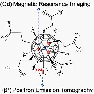
<?xml version="1.0" encoding="UTF-8"?>
<!DOCTYPE article PUBLIC "-//NLM//DTD Journal Publishing DTD v2.3 20070202//EN" "journalpublishing.dtd">
<article xmlns:mml="http://www.w3.org/1998/Math/MathML" xmlns:xlink="http://www.w3.org/1999/xlink" xml:lang="en" article-type="rapid-communication">
  <front>
    <journal-meta>
      <journal-id journal-id-type="publisher-id">applsci</journal-id>
      <journal-title>Applied Sciences</journal-title>
      <abbrev-journal-title abbrev-type="publisher">Appl. Sci.</abbrev-journal-title>
      <abbrev-journal-title abbrev-type="pubmed">Appl. Sci.</abbrev-journal-title>
      <issn pub-type="epub">2076-3417</issn>
      <publisher>
        <publisher-name>MDPI</publisher-name>
      </publisher>
    </journal-meta>
    <article-meta>
      <article-id pub-id-type="doi">10.3390/app2020465</article-id>
      <article-id pub-id-type="publisher-id">applsci-02-00465</article-id>
      <article-categories>
        <subj-group>
          <subject>Communication</subject>
        </subj-group>
      </article-categories>
      <title-group>
        <article-title>A Dual PET/MR Imaging Nanoprobe: <sup>124</sup>I Labeled Gd<sub>3</sub>N@C<sub>80</sub></article-title>
      </title-group>
      <contrib-group>
        <contrib contrib-type="author">
          <name>
            <surname>Luo</surname>
            <given-names>Jianqiao</given-names>
          </name>
          <xref rid="af1-applsci-02-00465" ref-type="aff">1</xref>
        </contrib>
        <contrib contrib-type="author">
          <name>
            <surname>Wilson</surname>
            <given-names>John D.</given-names>
          </name>
          <xref rid="af1-applsci-02-00465" ref-type="aff">1</xref>
        </contrib>
        <contrib contrib-type="author">
          <name>
            <surname>Zhang</surname>
            <given-names>Jianyuan</given-names>
          </name>
          <xref rid="af2-applsci-02-00465" ref-type="aff">2</xref>
        </contrib>
        <contrib contrib-type="author">
          <name>
            <surname>Hirsch</surname>
            <given-names>Jerry I.</given-names>
          </name>
          <xref rid="af1-applsci-02-00465" ref-type="aff">1</xref>
        </contrib>
        <contrib contrib-type="author">
          <name>
            <surname>Dorn</surname>
            <given-names>Harry C.</given-names>
          </name>
          <xref rid="af2-applsci-02-00465" ref-type="aff">2</xref>
        </contrib>
        <contrib contrib-type="author">
          <name>
            <surname>Fatouros</surname>
            <given-names>Panos P.</given-names>
          </name>
          <xref rid="af1-applsci-02-00465" ref-type="aff">1</xref>
        </contrib>
        <contrib contrib-type="author">
          <name>
            <surname>Shultz</surname>
            <given-names>Michael D.</given-names>
          </name>
          <xref rid="af3-applsci-02-00465" ref-type="aff">3</xref>
          <xref rid="c1-applsci-02-00465" ref-type="corresp">*</xref>
        </contrib>
      </contrib-group>
      <aff id="af1-applsci-02-00465"><label>1 </label>Department of Radiology, Virginia Commonwealth University, 1101 East Marshall Street, Richmond, VA 23298, USA; Email: <email>jluo@vcu.edu</email> (J.L.); <email>wilsonjd@vcu.edu</email> (J.D.W.); <email>jihirsch@vcu.edu</email> (J.I.H.); <email>ppfatouros@vcu.edu</email> (P.P.F.)</aff>
      <aff id="af2-applsci-02-00465"><label>2 </label>Department of Chemistry, Virginia Tech, 1109 Hahn Hall, Blacksburg, VA 24061, USA; Email: <email>zhangjy@vt.edu</email> (J.Z.); <email>hdorn@vt.edu</email> (H.C.D.)</aff>
      <aff id="af3-applsci-02-00465"><label>3 </label>Department of Biochemistry and Molecular Biology, Virginia Commonwealth University, 1101 East Marshall Street, Richmond, VA 23298, USA</aff>
      <author-notes>
        <corresp id="c1-applsci-02-00465"><label>*</label> Author to whom correspondence should be addressed; Email: <email>shultzmd@vcu.edu</email>; Tel.: +1-804-828-7943; Fax: +1-804-828-1473.</corresp>
      </author-notes>
      <pub-date pub-type="epub">
        <day>10</day>
        <month>05</month>
        <year>2012</year>
      </pub-date>
      <pub-date pub-type="collection"><month>05</month>
        <year>2012</year>
      </pub-date>
      <volume>2</volume>
      <issue>2</issue>
      <fpage>465</fpage>
      <lpage>478</lpage>
      <history>
        <date date-type="received">
          <day>09</day>
          <month>02</month>
          <year>2012</year>
        </date>
        <date date-type="rev-recd">
          <day>02</day>
          <month>05</month>
          <year>2012</year>
        </date>
        <date date-type="accepted">
          <day>03</day>
          <month>05</month>
          <year>2012</year>
        </date>
      </history>
      <permissions>
        <copyright-statement>© 2012 by the authors; licensee MDPI, Basel, Switzerland.</copyright-statement>
        <copyright-year>2012</copyright-year>
        <license xmlns:xlink="http://www.w3.org/1999/xlink" license-type="open-access" xlink:href="http://creativecommons.org/licenses/by/3.0/">
          <p>This article is an open-access article distributed under the terms and conditions of the Creative Commons Attribution license (http://creativecommons.org/licenses/by/3.0/).</p>
        </license>
      </permissions>
      <abstract>
        <p> The current report describes the development of a dual modality tomographic agent for both positron emission tomography and magnetic resonance imaging (PET/MRI). The dual-modality agent in this study was based on a <sup>124</sup>I (PET) radiolabeled tri-gadolinium endohedral metallofullerene Gd<sub>3</sub>N@C<sub>80</sub> (MRI) nanoprobe platform. The outer surface of the fullerene cage of the Gd<sub>3</sub>N@C<sub>80</sub> metallofullerenes was surface functionalized with carboxyl and hydroxyl groups (f-Gd<sub>3</sub>N@C<sub>80</sub>) using previously developed procedures and subsequently iodinated with <sup>124</sup>I to produce <sup>124</sup>I-f-Gd<sub>3</sub>N@C<sub>80</sub> nanoprobe. Orthotopic tumor-bearing rats were infused intratumorally by convection-enhanced delivery (CED) with the <sup>124</sup>I-f-Gd<sub>3</sub>N@C<sub>80</sub> agent and imaged by MRI or micro PET. The anatomical positioning and distribution of the <sup>124</sup>I-f-Gd<sub>3</sub>N@C<sub>80</sub> agent werecomparable between the MRI and PET scans. The <sup>124</sup>I-f-Gd<sub>3</sub>N@C<sub>80­</sub> dual-agent distribution and infusion site within the tumor was clearly evident in both T<sub>1</sub>- and T<sub>2</sub>-weighted MR images. The results demonstrate the successful preparation of a dual-modality imaging agent, <sup>124</sup>I-f-Gd<sub>3</sub>N@C<sub>80</sub>, which could ultimately be used for simultaneous PET/MR imaging.</p>
      </abstract>
      <kwd-group>
        <kwd>PET</kwd>
        <kwd>MRI</kwd>
        <kwd>metallofullerene</kwd>
        <kwd>iodination</kwd>
      </kwd-group>
    </article-meta>
  </front>
  <body>
    <sec sec-type="intro">
      <title>1. Introduction</title>
      <p>The ability to perform comprehensive evaluations of anatomical, functional and molecular imaging data is important for early detection and accurate prognosis of cancer. In many cases, this information is acquired using several imaging modalities with various contrast agents being administered during multiple hospital visits. The assessment of these images is also performed separately because of difficulties in image registration, primarily due to patient repositioning and variations in organ position and shape. Also, detection and measurement of low amplitude signals in noisy, heterogeneous biological environments is a fundamental challenge in diagnostic tomographic applications. Combined imaging during a single examination would solve some of these difficulties and has been realized using x-ray computed tomography (CT) and positron emission tomography (PET), with PET/CT now fully integrated into the clinical environment. PET/CT was one of the first multi-modal diagnostic approaches that made significant strides towards meeting new tomographic challenges [<xref ref-type="bibr" rid="B1-applsci-02-00465">1</xref>,<xref ref-type="bibr" rid="B2-applsci-02-00465">2</xref>,<xref ref-type="bibr" rid="B3-applsci-02-00465">3</xref>,<xref ref-type="bibr" rid="B4-applsci-02-00465">4</xref>,<xref ref-type="bibr" rid="B5-applsci-02-00465">5</xref>]. Clearly, tomography will continue to evolve beyond PET/CT and many other omni-tomography/multi-tomography approaches are envisioned [<xref ref-type="bibr" rid="B6-applsci-02-00465">6</xref>].</p>
      <p>Over the last several years a number of dual modality diagnostic approaches in addition to PET/CT have been reported including SPECT/CT, SPECT/MRI and PET/MRI, the latter which appears to have distinct advantages [<xref ref-type="bibr" rid="B7-applsci-02-00465">7</xref>,<xref ref-type="bibr" rid="B8-applsci-02-00465">8</xref>,<xref ref-type="bibr" rid="B9-applsci-02-00465">9</xref>,<xref ref-type="bibr" rid="B10-applsci-02-00465">10</xref>,<xref ref-type="bibr" rid="B11-applsci-02-00465">11</xref>,<xref ref-type="bibr" rid="B12-applsci-02-00465">12</xref>,<xref ref-type="bibr" rid="B13-applsci-02-00465">13</xref>,<xref ref-type="bibr" rid="B14-applsci-02-00465">14</xref>,<xref ref-type="bibr" rid="B15-applsci-02-00465">15</xref>,<xref ref-type="bibr" rid="B16-applsci-02-00465">16</xref>]. The dual imaging merger of PET/MRI has recently gained interest due to the combination of PET functional scanning with the anatomical and functional information provided by magnetic resonance imaging (MRI). For example, the PET/MRI combination has a distinct clinical advantage over CT because a lower level of ionizing radiation is utilized. In addition, the PET/MRI has the added advantage of combining biochemical soft tissue information (MRI) while the PET approach provides higher sensitivity with specific targeting dependent on the spatial location of the radiolabel. Unlike PET/CT, which requires sequential acquisitions involving patient table movement, current technological advances have led to a PET detector that is unaffected by magnetic fields allowing for true simultaneous image acquisition [<xref ref-type="bibr" rid="B16-applsci-02-00465">16</xref>,<xref ref-type="bibr" rid="B17-applsci-02-00465">17</xref>,<xref ref-type="bibr" rid="B18-applsci-02-00465">18</xref>,<xref ref-type="bibr" rid="B19-applsci-02-00465">19</xref>,<xref ref-type="bibr" rid="B20-applsci-02-00465">20</xref>]. The feasibility of a dual-modality PET/MRI system has proven successful in both small animal studies and in imaging the human brain [<xref ref-type="bibr" rid="B16-applsci-02-00465">16</xref>,<xref ref-type="bibr" rid="B20-applsci-02-00465">20</xref>]. As simultaneous PET/MR imaging progresses toward full clinical integration and application, the development of imaging agents with dual-modality properties will provide an important component to advancements in research and clinical applications in this area. Based on the variety of information that these imaging modalities can provide, it can be anticipated that simultaneous PET/MRI acquisition may yield complementary multiparametric imaging leading to new diagnostic paradigms. Therefore, the interest in development and characterization of dual imaging agents is apparent [<xref ref-type="bibr" rid="B21-applsci-02-00465">21</xref>].</p>
      <p>Fullerenes of various carbon content (e.g., C<sub>60</sub>/C<sub>80</sub>/C<sub>82</sub>) with an assortment of endohedral components including empty cages, single metal ions, and tri-metallic nitrides, have been increasingly employed in investigational animal studies as imaging contrast agents. Isolating the metal component from the biological system by placing it inside the carbon cage is a primary benefit of this nanoplatform. In regards to undesired biological effects, the toxicity of nanoparticles in general is more complex than with traditional bulk materials and highly variable, dependent on multiple parameters including size, shape, surface functionality, aggregation, surface charge, <italic>etc.</italic> [<xref ref-type="bibr" rid="B22-applsci-02-00465">22</xref>,<xref ref-type="bibr" rid="B23-applsci-02-00465">23</xref>,<xref ref-type="bibr" rid="B24-applsci-02-00465">24</xref>,<xref ref-type="bibr" rid="B25-applsci-02-00465">25</xref>]. In regard to the fullerene nanoparticles utilized in this application, the toxicities <italic>in vitro</italic> and <italic>in vivo</italic> have been studied extensively [<xref ref-type="bibr" rid="B22-applsci-02-00465">22</xref>,<xref ref-type="bibr" rid="B23-applsci-02-00465">23</xref>,<xref ref-type="bibr" rid="B24-applsci-02-00465">24</xref>,<xref ref-type="bibr" rid="B25-applsci-02-00465">25</xref>,<xref ref-type="bibr" rid="B26-applsci-02-00465">26</xref>,<xref ref-type="bibr" rid="B27-applsci-02-00465">27</xref>,<xref ref-type="bibr" rid="B28-applsci-02-00465">28</xref>,<xref ref-type="bibr" rid="B29-applsci-02-00465">29</xref>,<xref ref-type="bibr" rid="B30-applsci-02-00465">30</xref>,<xref ref-type="bibr" rid="B31-applsci-02-00465">31</xref>,<xref ref-type="bibr" rid="B32-applsci-02-00465">32</xref>]. Recent studies have also demonstrated the low cytotoxicity of the hydroxylated Gd<sub>3</sub>N@C<sub>80</sub>(OH)<sub>n</sub> system in a cell viability study of hen brain and human neuroblastoma SH-SY5Y cells [<xref ref-type="bibr" rid="B33-applsci-02-00465">33</xref>]. Overall, these reports demonstrated that fullerenes have low systemic toxicity and ameliorate inflammatory responses due to radical and ROS scavenging properties [<xref ref-type="bibr" rid="B22-applsci-02-00465">22</xref>,<xref ref-type="bibr" rid="B23-applsci-02-00465">23</xref>,<xref ref-type="bibr" rid="B24-applsci-02-00465">24</xref>,<xref ref-type="bibr" rid="B29-applsci-02-00465">29</xref>,<xref ref-type="bibr" rid="B31-applsci-02-00465">31</xref>,<xref ref-type="bibr" rid="B32-applsci-02-00465">32</xref>]. Furthermore, the ease of surface functionalization and ability to use the versatile metallofullerene platform as the basis for multiple imaging agents makes it an ideal candidate for multi-modality imaging applications. Altering the endohedral metal is the basic method for endowing additional imaging properties to the fullerene. Examples of this include encapsulation of Gd or Gd<sub>3</sub>N for superior MRI contrast [<xref ref-type="bibr" rid="B34-applsci-02-00465">34</xref>,<xref ref-type="bibr" rid="B35-applsci-02-00465">35</xref>], Lu<sub>3</sub>N for x-ray contrast [<xref ref-type="bibr" rid="B36-applsci-02-00465">36</xref>], and <sup>177</sup>Lu for SPECT detection [<xref ref-type="bibr" rid="B37-applsci-02-00465">37</xref>]. External ligands have also been added to empty C<sub>60</sub> cages to provide x-ray contrast [<xref ref-type="bibr" rid="B38-applsci-02-00465">38</xref>,<xref ref-type="bibr" rid="B39-applsci-02-00465">39</xref>] and radiolabeling [<xref ref-type="bibr" rid="B40-applsci-02-00465">40</xref>]. Recent work has shown the imaging and therapeutic, or theranostic potential of f-Gd<sub>3</sub>N@C<sub>80</sub> nanoparticles externally labeled with <sup>177</sup>Lu in an orthotopic xenograft murine brain tumor model [<xref ref-type="bibr" rid="B41-applsci-02-00465">41</xref>]. Therefore, there is clear potential for combining endohedral and exohedral components to yield a desired multi-modal nanomaterial imaging agent. </p>
      <p>The current study describes the synthesis and initial <italic>in vivo</italic> imaging demonstration of a metallofullerene-based nanoplatform for combined PET/MRI. Two other dual probes for PET/MRI have recently been reported, both of which utilize T<sub>2</sub> MRI contrast agents (iron oxide based) [<xref ref-type="bibr" rid="B13-applsci-02-00465">13</xref>,<xref ref-type="bibr" rid="B14-applsci-02-00465">14</xref>,<xref ref-type="bibr" rid="B42-applsci-02-00465">42</xref>]. The agent presented in this work offers T<sub>1</sub> MRI contrast and affects T<sub>2</sub> sufficiently to provide T<sub>2</sub>-weighted imaging as well.</p>
    </sec>
    <sec>
      <title>2. Experimental Section</title>
      <sec>
        <title>
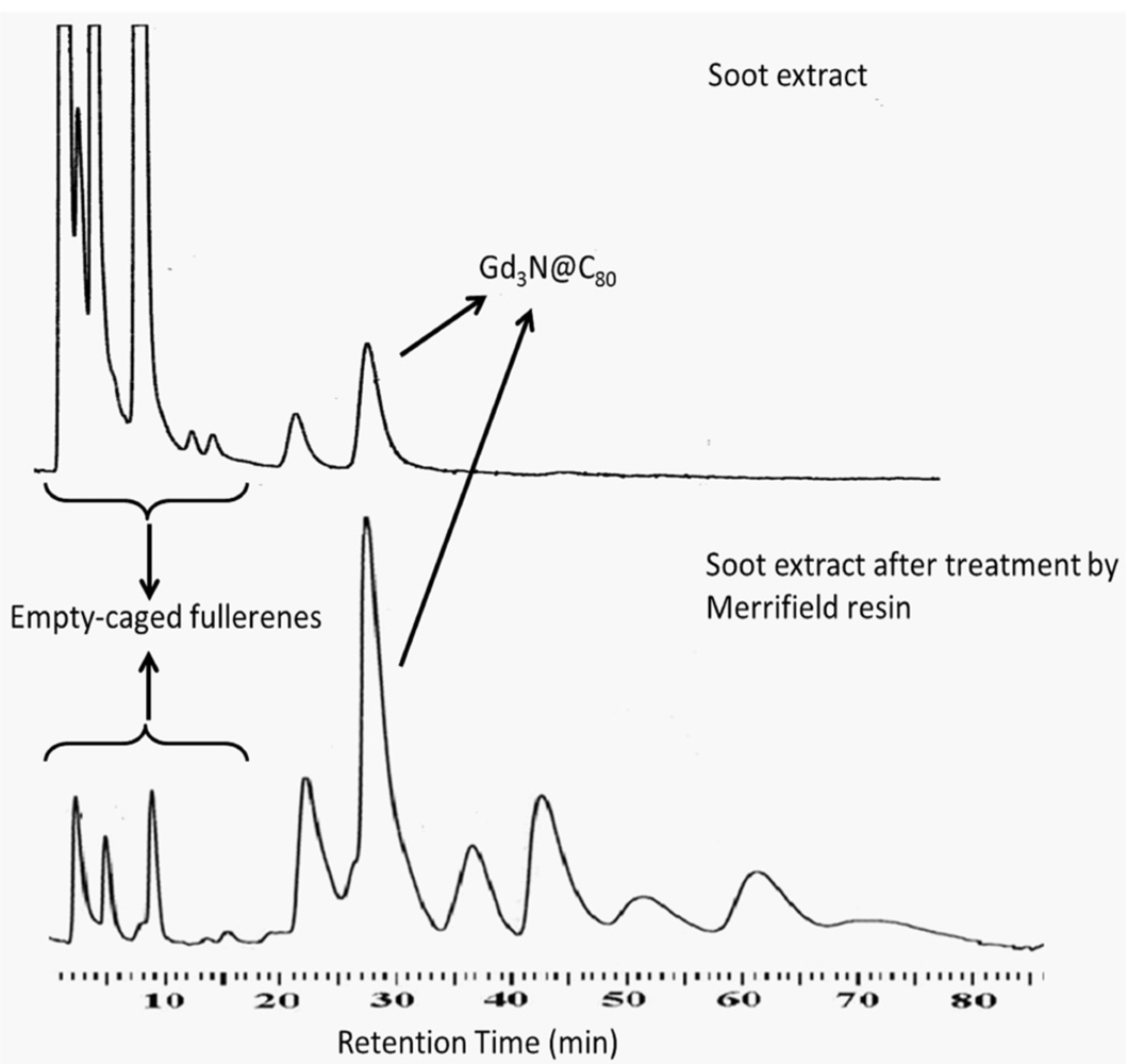
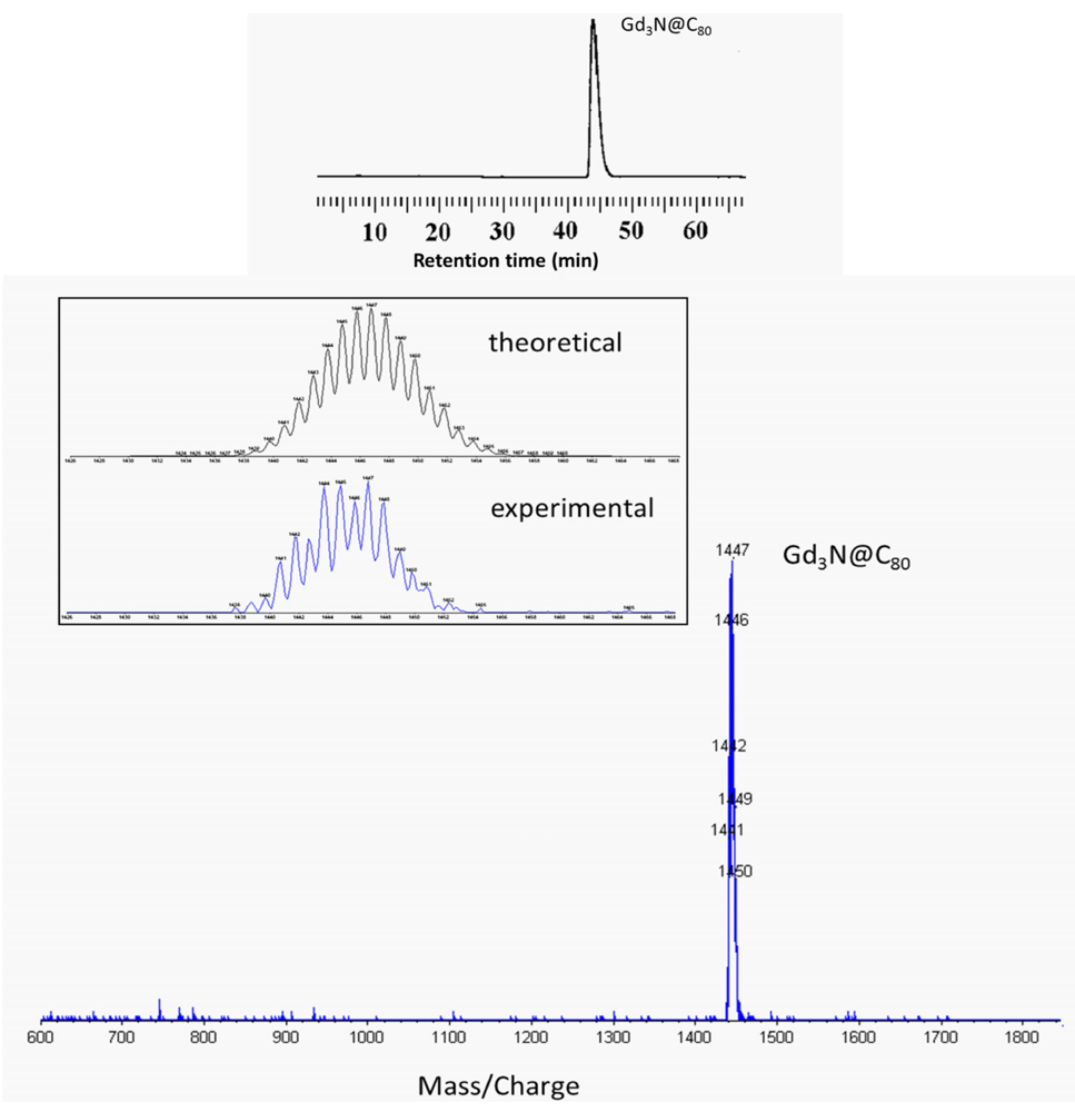
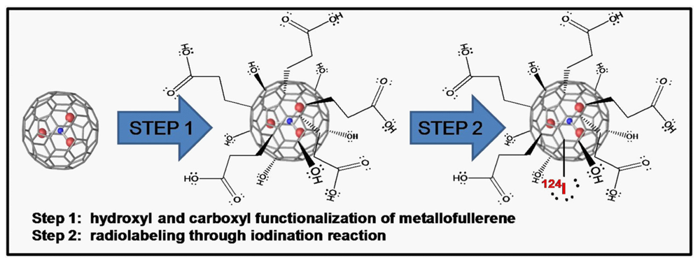
2.1. Synthesis and Surface Functionalization of Gd<sub>3</sub>N@C<sub>80</sub></title>
        <p>Gadolinium containing fullerenes, Gd<sub>3</sub>N@C<sub>80</sub>, were prepared by vaporization of graphite rods packed with a mixture of Gd<sub>2</sub>O<sub>3</sub>, graphite powder and metallic copper with a weight ratio of 2.0/1.0/2.1 under a dynamic flow of He and N<sub>2</sub> (flow rate ratio of N<sub>2</sub>:He = 3:100) in a Krätschmer-Huffman generator as previously reported [<xref ref-type="bibr" rid="B43-applsci-02-00465">43</xref>]. Fullerene products were extracted from the soot with xylene in a Soxlet extractor and the soluble empty-caged fullerenes and endohedral metallofullerenes are dissolved in the solvent. The Gd<sub>3</sub>N@C<sub>80</sub> product was purified by loading the solution containing fullerenes and metallofullerenes onto a cyclopentadiene functionalized Merrifield resin column eluted with toluene, which selectively removes empty-caged fullerenes [<xref ref-type="bibr" rid="B44-applsci-02-00465">44</xref>]. Removal of the empty cage fullerenes was verified by HPLC (column: Cosmosil 5-PBB; flow-rate: 1mL/min; mobile phase: toluene). As can be seen in <xref ref-type="fig" rid="applsci-02-00465-f001">Figure 1</xref> (top trace), the empty cage fullerenes are a predominant product in the direct soot extraction with the endohedral Gd<sub>3</sub>N@C<sub>80</sub> species having a retention time around 28 min. After elution through the Merrifield resin column [<xref ref-type="fig" rid="applsci-02-00465-f001">Figure 1</xref> (bottom trace)], the empty cages are greatly reduced and further endohedral products become evident beyond the Gd<sub>3</sub>N@C<sub>80</sub> peak. In order to obtain only the Gd<sub>3</sub>N@C<sub>80</sub> product, further purification was performed by fraction collecting with multi-stage HPLC (column: Cosmosil 5-PYE; flow-rate: 2 mL/min; mobile phase: toluene), which separates different species of metallofullerenes. Verification of the pure Gd<sub>3</sub>N@C<sub>80</sub> product was performed by HPLC and laser desorption time-of-flight mass spectrometry (LD-TOF-MS). <xref ref-type="fig" rid="applsci-02-00465-f002">Figure 2</xref> (top) shows only one peak for the fraction-collection purified product. The experimental isotopic distribution obtained for this product by LD-TOF-MS matched the theoretical isotopic distribution calculated for Gd<sub>3</sub>NC<sub>80</sub> as shown in <xref ref-type="fig" rid="applsci-02-00465-f002">Figure 2</xref> (bottom) giving verification of the product being pure Gd<sub>3</sub>N@C<sub>80</sub>.</p>
        <fig id="applsci-02-00465-f001" position="anchor">
          <label>Figure 1</label>
          <caption>
            <p> HPLC traces of the fullerene and metallofullerene solution before (<bold>top</bold>) and after (<bold>bottom</bold>) selective removal of empty-caged fullerenes with cyclopentadiene functionalized Merrifield resin column. </p>
          </caption>
          <graphic xmlns:xlink="http://www.w3.org/1999/xlink" xlink:href="applsci-02-00465-g001.tif"/>
        </fig>
        <fig id="applsci-02-00465-f002" position="anchor">
          <label>Figure 2</label>
          <caption>
            <p> (<bold>top</bold>) HPLC trace of purified Gd<sub>3</sub>N@C<sub>80</sub> and (<bold>bottom</bold>) laser desorption time-of-flight (LD-TOF) mass spectrometry of the purified Gd<sub>3</sub>N@C<sub>80</sub>. Inset: expansion of the spectrum showing theoretical and experimental isotopic distribution. </p>
          </caption>
          <graphic xmlns:xlink="http://www.w3.org/1999/xlink" xlink:href="applsci-02-00465-g002.tif"/>
        </fig>
        <p>Toluene was then removed by rotovap to yield the dry purified Gd<sub>3</sub>N@C<sub>80</sub> product, and functionalization of the Gd<sub>3</sub>N@C<sub>80</sub> with carboxyl and hydroxyl groups was carried out by methods detailed previously [<xref ref-type="bibr" rid="B34-applsci-02-00465">34</xref>]. Briefly, succinic acid acyl peroxide (3.2 mg) was added to Gd<sub>3</sub>N@C<sub>80</sub> (4 mg) dissolved in 5 mL of o-dichlorobenzene. The solution was de-oxygenated with flowing argon and heated at 84 °C for 48 h. At 12 h intervals, additional succinic acid acyl peroxide (3.2 mg) was added to ensure adequate carboxyl functionalization for aqueous solubility. After 48 h, a brown precipitant was formed from which the water soluble product was extracted and endowed with hydroxyl groups by addition of 8 mL of 0.2 M NaOH. The aqueous solution was concentrated and purified using a Sephadex G-25 (Parmacia) size-exclusion gel column. The product was eluted using deionized water as a narrow brown band with pH = 7–7.5. Following the purification steps, the Gd<sup>3+</sup> concentration was determined by inductively coupled plasma mass spectrometry (ICP-MS). This was performed by digesting triplicate samples in Optima grade nitric acid and measuring for Gd against a standard curve.</p>
      </sec>
      <sec>
        <title>2.2. <sup>124</sup>I Labeling of Carboxyl and Hydroxyl Functionalized Gd<sub>3</sub>N@C<sub>80</sub></title>
        <p>The functionalized metallofullerene sample (f-Gd<sub>3</sub>N@C<sub>80</sub>) was then iodinated with <sup>124</sup>I by methods similar to those reported for <sup>125</sup>I radio-iodination of hydroxylated C<sub>60</sub> fullerene derivatives [<xref ref-type="bibr" rid="B40-applsci-02-00465">40</xref>] with modifications. The <sup>124</sup>I was activated by placing 100 µL (4.3 MBq) of a Na<sup>124</sup>I solution into an iodogen-coated tube (Pierce). Then 100 µL of 0.08 mM f-Gd<sub>3</sub>N@C<sub>80</sub> was added to the activated <sup>124</sup>I solution and allowed to react for 18 min with periodic vortexing. The <sup>124</sup>I-labeled fullerenes were subsequently purified by gel chromatography using a 30 × 10 mL Bio-Rad Econo-Pac 10 DG desalting column eluted with water (20 × 0.5 mL fractions). The <sup>124</sup>I activity of each fraction was determined using a dose calibrator and the UV absorbance was measured at 263 nm, the absorption peak for the metallofullerenes. Twenty microliter aliquots from the peak fraction of the elution profile were subjected to analytical HPLC to determine radiochemical purity (Waters, Ultrahydrogel<sup>TM</sup>, 7.8 × 300 mm, GPC-1000 Å; isocratic, 90% 0.05 M ammonium acetate, 10% methanol; 0.7 mL/min).</p>
      </sec>
      <sec>
        <title>2.3. Cell Culture and Animal Model</title>
        <p>T9 rat glioblastoma cells were grown in Dulbecco’s Modified Eagle’s Medium supplemented with 10% FBS at 37 °C in a 5% CO<sub>2</sub>/95% air atmosphere. To prepare cell inocula, cells were harvested from log phase cultures and resuspended in PBS at a concentration of 5 × 10<sup>4</sup> cells/5 µL.</p>
        <p>All experiments involving the use of rats were performed in accordance with protocols approved by the Virginia Commonwealth University Institutional Animal Care and Use Committee. Female Fisher 344 rats (~150 g) were anesthetized by intraperitoneal (ip) injection of ketamine/xylazine (70–100/7.5–10 mg/kg) and placed in a stereotactic frame (David Kopf Instruments). An incision site on the top of the head was shaved with clippers and disinfected with betadine. After making a midline incision, a 0.9 mm burr hole was drilled in the skull 1.5 mm anterior to the bregma and 3 mm laterally to the right. A 22 gauge guide cannula (Plastics ONE, Inc.) was mounted stereotactically, centered on the burr hole and held in place with surgical adhesive. The guide cannula was then anchored securely to the skull by application of dental cement encompassing both the guide cannula and an implanted MRI compatible screw. Subsequently, the incision was sutured and a 25 μL Hamilton Syringe with a 28 G removable needle was used to inject 5 × 10<sup>4</sup> T9 cells in 5 μL of phosphate buffered saline (PBS) at a depth of 6.5 mm from the surface of the skull in the caudoputamen. Animals were allowed to recover and then placed in the vivarium. Starting 7 days post tumor implantation, tumor size was monitored by magnetic resonance imaging (MRI) with gadodiamide (OMNISCAN<sup>TM</sup>) administered by tail vein. On day 12 post tumor implantation, 24 µL of the <sup>124</sup>I labeled f-Gd<sub>3</sub>N@C<sub>80</sub> (7.77 × 10<sup>−2</sup> MBq, 0.04 mM Gd<sub>3</sub>N@C<sub>80</sub>) were infused into the developed intracerebral tumor via convection enhanced delivery (CED) using a micro-injection pump (Bioanalytical Systems, West Lafayette, IN) at a flow rate of 0.2 µL/min. Rats were then imaged by either MRI or microPET separately.</p>
      </sec>
      <sec>
        <title>2.4. <italic>In Vivo</italic> Imaging Methods: MRI and microPET</title>
        <p>Rats were intubated and anesthetized using 0.5–1% isoflurane with 1:2 N<sub>2</sub>O:O<sub>2</sub> carrier gas delivered through a ventilator system. Magnetic resonance imaging was performed on a 2.35 T, 40 cm bore magnet (Bruker Instruments, Billerica, MA). The animals were imaged by MRI (T<sub>1</sub>- and T<sub>2</sub>-weighted images) to obtain baseline data immediately prior to intratumoral infusion, and at time points of 1, 24, and up to 72 h post infusion. The rats for microPET imaging were anesthetized using 2–3% isoflurane with O<sub>2</sub> carrier via nose cone and a 2 h post infusion PET scan was performed using a microPET camera (Rodent R4, Concorde Microsystems) to visualize the <sup>124</sup>I distribution. Immediately following <sup>124</sup>I imaging, 6.7 MBq of <sup>18</sup>F FDG was administered by ip injection to highlight the brain anatomy. Five minute static images utilizing a 350–550 keV energy window were obtained 5 and 45 min following <sup>18</sup>F-FDG administration without transmission scanning. Histogramming of list mode data and image reconstruction were conducted using ASIPro VM software. </p>
      </sec>
    </sec>
    <sec sec-type="results">
      <title>
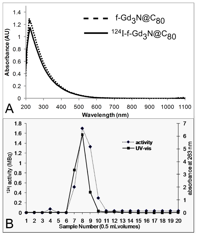
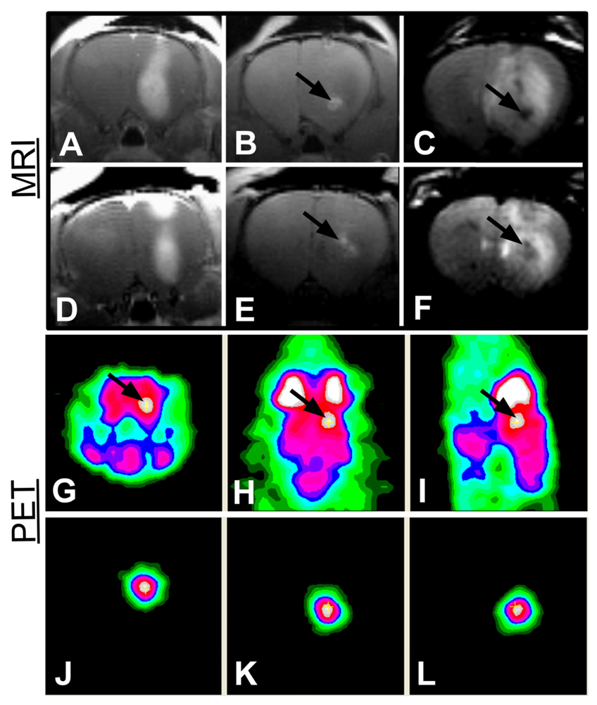
3. Results and Discussion</title>
      <p>In a typical iodination reaction involving the radiolabeling of proteins or antibodies, the benzene ring of tyrosine is labeled at the ortho position relative to the hydroxyl group [<xref ref-type="bibr" rid="B45-applsci-02-00465">45</xref>]. Given the average number of hydroxyl groups per f-Gd<sub>3</sub>N@C<sub>80</sub> (~26) following the functionalization procedure [<xref ref-type="bibr" rid="B34-applsci-02-00465">34</xref>], it is likely that the <sup>124</sup>I attaches directly to the fullerene cage but the position of attachment is unclear. A possible iodination position is illustrated in the reaction schematic (<xref ref-type="fig" rid="applsci-02-00465-f003">Figure 3</xref>); however further studies involving iodination with non-radioactive NaI at higher I to f-Gd<sub>3</sub>N@C<sub>80</sub> ratios will be required to fully elucidate the iodination position. In the radiolabeled product prepared in this study, the I to f-Gd<sub>3</sub>N@C<sub>80</sub> ratio was low due to the high specific activity of <sup>124</sup>I used, and the low radioactivity (or number of radioactive molecules) required for PET detection relative to the concentration of f-Gd<sub>3</sub>N@C<sub>80</sub> required for MRI. Therefore, at these low ratios, I labeling would not be expected to significantly alter the physical and chemical characteristics of the f-Gd<sub>3</sub>N@C<sub>80</sub>. Indeed, <xref ref-type="fig" rid="applsci-02-00465-f004">Figure 4</xref> (A) shows no detectable differences in the UV-vis absorption spectra; likewise, MRI relaxivity was unchanged following iodination. Following the iodination reaction, the radiolabeled product (<sup>124</sup>I-f-Gd<sub>3</sub>N@C<sub>80</sub>) was purified by gel chromatography during which 0.5 mL fractions were collected and radioactivity measured using a dose calibrator. UV absorbance measurements were also collected for each fraction at 263 nm to monitor the elution of the f-Gd<sub>3</sub>N@C<sub>80</sub>. Radioactivity and UV absorption elution profiles are presented in <xref ref-type="fig" rid="applsci-02-00465-f004">Figure 4</xref> (B). In order to determine the labeling efficiency, the total activity of the 3 fractions containing the <sup>124</sup>I-f-Gd<sub>3</sub>N@C<sub>80</sub> was divided by the activity of <sup>124</sup>I placed in the iodogen tube at the beginning of the synthesis. The results from 7 independent radiolabeling syntheses yielded 87 ± 5% labeling efficiency. </p>
      <fig id="applsci-02-00465-f003" position="anchor">
        <label>Figure 3</label>
        <caption>
          <p> Illustration of the reaction scheme starting with the endohedral metallofullerene and resulting in the aqueous soluble, iodinated, dual-modality imaging probe.</p>
        </caption>
        <graphic xmlns:xlink="http://www.w3.org/1999/xlink" xlink:href="applsci-02-00465-g003.tif"/>
      </fig>
      <fig id="applsci-02-00465-f004" position="anchor">
        <label>Figure 4</label>
        <caption>
          <p> Analysis of 0.5 mL fractions collected post iodination during elution through the desalting column by UV absorption (solid line—right axis) and activity measurements (dashed line—left axis). </p>
        </caption>
        <graphic xmlns:xlink="http://www.w3.org/1999/xlink" xlink:href="applsci-02-00465-g004.tif"/>
      </fig>
      <p>Fraction #8 contained both the highest <sup>124</sup>I activity and f-Gd<sub>3</sub>N@C<sub>80</sub> concentration and thus was further subjected to HPLC analysis with UV/radioactive detection in order to determine the radiochemical purity. As the f-Gd<sub>3</sub>N@C<sub>80</sub> nanoplatform has been extensively characterized in the literature [<xref ref-type="bibr" rid="B34-applsci-02-00465">34</xref>,<xref ref-type="bibr" rid="B35-applsci-02-00465">35</xref>,<xref ref-type="bibr" rid="B36-applsci-02-00465">36</xref>,<xref ref-type="bibr" rid="B37-applsci-02-00465">37</xref>,<xref ref-type="bibr" rid="B41-applsci-02-00465">41</xref>,<xref ref-type="bibr" rid="B43-applsci-02-00465">43</xref>,<xref ref-type="bibr" rid="B44-applsci-02-00465">44</xref>], it was possible to obtain the radiochemical purity by HPLC separation. This was due to the high purity of the f-Gd<sub>3</sub>N@C<sub>80</sub> starting material which would only yield three possible products following the iodination reaction, namely f-Gd<sub>3</sub>N@C<sub>80</sub>, <sup>124</sup>I-f-Gd<sub>3</sub>N@C<sub>80</sub>, and free <sup>124</sup>I. Using size exclusion separation methods, the radiolabeled and non-labeled f-Gd<sub>3</sub>N@C<sub>80</sub> will have similar retention, leaving the free <sup>124</sup>I to be the only separable species. As seen in <xref ref-type="fig" rid="applsci-02-00465-f005">Figure 5</xref>, the peak in the UV trace at 14.5 min (red line—right axis), due to UV absorbance by the f-Gd<sub>3</sub>N@C<sub>80</sub>, was accompanied by a corresponding peak in the radioactive trace at 14.9 min (black line—left axis) which indicated the formation of the labeled <sup>124</sup>I-f-Gd<sub>3</sub>N@C<sub>80</sub> product. The 0.4 min offset in retention time is due to the placement of the radioactive detector in series after the UV detector in our HPLC setup and is a factor of tube length and flow rate. The radioactive peak in the iodination product at 19.5 min is free <sup>124</sup>I. This is corroborated by the 19.5 min retention time of <sup>124</sup>I alone in solution (black dashed line—left axis). Integration of the peak areas of the iodination product yields 95% radiochemical purity. </p>
      <fig id="applsci-02-00465-f005" position="anchor">
        <label>Figure 5</label>
        <caption>
          <p> Analysis by HPLC of the iodinated product <sup>124</sup>I-f-Gd<sub>3</sub>N@C<sub>80</sub> (solid lines) showing detection by UV absorption (red trace—right axis) and radioactivity (black trace—left axis). The slight offset in the two traces is due to the detectors being connected in series. The trailing peak in the radioactive trace for the <sup>124</sup>I-f-Gd<sub>3</sub>N@C<sub>80</sub> product was confirmed to be free <sup>124</sup>I as evidenced in the matching retention time for <sup>124</sup>I alone injection (dashed line—black trace).</p>
        </caption>
        <graphic xmlns:xlink="http://www.w3.org/1999/xlink" xlink:href="applsci-02-00465-g005.tif"/>
      </fig>
      <p>Rats bearing orthotopic T9 brain tumors were imaged day 10 post tumor implantation with commercial gadodiamide in order to determine the proper depth of cannula placement for infusion (images from two rats: <xref ref-type="fig" rid="applsci-02-00465-f006">Figure 6</xref>(A,D)). On day 12, 24 µL of <sup>124</sup>I-f-Gd<sub>3</sub>N@C<sub>80</sub> (7.77 × 10<sup>−2</sup> MBq <sup>124</sup>I, 0.04 mM Gd<sub>3</sub>N@C<sub>80</sub>) was infused intratumorally; bright contrast (due to the endohedral Gd) can be seen around the infusion site on the T<sub>1W</sub> image (<xref ref-type="fig" rid="applsci-02-00465-f006">Figure 6</xref>(B,E)). In both cases shown, the infused agent is located within the tumor. This is further evidenced by the T<sub>2W</sub> images in <xref ref-type="fig" rid="applsci-02-00465-f006">Figure 6</xref>(C,F) which show dark contrast (due to high T<sub>2</sub> relaxivity) at the infusion site in the center of the tumor outlined by bright contrasting peritumoral edema. Other rats infused identically to those above were subjected to microPET imaging; the resulting images from a representative rat are shown in <xref ref-type="fig" rid="applsci-02-00465-f006">Figure 6</xref>(G–L). <xref ref-type="fig" rid="applsci-02-00465-f006">Figure 6</xref>(J–L) was collected 2 h post <sup>124</sup>I-f-Gd<sub>3</sub>N@C<sub>80</sub> infusion by imaging for 5 min on the microPET scanner and illustrates the intratumoral distribution of the <sup>124</sup>I. The <sup>18</sup>F-FDG image obtained subsequently (<xref ref-type="fig" rid="applsci-02-00465-f006">Figure 6</xref>(G–I)) provides an anatomical reference for the location of the tumor and infusion site. These results show that the position of the infusion site seen in the microPET image is comparable with the positions evident in the MR images. </p>
      <fig id="applsci-02-00465-f006" position="anchor">
        <label>Figure 6</label>
        <caption>
          <p> Magnetic resonance (MR) images from two representative orthotopic tumor bearing rat brains and microPET images from one representative rat infused with the <sup>124</sup>I-f-Gd<sub>3</sub>N@C<sub>80</sub> product. (<bold>A</bold>, <bold>D</bold>) T<sub>1</sub>-weighted images with gadodiamide contrast. (<bold>B</bold>, <bold>E</bold>) T<sub>1</sub>-weighted images showing bright contrast at the infusion site due to the <sup>124</sup>I-f-Gd<sub>3</sub>N@C<sub>80</sub>. (<bold>C</bold>, <bold>F</bold>) T<sub>2</sub>-weighted images with dark contrast due to the <sup>124</sup>I-f-Gd<sub>3</sub>N@C<sub>80</sub>. (J-L) microPET images showing signal from the <sup>124</sup>I-f-Gd<sub>3</sub>N@C<sub>80</sub>. (<bold>G</bold>) Coronal, (<bold>H</bold>) axial, and (<bold>I</bold>) sagittal microPET images following <sup>18</sup>F-FDG injection with the additive image signal allowing for localization of the <sup>124</sup>I-f-Gd<sub>3</sub>N@C<sub>80</sub> within the right hemisphere of the rat brain (arrows point to infusion sites).</p>
        </caption>
        <graphic xmlns:xlink="http://www.w3.org/1999/xlink" xlink:href="applsci-02-00465-g006.tif"/>
      </fig>
      <p>These studies have produced a dual-modality agent through the combination of a nanoplatform based MR T<sub>1</sub> agent with high relaxivity (Gd<sub>3</sub>N@C<sub>80</sub>) and a <sup>124</sup>I label for PET detection. The use of <sup>124</sup>I is advantageous due to the facile and rapid radiolabeling process that yields a product having high radiochemical purity. In addition, relaxivity r<sub>1</sub> values of 70 s<sup>−1</sup>·mM<sup>−1</sup> with respect to Gd concentration have been reported for the carboxyl and hydroxyl functionalized Gd<sub>3</sub>N@C<sub>80</sub> (f-Gd<sub>3</sub>N@C<sub>80</sub>), which is over an order of magnitude greater than most chelate-based contrast agents [<xref ref-type="bibr" rid="B34-applsci-02-00465">34</xref>]. The high stability of the metallofullerene cage also provides a robust means for isolating the free metal from the biological system thus suppressing toxic side effects [<xref ref-type="bibr" rid="B46-applsci-02-00465">46</xref>,<xref ref-type="bibr" rid="B47-applsci-02-00465">47</xref>]. Although the stability of the iodinated form of the f-Gd<sub>3</sub>N@C<sub>80</sub> (<sup>124</sup>I-f-Gd<sub>3</sub>N@C<sub>80</sub>) will require further investigation, our preliminary study suggested a negligible amount of free <sup>124</sup>I as indicated by a lack of thyroid uptake in the PET scans following infusion of the radio-iodinated agent. </p>
      <p>The <italic>in vivo</italic> imaging investigations have clearly demonstrated the dual properties of the <sup>124</sup>I-f-Gd<sub>3</sub>N@C<sub>80</sub> agent through visualization in orthotopic T9 tumors with both MRI and microPET imaging with confirmation of anatomical location using subsequent <sup>18</sup>F-FDG microPET scanning. The use of CED in this study to deliver the agent directly into the tumor was chosen to bypass the blood brain barrier (BBB), in order to sufficiently demonstrate image acquisition orthotopically. Although CED administrations are currently used selectively in the clinic to deliver high doses of therapeutic agents directly into malignant tumors, ultimately it would be desirable to have these imaging agents administered intravenously as well. Surface modifications to metallofullerenes have been attempted to allow for limited opsonization and diffusion across the BBB (e.g., polyethylene glycols) [<xref ref-type="bibr" rid="B46-applsci-02-00465">46</xref>]; however, further studies are needed to elucidate the most favorable surface coating. Additionally, the optimal use of the agent will be realized with investigations using integrated and simultaneous PET/MR scanners now under development and testing. </p>
    </sec>
    <sec sec-type="conclusions">
      <title>4. Conclusions</title>
      <p>In summary, this work has successfully demonstrated the production of <sup>124</sup>I-f-Gd<sub>3</sub>N@C<sub>80</sub>, a dual modality imaging agent designed for simultaneous PET/MRI examinations. MR contrast is provided by the Gd, while the <sup>124</sup>I annihilation photons allow for PET detection. The facile and efficient iodination process is capable of yielding products with high specific activity. Since sensitivity differences between the two imaging modalities could result in various ratios being required, future work will address optimization of the reaction conditions to allow for control of the radiolabeled <sup>124</sup>I (PET)/Gd (MRI) molar ratio in the <sup>124</sup>I-f-Gd<sub>3</sub>N@C<sub>80</sub> agent. The employment of a single agent is important in studies requiring close correspondence of tissue distribution profiles of the signals acquired in both imaging modalities. As clinical PET/MRI research advances, the current <sup>124</sup>I-f-Gd<sub>3</sub>N@C<sub>80</sub> agent is promising because it will have identical biodistribution in both imaging modalities. Additionally, the substitution of <sup>124</sup>I with <sup>125</sup>I or <sup>131</sup>I utilizing the same synthetic methods in this work will produce a dual MR diagnostic/therapeutic (theranostic) nanoprobe.</p>
    </sec>
    </body>
  <back>
    <ack>
      <title>Acknowledgments</title>
      <p>This work was supported by the National Institutes of Health, National Cancer Institute R01 CA119371, National Science Foundation, Nanoscale Interdisciplinary Research Team DMR-0507083 and the Center for Innovative Technology, Commonwealth of Virginia, Virginia Commonwealth Technology Research Fund. We would like to thank Chun-Ying Shu, Virginia Polytechnic Institute &amp; State University, for assistance in aqueous functionalization of the fullerenes and Frank D. Corwin, Virginia Commonwealth University for aiding in collection of MR images. </p>
    </ack>
    <notes>
      <title>Conflict of Interest</title>
      <p>The authors declare no conflict of interest. </p>
    </notes>
  <ref-list>
      <title>References</title>
      <ref id="B1-applsci-02-00465">
        <label>1.</label>
        <citation citation-type="journal">
          <person-group person-group-type="author">
            <name>
              <surname>Ahn</surname>
              <given-names>P.H.</given-names>
            </name>
            <name>
              <surname>Garg</surname>
              <given-names>M.K.</given-names>
            </name>
          </person-group>
          <article-title>Positron emission tomography/computed tomography for target delineation in head and neck cancers</article-title>
          <source>Semin. Nuclear Med.</source>
          <year>2008</year>
          <volume>38</volume>
          <fpage>141</fpage>
          <lpage>148</lpage>
          <pub-id pub-id-type="doi">10.1053/j.semnuclmed.2007.11.002</pub-id>
        </citation>
      </ref>
      <ref id="B2-applsci-02-00465">
        <label>2.</label>
        <citation citation-type="journal">
          <person-group person-group-type="author">
            <name>
              <surname>Bockisch</surname>
              <given-names>A.</given-names>
            </name>
            <name>
              <surname>Freudenberg</surname>
              <given-names>L.S.</given-names>
            </name>
            <name>
              <surname>Schmidt</surname>
              <given-names>D.</given-names>
            </name>
            <name>
              <surname>Kuwert</surname>
              <given-names>T.</given-names>
            </name>
          </person-group>
          <article-title>Hybrid imaging by SPECT/CT and PET/CT: Proven outcomes in cancer imaging</article-title>
          <source>Semin. Nuclear Med.</source>
          <year>2009</year>
          <volume>39</volume>
          <fpage>276</fpage>
          <lpage>289</lpage>
          <pub-id pub-id-type="doi">10.1053/j.semnuclmed.2009.03.003</pub-id>
        </citation>
      </ref>
      <ref id="B3-applsci-02-00465">
        <label>3.</label>
        <citation citation-type="journal">
          <person-group person-group-type="author">
            <name>
              <surname>Delbeke</surname>
              <given-names>D.</given-names>
            </name>
            <name>
              <surname>Schöder</surname>
              <given-names>H.</given-names>
            </name>
            <name>
              <surname>Martin</surname>
              <given-names>W.H.</given-names>
            </name>
            <name>
              <surname>Wahl</surname>
              <given-names>R.L.</given-names>
            </name>
          </person-group>
          <article-title>Hybrid imaging (SPECT/CT and PET/CT): Improving therapeutic decisions</article-title>
          <source>Semin. Nuclear Med.</source>
          <year>2009</year>
          <volume>39</volume>
          <fpage>308</fpage>
          <lpage>340</lpage>
          <pub-id pub-id-type="doi">10.1053/j.semnuclmed.2009.03.002</pub-id>
        </citation>
      </ref>
      <ref id="B4-applsci-02-00465">
        <label>4.</label>
        <citation citation-type="journal">
          <person-group person-group-type="author">
            <name>
              <surname>Even-Sapir</surname>
              <given-names>E.</given-names>
            </name>
            <name>
              <surname>Keidar</surname>
              <given-names>Z.</given-names>
            </name>
            <name>
              <surname>Bar-Shalom</surname>
              <given-names>R.</given-names>
            </name>
          </person-group>
          <article-title>Hybrid imaging (SPECT/CT and PET/CT)—Improving the diagnostic accuracy of functional/metabolic and anatomic imaging</article-title>
          <source>Semin. Nuclear Med.</source>
          <year>2009</year>
          <volume>39</volume>
          <fpage>264</fpage>
          <lpage>275</lpage>
          <pub-id pub-id-type="doi">10.1053/j.semnuclmed.2009.03.004</pub-id>
        </citation>
      </ref>
      <ref id="B5-applsci-02-00465">
        <label>5.</label>
        <citation citation-type="journal">
          <person-group person-group-type="author">
            <name>
              <surname>Kaufmann</surname>
              <given-names>P.A.</given-names>
            </name>
            <name>
              <surname>di Carli</surname>
              <given-names>M.F.</given-names>
            </name>
          </person-group>
          <article-title>Hybrid SPECT/CT and PET/CT imaging: The next step in noninvasive cardiac imaging</article-title>
          <source>Semin. Nuclear Med.</source>
          <year>2009</year>
          <volume>39</volume>
          <fpage>341</fpage>
          <lpage>347</lpage>
          <pub-id pub-id-type="doi">10.1053/j.semnuclmed.2009.03.007</pub-id>
        </citation>
      </ref>
      <ref id="B6-applsci-02-00465">
        <label>6.</label>
        <citation citation-type="other">
          <person-group person-group-type="author">
            <name>
              <surname>Wang</surname>
              <given-names>G.</given-names>
            </name>
            <name>
              <surname>Zhang</surname>
              <given-names>J.</given-names>
            </name>
            <name>
              <surname>Gao</surname>
              <given-names>H.</given-names>
            </name>
            <name>
              <surname>Weir</surname>
              <given-names>V.</given-names>
            </name>
            <name>
              <surname>Yu</surname>
              <given-names>H.</given-names>
            </name>
            <name>
              <surname>Cong</surname>
              <given-names>W.</given-names>
            </name>
            <name>
              <surname>Xu</surname>
              <given-names>X.</given-names>
            </name>
            <name>
              <surname>Shen</surname>
              <given-names>H.</given-names>
            </name>
            <name>
              <surname>Bennett</surname>
              <given-names>J.</given-names>
            </name>
            <name>
              <surname> Wang</surname>
              <given-names>Y.</given-names>
            </name>
            <name>
              <surname>Vannier</surname>
              <given-names>M.</given-names>
            </name>
          </person-group>
          <article-title>Omni-tomography/multi—tomography-Integrating multiple modalities for simultaneous imaging</article-title>
          <year>2011</year>
          <comment><italic>arXiv:11062124v1 [physicsmed-ph]</italic></comment>
        </citation>
      </ref>
      <ref id="B7-applsci-02-00465">
        <label>7.</label>
        <citation citation-type="journal">
          <person-group person-group-type="author">
            <name>
              <surname>Shao</surname>
              <given-names>Y.</given-names>
            </name>
            <name>
              <surname>Cherry</surname>
              <given-names>S.R.</given-names>
            </name>
            <name>
              <surname>Farahani</surname>
              <given-names>K.</given-names>
            </name>
            <name>
              <surname>Meadors</surname>
              <given-names>K.</given-names>
            </name>
            <name>
              <surname>Siegel</surname>
              <given-names>S.</given-names>
            </name>
            <name>
              <surname>Silverman</surname>
              <given-names>R.W.</given-names>
            </name>
            <name>
              <surname>Marsden</surname>
              <given-names>P.K.</given-names>
            </name>
          </person-group>
          <article-title>Simultaneous PET and MR imaging</article-title>
          <source>Phys. Med. Biol.</source>
          <year>1997</year>
          <volume>42</volume>
          <fpage>1965</fpage>
          <lpage>1970</lpage>
          <pub-id pub-id-type="doi">10.1088/0031-9155/42/10/010</pub-id>
        </citation>
      </ref>
      <ref id="B8-applsci-02-00465">
        <label>8.</label>
        <citation citation-type="journal">
          <person-group person-group-type="author">
            <name>
              <surname>Beyer</surname>
              <given-names>T.</given-names>
            </name>
            <name>
              <surname>Townsend</surname>
              <given-names>D.W.</given-names>
            </name>
            <name>
              <surname>Brun</surname>
              <given-names>T.</given-names>
            </name>
            <name>
              <surname>Kinahan</surname>
              <given-names>P.E.</given-names>
            </name>
            <name>
              <surname>Charron</surname>
              <given-names>M.</given-names>
            </name>
            <name>
              <surname>Roddy</surname>
              <given-names>R.</given-names>
            </name>
            <name>
              <surname>Jerin</surname>
              <given-names>J.</given-names>
            </name>
            <name>
              <surname>Young</surname>
              <given-names>J.</given-names>
            </name>
            <name>
              <surname>Byars</surname>
              <given-names>L.</given-names>
            </name>
            <name>
              <surname>Nutt</surname>
              <given-names>R.</given-names>
            </name>
          </person-group>
          <article-title>A combined PET/CT scanner for clinical oncology</article-title>
          <source>J. Nuclear Med.</source>
          <year>2000</year>
          <volume>41</volume>
          <fpage>1369</fpage>
          <lpage>1379</lpage>
        </citation>
      </ref>
      <ref id="B9-applsci-02-00465">
        <label>9.</label>
        <citation citation-type="journal">
          <person-group person-group-type="author">
            <name>
              <surname>Liang</surname>
              <given-names>H.</given-names>
            </name>
            <name>
              <surname>Yang</surname>
              <given-names>Y.</given-names>
            </name>
            <name>
              <surname>Yang</surname>
              <given-names>K.</given-names>
            </name>
            <name>
              <surname>Wu</surname>
              <given-names>Y.</given-names>
            </name>
            <name>
              <surname>Boone</surname>
              <given-names>J.M.</given-names>
            </name>
            <name>
              <surname>Cherry</surname>
              <given-names>S.R.</given-names>
            </name>
          </person-group>
          <article-title>A microPET/CT system for <italic>in vivo</italic> small animal imaging</article-title>
          <source>Phys. Med. Biol.</source>
          <year>2007</year>
          <volume>52</volume>
          <fpage>3881</fpage>
          <lpage>3894</lpage>
          <pub-id pub-id-type="doi">10.1088/0031-9155/52/13/015</pub-id>
        </citation>
      </ref>
      <ref id="B10-applsci-02-00465">
        <label>10.</label>
        <citation citation-type="journal">
          <person-group person-group-type="author">
            <name>
              <surname>Pichler</surname>
              <given-names>B.J.</given-names>
            </name>
            <name>
              <surname>Kolb</surname>
              <given-names>A.</given-names>
            </name>
            <name>
              <surname>Nägele</surname>
              <given-names>T.</given-names>
            </name>
            <name>
              <surname>Schlemmer</surname>
              <given-names>H.P.</given-names>
            </name>
          </person-group>
          <article-title>PET/MRI: Paving the way for the next generation of clinical multimodality imaging applications</article-title>
          <source>J. Nuclear Med.</source>
          <year>2010</year>
          <volume>51</volume>
          <fpage>333</fpage>
          <lpage>336</lpage>
          <pub-id pub-id-type="doi">10.2967/jnumed.109.061853</pub-id>
        </citation>
      </ref>
      <ref id="B11-applsci-02-00465">
        <label>11.</label>
        <citation citation-type="journal">
          <person-group person-group-type="author">
            <name>
              <surname>Cho</surname>
              <given-names>Z.H.</given-names>
            </name>
            <name>
              <surname>Son</surname>
              <given-names>Y.D.</given-names>
            </name>
            <name>
              <surname>Kim</surname>
              <given-names>H.K.</given-names>
            </name>
            <name>
              <surname>Kim</surname>
              <given-names>N.B.</given-names>
            </name>
            <name>
              <surname>Choi</surname>
              <given-names>E.J.</given-names>
            </name>
            <name>
              <surname>Lee</surname>
              <given-names>S.Y.</given-names>
            </name>
            <name>
              <surname>Chi</surname>
              <given-names>J.G.</given-names>
            </name>
            <name>
              <surname>Park</surname>
              <given-names>C.W.</given-names>
            </name>
            <name>
              <surname>Kim</surname>
              <given-names>Y.B.</given-names>
            </name>
            <name>
              <surname>Ogawa</surname>
              <given-names>S.</given-names>
            </name>
          </person-group>
          <article-title>Observation of glucose metabolism in the thalamic nuclei by fusion PET/MRI</article-title>
          <source>J. Nuclear Med.</source>
          <year>2011</year>
          <volume>52</volume>
          <fpage>401</fpage>
          <lpage>404</lpage>
          <pub-id pub-id-type="doi">10.2967/jnumed.110.081281</pub-id>
        </citation>
      </ref>
      <ref id="B12-applsci-02-00465">
        <label>12.</label>
        <citation citation-type="journal">
          <person-group person-group-type="author">
            <name>
              <surname>Choi</surname>
              <given-names>J.S.</given-names>
            </name>
            <name>
              <surname>Park</surname>
              <given-names>J.C.</given-names>
            </name>
            <name>
              <surname>Nah</surname>
              <given-names>H.</given-names>
            </name>
            <name>
              <surname>Woo</surname>
              <given-names>S</given-names>
            </name>
            <name>
              <surname>Oh</surname>
              <given-names>J.</given-names>
            </name>
            <name>
              <surname>Kim</surname>
              <given-names>K.M.</given-names>
            </name>
            <name>
              <surname>Cheon</surname>
              <given-names>G.J.</given-names>
            </name>
            <name>
              <surname>Chang</surname>
              <given-names>Y.</given-names>
            </name>
            <name>
              <surname>Yoo</surname>
              <given-names>J.</given-names>
            </name>
            <name>
              <surname>Cheon</surname>
              <given-names>J.</given-names>
            </name>
          </person-group>
          <article-title>A hybrid nanoparticle probe for dual-modality positron emission tomography and magnetic resonance imaging</article-title>
          <source>Angew. Chem. Int. Ed.</source>
          <year>2008</year>
          <volume>47</volume>
          <fpage>6259</fpage>
          <lpage>6262</lpage>
        <pub-id pub-id-type="doi">10.1002/anie.200801369</pub-id></citation>
      </ref>
      <ref id="B13-applsci-02-00465">
        <label>13.</label>
        <citation citation-type="journal">
          <person-group person-group-type="author">
            <name>
              <surname>Glaus</surname>
              <given-names>C.</given-names>
            </name>
            <name>
              <surname>Rossin</surname>
              <given-names>R.</given-names>
            </name>
            <name>
              <surname>Welch</surname>
              <given-names>M.J.</given-names>
            </name>
            <name>
              <surname>Bao</surname>
              <given-names>G.</given-names>
            </name>
          </person-group>
          <article-title><italic>In vivo</italic> evaluation of Cu-64-labeled magnetic nanoparticles as a dual-modality PET/MR imaging agent</article-title>
          <source>Bioconjugate Chem.</source>
          <year>2010</year>
          <volume>21</volume>
          <fpage>715</fpage>
          <lpage>722</lpage>
          <pub-id pub-id-type="doi">10.1021/bc900511j</pub-id>
        </citation>
      </ref>
      <ref id="B14-applsci-02-00465">
        <label>14.</label>
        <citation citation-type="journal">
          <person-group person-group-type="author">
            <name>
              <surname>Lee</surname>
              <given-names>H.Y.</given-names>
            </name>
            <name>
              <surname>Li</surname>
              <given-names>Z.</given-names>
            </name>
            <name>
              <surname>Chen</surname>
              <given-names>K.</given-names>
            </name>
            <name>
              <surname>Hsu</surname>
              <given-names>A.R.</given-names>
            </name>
            <name>
              <surname>Xu</surname>
              <given-names>C.J.</given-names>
            </name>
            <name>
              <surname>Xie</surname>
              <given-names>J.</given-names>
            </name>
            <name>
              <surname>Sun</surname>
              <given-names>S.H.</given-names>
            </name>
            <name>
              <surname>Chen</surname>
              <given-names>X.Y.</given-names>
            </name>
          </person-group>
          <article-title>PET/MRI dual-modality tumor imaging using arginine-glycine-aspartic (RGD)—Conjugated radiolabeled iron oxide nanoparticles</article-title>
          <source>J. Nuclear Med.</source>
          <year>2008</year>
          <volume>49</volume>
          <fpage>1371</fpage>
          <lpage>1379</lpage>
          <pub-id pub-id-type="doi">10.2967/jnumed.108.051243</pub-id>
        </citation>
      </ref>
      <ref id="B15-applsci-02-00465">
        <label>15.</label>
        <citation citation-type="journal">
          <person-group person-group-type="author">
            <name>
              <surname>Uppal</surname>
              <given-names>R.</given-names>
            </name>
            <name>
              <surname>Catana</surname>
              <given-names>C.</given-names>
            </name>
            <name>
              <surname>Ay</surname>
              <given-names>I.</given-names>
            </name>
            <name>
              <surname>Benner</surname>
              <given-names>T.</given-names>
            </name>
            <name>
              <surname>Sorensen</surname>
              <given-names>A.G.</given-names>
            </name>
            <name>
              <surname>Caravan</surname>
              <given-names>P.</given-names>
            </name>
          </person-group>
          <article-title>Bimodal thrombus imaging: Simultaneous PET/MR imaging with a fibrin-targeted dual PET/MR probe—Feasibility study in rat model</article-title>
          <source>Radiology</source>
          <year>2011</year>
          <volume>258</volume>
          <fpage>812</fpage>
          <lpage>820</lpage>
        <pub-id pub-id-type="doi">10.1148/radiol.10100881</pub-id><pub-id pub-id-type="pmid">21177389</pub-id></citation>
      </ref>
      <ref id="B16-applsci-02-00465">
        <label>16.</label>
        <citation citation-type="journal">
          <person-group person-group-type="author">
            <name>
              <surname>Catana</surname>
              <given-names>C.</given-names>
            </name>
            <name>
              <surname>Procissi</surname>
              <given-names>D.</given-names>
            </name>
            <name>
              <surname>Wu</surname>
              <given-names>Y.B.</given-names>
            </name>
            <name>
              <surname>Judenhofer</surname>
              <given-names>M.S.</given-names>
            </name>
            <name>
              <surname>Qi</surname>
              <given-names>J.Y.</given-names>
            </name>
            <name>
              <surname>Pichler</surname>
              <given-names>B.J.</given-names>
            </name>
            <name>
              <surname>Jacobs</surname>
              <given-names>R.E.</given-names>
            </name>
            <name>
              <surname>Cherry</surname>
              <given-names>S.R.</given-names>
            </name>
          </person-group>
          <article-title>Simultaneous in vivo positron emission tomography and magnetic resonance imaging</article-title>
          <source>Proc. Natl. Acad. Sci. USA</source>
          <year>2008</year>
          <volume>105</volume>
          <fpage>3705</fpage>
          <lpage>3710</lpage>
        <pub-id pub-id-type="doi">10.1073/pnas.0711622105</pub-id><pub-id pub-id-type="pmid">18319342</pub-id></citation>
      </ref>
      <ref id="B17-applsci-02-00465">
        <label>17.</label>
        <citation citation-type="journal">
          <person-group person-group-type="author">
            <name>
              <surname>Farahani</surname>
              <given-names>K.</given-names>
            </name>
            <name>
              <surname>Slates</surname>
              <given-names>R.</given-names>
            </name>
            <name>
              <surname>Shao</surname>
              <given-names>Y.P.</given-names>
            </name>
            <name>
              <surname>Silverman</surname>
              <given-names>R.</given-names>
            </name>
            <name>
              <surname>Cherry</surname>
              <given-names>S.</given-names>
            </name>
          </person-group>
          <article-title>Contemporaneous positron emission tomography and MR imaging at 1.5 T</article-title>
          <source> J. Magn. Reson. Imaging</source>
          <year>1999</year>
          <volume>9</volume>
          <fpage>497</fpage>
          <lpage>500</lpage>
          <pub-id pub-id-type="doi">10.1002/(SICI)1522-2586(199903)9:3&lt;497::AID-JMRI21&gt;3.0.CO;2-6</pub-id>
        </citation>
      </ref>
      <ref id="B18-applsci-02-00465">
        <label>18.</label>
        <citation citation-type="journal">
          <person-group person-group-type="author">
            <name>
              <surname>Judenhofer</surname>
              <given-names>M.S.</given-names>
            </name>
            <name>
              <surname>Wehrl</surname>
              <given-names>H.F.</given-names>
            </name>
            <name>
              <surname>Newport</surname>
              <given-names>D.F.</given-names>
            </name>
            <name>
              <surname>Catana</surname>
              <given-names>C.</given-names>
            </name>
            <name>
              <surname>Siegel</surname>
              <given-names>S.B.</given-names>
            </name>
            <name>
              <surname>Becker</surname>
              <given-names>M.</given-names>
            </name>
            <name>
              <surname>Thielscher</surname>
              <given-names>A.</given-names>
            </name>
            <name>
              <surname>Kneilling</surname>
              <given-names>M.</given-names>
            </name>
            <name>
              <surname>Lichy</surname>
              <given-names>M.P.</given-names>
            </name>
            <name>
              <surname>Eichner</surname>
              <given-names>M.</given-names>
            </name>
            <name>
              <surname>Klingel</surname>
              <given-names>K.</given-names>
            </name>
            <name>
              <surname>Reischl</surname>
              <given-names>G.</given-names>
            </name>
            <name>
              <surname>Widmaier</surname>
              <given-names>S.</given-names>
            </name>
            <name>
              <surname>Rocken</surname>
              <given-names>M.</given-names>
            </name>
            <name>
              <surname>Nutt</surname>
              <given-names>R.E.</given-names>
            </name>
            <name>
              <surname>Machulla</surname>
              <given-names>H.J.</given-names>
            </name>
            <name>
              <surname>Uludag</surname>
              <given-names>K.</given-names>
            </name>
            <name>
              <surname>Cherry</surname>
              <given-names>S.R.</given-names>
            </name>
            <name>
              <surname>Claussen</surname>
              <given-names>C.D.</given-names>
            </name>
            <name>
              <surname>Pichler</surname>
              <given-names>B.J.</given-names>
            </name>
          </person-group>
          <article-title>Simultaneous PET-MRI: A new approach for functional and morphological imaging</article-title>
          <source>Nat. Med.</source>
          <year>2008</year>
          <volume>14</volume>
          <fpage>459</fpage>
          <lpage>465</lpage>
        <pub-id pub-id-type="doi">10.1038/nm1700</pub-id><pub-id pub-id-type="pmid">18376410</pub-id></citation>
      </ref>
      <ref id="B19-applsci-02-00465">
        <label>19.</label>
        <citation citation-type="journal">
          <person-group person-group-type="author">
            <name>
              <surname>Pichler</surname>
              <given-names>B.J.</given-names>
            </name>
            <name>
              <surname>Judenhofer</surname>
              <given-names>M.S.</given-names>
            </name>
            <name>
              <surname>Catana</surname>
              <given-names>C.</given-names>
            </name>
            <name>
              <surname>Walton</surname>
              <given-names>J.H.</given-names>
            </name>
            <name>
              <surname>Kneilling</surname>
              <given-names>M.</given-names>
            </name>
            <name>
              <surname>Nutt</surname>
              <given-names>R.E.</given-names>
            </name>
            <name>
              <surname>Siegel</surname>
              <given-names>S.B.</given-names>
            </name>
            <name>
              <surname>Claussen</surname>
              <given-names>C.D.</given-names>
            </name>
            <name>
              <surname>Cherry</surname>
              <given-names>S.R.</given-names>
            </name>
          </person-group>
          <article-title>Performance test of an LSO-APD detector in a 7-T MRI scanner for simultaneous PET/MRI</article-title>
          <source>J. Nuclear Med.</source>
          <year>2006</year>
          <volume>47</volume>
          <fpage>639</fpage>
          <lpage>647</lpage>
        </citation>
      </ref>
      <ref id="B20-applsci-02-00465">
        <label>20.</label>
        <citation citation-type="journal">
          <person-group person-group-type="author">
            <name>
              <surname>Schlemmer</surname>
              <given-names>H.P.W.</given-names>
            </name>
            <name>
              <surname>Pichler</surname>
              <given-names>B.J.</given-names>
            </name>
            <name>
              <surname>Schmand</surname>
              <given-names>M.</given-names>
            </name>
            <name>
              <surname>Burbar</surname>
              <given-names>Z.</given-names>
            </name>
            <name>
              <surname>Michel</surname>
              <given-names>C.</given-names>
            </name>
            <name>
              <surname>Ladebeck</surname>
              <given-names>R.</given-names>
            </name>
            <name>
              <surname>Jattke</surname>
              <given-names>K.</given-names>
            </name>
            <name>
              <surname>Townsend</surname>
              <given-names>D.</given-names>
            </name>
            <name>
              <surname>Nahmias</surname>
              <given-names>C.</given-names>
            </name>
            <name>
              <surname>Jacob</surname>
              <given-names>P.K.</given-names>
            </name>
            <name>
              <surname>Heiss</surname>
              <given-names>W.D.</given-names>
            </name>
            <name>
              <surname>Claussen</surname>
              <given-names>C.D.</given-names>
            </name>
          </person-group>
          <article-title>Simultaneous MR/PET imaging of the human brain: Feasibility study</article-title>
          <source>Radiology</source>
          <year>2008</year>
          <volume>248</volume>
          <fpage>1028</fpage>
          <lpage>1035</lpage>
          <pub-id pub-id-type="doi">10.1148/radiol.2483071927</pub-id>
        </citation>
      </ref>
      <ref id="B21-applsci-02-00465">
        <label>21.</label>
        <citation citation-type="journal">
          <person-group person-group-type="author">
            <name>
              <surname>Jennings</surname>
              <given-names>L.E.</given-names>
            </name>
            <name>
              <surname>Long</surname>
              <given-names>N.J.</given-names>
            </name>
          </person-group>
          <article-title>‘Two is better than one’-probes for dual-modality molecular imaging</article-title>
          <source>Chem. Commun.</source>
          <year>2009</year>
          <fpage>3511</fpage>
          <lpage>3524</lpage>
          <pub-id pub-id-type="doi">10.1039/b821903f</pub-id>
        </citation>
      </ref>
      <ref id="B22-applsci-02-00465">
        <label>22.</label>
        <citation citation-type="journal">
          <person-group person-group-type="author">
            <name>
              <surname>Johnston</surname>
              <given-names>H.J.</given-names>
            </name>
            <name>
              <surname>Hutchison</surname>
              <given-names>G.R.</given-names>
            </name>
            <name>
              <surname>Christensen</surname>
              <given-names>F.M.</given-names>
            </name>
            <name>
              <surname>Aschberger</surname>
              <given-names>K.</given-names>
            </name>
            <name>
              <surname>Stone</surname>
              <given-names>V.</given-names>
            </name>
          </person-group>
          <article-title>The biological mechanisms and physicochemical characteristics responsible for driving fullerene toxicity</article-title>
          <source>Toxicol. Sci.</source>
          <year>2010</year>
          <volume>114</volume>
          <fpage>162</fpage>
          <lpage>182</lpage>
          <pub-id pub-id-type="doi">10.1093/toxsci/kfp265</pub-id>
        </citation>
      </ref>
      <ref id="B23-applsci-02-00465">
        <label>23.</label>
        <citation citation-type="journal">
          <person-group person-group-type="author">
            <name>
              <surname>Levi</surname>
              <given-names>N.</given-names>
            </name>
            <name>
              <surname>Hantgan</surname>
              <given-names>R.R.</given-names>
            </name>
            <name>
              <surname>Lively</surname>
              <given-names>M.O.</given-names>
            </name>
            <name>
              <surname>Carroll</surname>
              <given-names>D.L.</given-names>
            </name>
            <name>
              <surname>Prasad</surname>
              <given-names>G.L.</given-names>
            </name>
          </person-group>
          <article-title>C60-fullerenes: Detection of intracellular photoluminescence and lack of cytotoxic effects</article-title>
          <source>J. Nanobiotechnol.</source>
          <year>2006</year>
          <volume>4</volume>
          <pub-id pub-id-type="doi">10.1186/1477-3155-4-14</pub-id>
        </citation>
      </ref>
      <ref id="B24-applsci-02-00465">
        <label>24.</label>
        <citation citation-type="journal">
          <person-group person-group-type="author">
            <name>
              <surname>Xia</surname>
              <given-names>X.R.</given-names>
            </name>
            <name>
              <surname>Monteiro-Riviere</surname>
              <given-names>N.A.</given-names>
            </name>
            <name>
              <surname>Riviere</surname>
              <given-names>J.E.</given-names>
            </name>
          </person-group>
          <article-title>Intrinsic biological property of colloidal fullerene nanoparticles (nC<sub>60</sub>): Lack of lethality after high dose exposure to human epidermal and bacterial cells</article-title>
          <source>Toxicol. Lett.</source>
          <year>2010</year>
          <volume>197</volume>
          <fpage>128</fpage>
          <lpage>134</lpage>
          <pub-id pub-id-type="doi">10.1016/j.toxlet.2010.05.010</pub-id>
        </citation>
      </ref>
      <ref id="B25-applsci-02-00465">
        <label>25.</label>
        <citation citation-type="journal">
          <person-group person-group-type="author">
            <name>
              <surname>Yan</surname>
              <given-names>L.A.</given-names>
            </name>
            <name>
              <surname>Zhao</surname>
              <given-names>F.</given-names>
            </name>
            <name>
              <surname>Li</surname>
              <given-names>S.J.</given-names>
            </name>
            <name>
              <surname>Hu</surname>
              <given-names>Z.B.</given-names>
            </name>
            <name>
              <surname>Zhao</surname>
              <given-names>Y.L.</given-names>
            </name>
          </person-group>
          <article-title>Low-toxic and safe nanomaterials by surface-chemical design, carbon nanotubes, fullerenes, metallofullerenes, and graphenes</article-title>
          <source>Nanoscale</source>
          <year>2011</year>
          <volume>3</volume>
          <fpage>362</fpage>
          <lpage>382</lpage>
          <pub-id pub-id-type="doi">10.1039/c0nr00647e</pub-id>
        </citation>
      </ref>
      <ref id="B26-applsci-02-00465">
        <label>26.</label>
        <citation citation-type="journal">
          <person-group person-group-type="author">
            <name>
              <surname>Baker</surname>
              <given-names>G.L.</given-names>
            </name>
            <name>
              <surname>Gupta</surname>
              <given-names>A.</given-names>
            </name>
            <name>
              <surname>Clark</surname>
              <given-names>M.L.</given-names>
            </name>
            <name>
              <surname>Valenzuela</surname>
              <given-names>B.R.</given-names>
            </name>
            <name>
              <surname>Staska</surname>
              <given-names>L.M.</given-names>
            </name>
            <name>
              <surname>Harbo</surname>
              <given-names>S.J.</given-names>
            </name>
            <name>
              <surname>Pierce</surname>
              <given-names>J.T.</given-names>
            </name>
            <name>
              <surname>Dill</surname>
              <given-names>J.A.</given-names>
            </name>
          </person-group>
          <article-title>Inhalation toxicity and lung toxicokinetics of C<sub>60</sub> fullerene nanoparticles and microparticles</article-title>
          <source>Toxicol. Sci.</source>
          <year>2008</year>
          <volume>101</volume>
          <fpage>122</fpage>
          <lpage>131</lpage>
        <pub-id pub-id-type="pmid">17878152</pub-id></citation>
      </ref>
      <ref id="B27-applsci-02-00465">
        <label>27.</label>
        <citation citation-type="journal">
          <person-group person-group-type="author">
            <name>
              <surname>Jia</surname>
              <given-names>G.</given-names>
            </name>
            <name>
              <surname>Wang</surname>
              <given-names>H.</given-names>
            </name>
            <name>
              <surname>Yan</surname>
              <given-names>L.</given-names>
            </name>
            <name>
              <surname>Wang</surname>
              <given-names>X.</given-names>
            </name>
            <name>
              <surname>Pei</surname>
              <given-names>R.</given-names>
            </name>
            <name>
              <surname>Yan</surname>
              <given-names>T.</given-names>
            </name>
            <name>
              <surname>Zhao</surname>
              <given-names>Y.</given-names>
            </name>
            <name>
              <surname>Guo</surname>
              <given-names>X.</given-names>
            </name>
          </person-group>
          <article-title>Cytotoxicity of carbon nanomaterials: Single-wall nanotube, multi-wall nanotube, and fullerene</article-title>
          <source>Environ. Sci. Technol.</source>
          <year>2005</year>
          <volume>39</volume>
          <fpage>1378</fpage>
          <lpage>1383</lpage>
          <pub-id pub-id-type="doi">10.1021/es048729l</pub-id>
        </citation>
      </ref>
      <ref id="B28-applsci-02-00465">
        <label>28.</label>
        <citation citation-type="journal">
          <person-group person-group-type="author">
            <name>
              <surname>Liang</surname>
              <given-names>X.J.</given-names>
            </name>
            <name>
              <surname>Meng</surname>
              <given-names>H.</given-names>
            </name>
            <name>
              <surname>Wang</surname>
              <given-names>Y.</given-names>
            </name>
            <name>
              <surname>He</surname>
              <given-names>H.</given-names>
            </name>
            <name>
              <surname>Meng</surname>
              <given-names>J.</given-names>
            </name>
            <name>
              <surname>Lu</surname>
              <given-names>J.</given-names>
            </name>
            <name>
              <surname>Wang</surname>
              <given-names>P.C.</given-names>
            </name>
            <name>
              <surname>Zhao</surname>
              <given-names>Y.</given-names>
            </name>
            <name>
              <surname>Gao</surname>
              <given-names>X.</given-names>
            </name>
            <name>
              <surname>Sun</surname>
              <given-names>B.</given-names>
            </name>
            <etal/>
          </person-group>
          <article-title>Metallofullerene nanoparticles circumvent tumor resistance to cisplatin by reactivating endocytosis</article-title>
          <source>Proc. Natl. Acad. Sci. USA</source>
          <year>2010</year>
          <volume>107</volume>
          <fpage>7449</fpage>
          <lpage>7454</lpage>
        <pub-id pub-id-type="doi">10.1073/pnas.0909707107</pub-id><pub-id pub-id-type="pmid">20368438</pub-id></citation>
      </ref>
      <ref id="B29-applsci-02-00465">
        <label>29.</label>
        <citation citation-type="journal">
          <person-group person-group-type="author">
            <name>
              <surname>Roursgaard</surname>
              <given-names>M.</given-names>
            </name>
            <name>
              <surname>Poulsen</surname>
              <given-names>S.S.</given-names>
            </name>
            <name>
              <surname>Kepley</surname>
              <given-names>C.L.</given-names>
            </name>
            <name>
              <surname>Hammer</surname>
              <given-names>M.</given-names>
            </name>
            <name>
              <surname>Nielsen</surname>
              <given-names>G.D.</given-names>
            </name>
            <name>
              <surname>Larsen</surname>
              <given-names>S.T.</given-names>
            </name>
          </person-group>
          <article-title>Polyhydroxylated C<sub>60</sub> fullerene (fullerenol) attenuates neutrophilic lung inflammation in mice</article-title>
          <source>Basic Clin. Pharmacol. Toxicol.</source>
          <year>2008</year>
          <volume>103</volume>
          <fpage>386</fpage>
          <lpage>388</lpage>
          <pub-id pub-id-type="doi">10.1111/j.1742-7843.2008.00315.x</pub-id>
        </citation>
      </ref>
      <ref id="B30-applsci-02-00465">
        <label>30.</label>
        <citation citation-type="journal">
          <person-group person-group-type="author">
            <name>
              <surname>Sayes</surname>
              <given-names>C.M.</given-names>
            </name>
            <name>
              <surname>Marchione</surname>
              <given-names>A.A.</given-names>
            </name>
            <name>
              <surname>Reed</surname>
              <given-names>K.L.</given-names>
            </name>
            <name>
              <surname>Warheit</surname>
              <given-names>D.B.</given-names>
            </name>
          </person-group>
          <article-title>Comparative pulmonary toxicity assessments of C60 water suspensions in rats: Few differences in fullerene toxicity <italic>in vivo</italic> in contrast to <italic>in vitro</italic> profiles</article-title>
          <source>Nano Lett.</source>
          <year>2007</year>
          <volume>7</volume>
          <fpage>2399</fpage>
          <lpage>2406</lpage>
          <pub-id pub-id-type="doi">10.1021/nl0710710</pub-id>
        </citation>
      </ref>
      <ref id="B31-applsci-02-00465">
        <label>31.</label>
        <citation citation-type="journal">
          <person-group person-group-type="author">
            <name>
              <surname>Tsao</surname>
              <given-names>N.</given-names>
            </name>
            <name>
              <surname>Kanakamma</surname>
              <given-names>P.P.</given-names>
            </name>
            <name>
              <surname>Luh</surname>
              <given-names>T.Y.</given-names>
            </name>
            <name>
              <surname>Chou</surname>
              <given-names>C.K.</given-names>
            </name>
            <name>
              <surname>Lei</surname>
              <given-names>H.Y.</given-names>
            </name>
          </person-group>
          <article-title>Inhibition of Escherichia coli-induced meningitis by carboxyfullerence</article-title>
          <source>Antimicrob. Agents Chemother.</source>
          <year>1999</year>
          <volume>43</volume>
          <fpage>2273</fpage>
          <lpage>2277</lpage>
        <pub-id pub-id-type="pmid">10471578</pub-id></citation>
      </ref>
      <ref id="B32-applsci-02-00465">
        <label>32.</label>
        <citation citation-type="journal">
          <person-group person-group-type="author">
            <name>
              <surname>Wang</surname>
              <given-names>J.</given-names>
            </name>
            <name>
              <surname>Chen</surname>
              <given-names>C.</given-names>
            </name>
            <name>
              <surname>Li</surname>
              <given-names>B.</given-names>
            </name>
            <name>
              <surname>Yu</surname>
              <given-names>H.</given-names>
            </name>
            <name>
              <surname>Zhao</surname>
              <given-names>Y.</given-names>
            </name>
            <name>
              <surname>Sun</surname>
              <given-names>J.</given-names>
            </name>
            <name>
              <surname>Li</surname>
              <given-names>Y.</given-names>
            </name>
            <name>
              <surname>Xing</surname>
              <given-names>G.</given-names>
            </name>
            <name>
              <surname>Yuan</surname>
              <given-names>H.</given-names>
            </name>
            <name>
              <surname>Tang</surname>
              <given-names>J.</given-names>
            </name>
            <etal/>
          </person-group>
          <article-title>Antioxidative function and biodistribution of [Gd@C<sub>82</sub>(OH)<sub>22</sub>]<sub>n</sub> nanoparticles in tumor-bearing mice</article-title>
          <source>Biochem. Pharmacol.</source>
          <year>2006</year>
          <volume>71</volume>
          <fpage>872</fpage>
          <lpage>881</lpage>
        <pub-id pub-id-type="doi">10.1016/j.bcp.2005.12.001</pub-id><pub-id pub-id-type="pmid">16436273</pub-id></citation>
      </ref>
      <ref id="B33-applsci-02-00465">
        <label>33.</label>
        <citation citation-type="journal">
          <person-group person-group-type="author">
            <name>
              <surname>Ehrich</surname>
              <given-names>M.</given-names>
            </name>
            <name>
              <surname>Van Tassell</surname>
              <given-names>R.</given-names>
            </name>
            <name>
              <surname>Li</surname>
              <given-names>Y.B.</given-names>
            </name>
            <name>
              <surname>Zhou</surname>
              <given-names>Z.G.</given-names>
            </name>
            <name>
              <surname>Kepley</surname>
              <given-names>C.L.</given-names>
            </name>
          </person-group>
          <article-title>Fullerene antioxidants decrease organophosphate-induced acetylcholinesterase inhibition <italic>in vitro</italic></article-title>
          <source>Toxicol. In Vitro</source>
          <year>2011</year>
          <volume>25</volume>
          <fpage>301</fpage>
          <lpage>307</lpage>
          <pub-id pub-id-type="doi">10.1016/j.tiv.2010.09.010</pub-id>
        </citation>
      </ref>
      <ref id="B34-applsci-02-00465">
        <label>34.</label>
        <citation citation-type="journal">
          <person-group person-group-type="author">
            <name>
              <surname>Shu</surname>
              <given-names>C.Y.</given-names>
            </name>
            <name>
              <surname>Corwin</surname>
              <given-names>F.D.</given-names>
            </name>
            <name>
              <surname>Zhang</surname>
              <given-names>J.F.</given-names>
            </name>
            <name>
              <surname>Chen</surname>
              <given-names>Z.J.</given-names>
            </name>
            <name>
              <surname>Reid</surname>
              <given-names>J.E.</given-names>
            </name>
            <name>
              <surname>Sun</surname>
              <given-names>M.H.</given-names>
            </name>
            <name>
              <surname>Xu</surname>
              <given-names>W.</given-names>
            </name>
            <name>
              <surname>Sim</surname>
              <given-names>J.H.</given-names>
            </name>
            <name>
              <surname>Wang</surname>
              <given-names>C.R.</given-names>
            </name>
            <name>
              <surname>Fatouros</surname>
              <given-names>P.P.</given-names>
            </name>
            <name>
              <surname>Esker</surname>
              <given-names>A.R.</given-names>
            </name>
            <name>
              <surname>Gibson</surname>
              <given-names>H.W.</given-names>
            </name>
            <name>
              <surname>Dorn</surname>
              <given-names>H.C.</given-names>
            </name>
          </person-group>
          <article-title>Facile preparation of a new gadofullerene-based magnetic resonance imaging contrast agent with high H-1 relaxivity</article-title>
          <source>Bioconjugate Chem.</source>
          <year>2009</year>
          <volume>20</volume>
          <fpage>1186</fpage>
          <lpage>1193</lpage>
          <pub-id pub-id-type="doi">10.1021/bc900051d</pub-id>
        </citation>
      </ref>
      <ref id="B35-applsci-02-00465">
        <label>35.</label>
        <citation citation-type="journal">
          <person-group person-group-type="author">
            <name>
              <surname>Zhang</surname>
              <given-names>J.F.</given-names>
            </name>
            <name>
              <surname>Fatouros</surname>
              <given-names>P.P.</given-names>
            </name>
            <name>
              <surname>Shu</surname>
              <given-names>C.Y.</given-names>
            </name>
            <name>
              <surname>Reid</surname>
              <given-names>J.</given-names>
            </name>
            <name>
              <surname>Owens</surname>
              <given-names>L.S.</given-names>
            </name>
            <name>
              <surname>Cai</surname>
              <given-names>T.</given-names>
            </name>
            <name>
              <surname>Gibson</surname>
              <given-names>H.W.</given-names>
            </name>
            <name>
              <surname>Long</surname>
              <given-names>G.L.</given-names>
            </name>
            <name>
              <surname>Corwin</surname>
              <given-names>F.D.</given-names>
            </name>
            <name>
              <surname>Chen</surname>
              <given-names>Z.J.</given-names>
            </name>
            <name>
              <surname>Dorn</surname>
              <given-names>H.C.</given-names>
            </name>
          </person-group>
          <article-title>High relaxivity trimetallic nitride (Gd<sub>3</sub>N) metallofullerene MRI contrast agents with optimized functionality</article-title>
          <source>Bioconjugate Chem.</source>
          <year>2010</year>
          <volume>21</volume>
          <fpage>610</fpage>
          <lpage>615</lpage>
        <pub-id pub-id-type="doi">10.1021/bc900375n</pub-id></citation>
      </ref>
      <ref id="B36-applsci-02-00465">
        <label>36.</label>
        <citation citation-type="journal">
          <person-group person-group-type="author">
            <name>
              <surname>Iezzi</surname>
              <given-names>E.B.</given-names>
            </name>
            <name>
              <surname>Duchamp</surname>
              <given-names>J.C.</given-names>
            </name>
            <name>
              <surname>Fletcher</surname>
              <given-names>K.R.</given-names>
            </name>
            <name>
              <surname>Glass</surname>
              <given-names>T.E.</given-names>
            </name>
            <name>
              <surname>Dorn</surname>
              <given-names>H.C.</given-names>
            </name>
          </person-group>
          <article-title>Lutetium-based trimetallic nitride endohedral metallofullerenes: New contrast agents</article-title>
          <source>Nano Lett.</source>
          <year>2002</year>
          <volume>2</volume>
          <fpage>1187</fpage>
          <lpage>1190</lpage>
          <pub-id pub-id-type="doi">10.1021/nl025643m</pub-id>
        </citation>
      </ref>
      <ref id="B37-applsci-02-00465">
        <label>37.</label>
        <citation citation-type="journal">
          <person-group person-group-type="author">
            <name>
              <surname>Shultz</surname>
              <given-names>M.D.</given-names>
            </name>
            <name>
              <surname>Duchamp</surname>
              <given-names>J.C.</given-names>
            </name>
            <name>
              <surname>Wilson</surname>
              <given-names>J.D.</given-names>
            </name>
            <name>
              <surname>Shu</surname>
              <given-names>C.Y.</given-names>
            </name>
            <name>
              <surname>Ge</surname>
              <given-names>J.C.</given-names>
            </name>
            <name>
              <surname>Zhang</surname>
              <given-names>J.Y.</given-names>
            </name>
            <name>
              <surname>Gibson</surname>
              <given-names>H.W.</given-names>
            </name>
            <name>
              <surname>Fillmore</surname>
              <given-names>H.L.</given-names>
            </name>
            <name>
              <surname>Hirsch</surname>
              <given-names>J.I.</given-names>
            </name>
            <name>
              <surname>Dorn</surname>
              <given-names>H.C.</given-names>
            </name>
            <name>
              <surname>Fatouros</surname>
              <given-names>P.P.</given-names>
            </name>
          </person-group>
          <article-title>Encapsulation of a radiolabeled cluster inside a fullerene cage, (LuxLu(3-x)N)-Lu-177@C-80: An interleukin-13-conjugated radiolabeled metallofullerene platform</article-title>
          <source>J. Am. Chem. Soc.</source>
          <year>2010</year>
          <volume>132</volume>
          <fpage>4980</fpage>
          <lpage>4981</lpage>
        <pub-id pub-id-type="doi">10.1021/ja9093617</pub-id><pub-id pub-id-type="pmid">20307100</pub-id></citation>
      </ref>
      <ref id="B38-applsci-02-00465">
        <label>38.</label>
        <citation citation-type="journal">
          <person-group person-group-type="author">
            <name>
              <surname>Wharton</surname>
              <given-names>T.</given-names>
            </name>
            <name>
              <surname>Wilson</surname>
              <given-names>L.J.</given-names>
            </name>
          </person-group>
          <article-title>Highly-iodinated fullerene as a contrast agent for X-ray imaging</article-title>
          <source>Bioorg. Med. Chem.</source>
          <year>2002</year>
          <volume>10</volume>
          <fpage>3545</fpage>
          <lpage>3554</lpage>
          <pub-id pub-id-type="doi">10.1016/S0968-0896(02)00252-3</pub-id>
        </citation>
      </ref>
      <ref id="B39-applsci-02-00465">
        <label>39.</label>
        <citation citation-type="journal">
          <person-group person-group-type="author">
            <name>
              <surname>Wharton</surname>
              <given-names>T.</given-names>
            </name>
            <name>
              <surname>Wilson</surname>
              <given-names>L.J.</given-names>
            </name>
          </person-group>
          <article-title>Toward fullerene-based X-ray contrast agents: Design and synthesis of non-ionic, highly-iodinated derivatives of C<sub>60</sub></article-title>
          <source>Tetrahedron Lett.</source>
          <year>2002</year>
          <volume>43</volume>
          <fpage>561</fpage>
          <lpage>564</lpage>
          <pub-id pub-id-type="doi">10.1016/S0040-4039(01)02119-0</pub-id>
        </citation>
      </ref>
      <ref id="B40-applsci-02-00465">
        <label>40.</label>
        <citation citation-type="journal">
          <person-group person-group-type="author">
            <name>
              <surname>Yuguo</surname>
              <given-names>L.</given-names>
            </name>
            <name>
              <surname>Xiaodong</surname>
              <given-names>Z.</given-names>
            </name>
            <name>
              <surname>Qingnuan</surname>
              <given-names>L.</given-names>
            </name>
            <name>
              <surname>Wenxin</surname>
              <given-names>L.</given-names>
            </name>
          </person-group>
          <article-title>Radioiodination of C<sub>60</sub> derivative C<sub>60</sub>(OH)<sub>x</sub>O<sub>y</sub></article-title>
          <source>J. Radioanal. Nuclear Chem.</source>
          <year>2001</year>
          <volume>250</volume>
          <fpage>363</fpage>
          <lpage>364</lpage>
          <pub-id pub-id-type="doi">10.1023/A:1017972318486</pub-id>
        </citation>
      </ref>
      <ref id="B41-applsci-02-00465">
        <label>41.</label>
        <citation citation-type="journal">
          <person-group person-group-type="author">
            <name>
              <surname>Shultz</surname>
              <given-names>M.D.</given-names>
            </name>
            <name>
              <surname>Wilson</surname>
              <given-names>J.D.</given-names>
            </name>
            <name>
              <surname>Fuller</surname>
              <given-names>C.E.</given-names>
            </name>
            <name>
              <surname>Zhang</surname>
              <given-names>J.</given-names>
            </name>
            <name>
              <surname>Dorn</surname>
              <given-names>H.C.</given-names>
            </name>
            <name>
              <surname>Fatouros</surname>
              <given-names>P.P.</given-names>
            </name>
          </person-group>
          <article-title>Metallofullerene-based nanoplatform for brain tumor brachytherapy and longitudinal imaging in a murine orthotopic xenograft model</article-title>
          <source>Radiology</source>
          <year>2011</year>
          <volume>261</volume>
          <fpage>136</fpage>
          <lpage>143</lpage>
          <pub-id pub-id-type="doi">10.1148/radiol.11102569</pub-id>
        </citation>
      </ref>
      <ref id="B42-applsci-02-00465">
        <label>42.</label>
        <citation citation-type="journal">
          <person-group person-group-type="author">
            <name>
              <surname>Choi</surname>
              <given-names>J.S.</given-names>
            </name>
            <name>
              <surname>Park</surname>
              <given-names>J.C.</given-names>
            </name>
            <name>
              <surname>Nah</surname>
              <given-names>H.</given-names>
            </name>
            <name>
              <surname>Woo</surname>
              <given-names>S.</given-names>
            </name>
            <name>
              <surname>Oh</surname>
              <given-names>J.</given-names>
            </name>
            <name>
              <surname>Kim</surname>
              <given-names>K.M.</given-names>
            </name>
            <name>
              <surname>Cheon</surname>
              <given-names>G.J.</given-names>
            </name>
            <name>
              <surname>Chang</surname>
              <given-names>Y.</given-names>
            </name>
            <name>
              <surname>Yoo</surname>
              <given-names>J.</given-names>
            </name>
            <name>
              <surname>Cheon</surname>
              <given-names>J.</given-names>
            </name>
          </person-group>
          <article-title>A hybrid nanoparticle probe for dual-modality positron emission tomography and magnetic resonance imaging</article-title>
          <source>Angew. Chem. Int. Ed.</source>
          <year>2008</year>
          <volume>47</volume>
          <fpage>6259</fpage>
          <lpage>6262</lpage>
        <pub-id pub-id-type="doi">10.1002/anie.200801369</pub-id></citation>
      </ref>
      <ref id="B43-applsci-02-00465">
        <label>43.</label>
        <citation citation-type="journal">
          <person-group person-group-type="author">
            <name>
              <surname>Stevenson</surname>
              <given-names>S.</given-names>
            </name>
            <name>
              <surname>Rice</surname>
              <given-names>G.</given-names>
            </name>
            <name>
              <surname>Glass</surname>
              <given-names>T.</given-names>
            </name>
            <name>
              <surname>Harich</surname>
              <given-names>K.</given-names>
            </name>
            <name>
              <surname>Cromer</surname>
              <given-names>F.</given-names>
            </name>
            <name>
              <surname>Jordan</surname>
              <given-names>M.R.</given-names>
            </name>
            <name>
              <surname>Craft</surname>
              <given-names>J.</given-names>
            </name>
            <name>
              <surname>Hadju</surname>
              <given-names>E.</given-names>
            </name>
            <name>
              <surname>Bible</surname>
              <given-names>R.</given-names>
            </name>
            <name>
              <surname>Olmstead</surname>
              <given-names>M.M.</given-names>
            </name>
            <name>
              <surname>Maitra</surname>
              <given-names>K.</given-names>
            </name>
            <name>
              <surname>Fisher</surname>
              <given-names>A.J.</given-names>
            </name>
            <name>
              <surname>Balch</surname>
              <given-names>A.L.</given-names>
            </name>
            <name>
              <surname>Dorn</surname>
              <given-names>H.C.</given-names>
            </name>
          </person-group>
          <article-title>Small-bandgap endohedral metallofullerenes in high yield and purity</article-title>
          <source>Nature</source>
          <year>1999</year>
          <volume>401</volume>
          <fpage>55</fpage>
          <lpage>57</lpage>
        <pub-id pub-id-type="doi">10.1038/43415</pub-id></citation>
      </ref>
      <ref id="B44-applsci-02-00465">
        <label>44.</label>
        <citation citation-type="journal">
          <person-group person-group-type="author">
            <name>
              <surname>Ge</surname>
              <given-names>Z.</given-names>
            </name>
            <name>
              <surname>Duchamp</surname>
              <given-names>J.</given-names>
            </name>
            <name>
              <surname>Cai</surname>
              <given-names>T.</given-names>
            </name>
            <name>
              <surname>Gibson</surname>
              <given-names>H.</given-names>
            </name>
            <name>
              <surname>Dorn</surname>
              <given-names>H.</given-names>
            </name>
          </person-group>
          <article-title>Purification of endohedral trimetallic nitride fullerenes in a single, facile step</article-title>
          <source>J. Am. Chem. Soc.</source>
          <year>2005</year>
          <volume>127</volume>
          <fpage>16292</fpage>
          <lpage>16298</lpage>
        <pub-id pub-id-type="doi">10.1021/ja055089t</pub-id><pub-id pub-id-type="pmid">16287323</pub-id></citation>
      </ref>
      <ref id="B45-applsci-02-00465">
        <label>45.</label>
        <citation citation-type="journal">
          <person-group person-group-type="author">
            <name>
              <surname>Vaidyanathan</surname>
              <given-names>G.</given-names>
            </name>
            <name>
              <surname>Zalutsky</surname>
              <given-names>M.R.</given-names>
            </name>
          </person-group>
          <article-title>Protein radiohalogenation: Observations on the design of N-succinimidyl ester acylation agents</article-title>
          <source>Bioconjugate Chem.</source>
          <year>1990</year>
          <volume>1</volume>
          <fpage>269</fpage>
          <lpage>273</lpage>
          <pub-id pub-id-type="doi">10.1021/bc00004a007</pub-id>
        </citation>
      </ref>
      <ref id="B46-applsci-02-00465">
        <label>46.</label>
        <citation citation-type="journal">
          <person-group person-group-type="author">
            <name>
              <surname>Fatouros</surname>
              <given-names>P.P.</given-names>
            </name>
            <name>
              <surname>Corwin</surname>
              <given-names>F.D.</given-names>
            </name>
            <name>
              <surname>Chen</surname>
              <given-names>Z.J.</given-names>
            </name>
            <name>
              <surname>Broaddus</surname>
              <given-names>W.C.</given-names>
            </name>
            <name>
              <surname>Tatum</surname>
              <given-names>J.L.</given-names>
            </name>
            <name>
              <surname>Kettenmann</surname>
              <given-names>B.</given-names>
            </name>
            <name>
              <surname>Ge</surname>
              <given-names>Z.</given-names>
            </name>
            <name>
              <surname>Gibson</surname>
              <given-names>H.W.</given-names>
            </name>
            <name>
              <surname>Russ</surname>
              <given-names>J.L.</given-names>
            </name>
            <name>
              <surname>Leonard</surname>
              <given-names>A.P.</given-names>
            </name>
            <name>
              <surname>Duchamp</surname>
              <given-names>J.C.</given-names>
            </name>
            <name>
              <surname>Dorn</surname>
              <given-names>H.C.</given-names>
            </name>
          </person-group>
          <article-title><italic>In vitro</italic> and <italic>in vivo</italic> imaging studies of a new endohedral metallofullerene nanoparticle</article-title>
          <source>Radiology</source>
          <year>2006</year>
          <volume>240</volume>
          <fpage>756</fpage>
          <lpage>764</lpage>
          <pub-id pub-id-type="doi">10.1148/radiol.2403051341</pub-id>
        </citation>
      </ref>
      <ref id="B47-applsci-02-00465">
        <label>47.</label>
        <citation citation-type="journal">
          <person-group person-group-type="author">
            <name>
              <surname>Qian</surname>
              <given-names>M.</given-names>
              <suffix>Ong</suffix>
            </name>
          </person-group>
          <article-title>Qian, M., Ong, S.V., Khanna, N., and Knickelbein, M.B</article-title>
          <source>Phys. Rev. B </source>
          <year>2007</year>
          <volume>75</volume>
          <fpage>104424:1</fpage>
          <lpage>104424:6</lpage>
        </citation>
      </ref>
    </ref-list>
  </back>
</article>
