Next Article in Journal
A Long-Term Study of Ecological Impacts of River Channelization on the Population of an Endangered Fish: Lessons Learned for Assessment and Restoration
Previous Article in Journal
A New Practical Method to Simulate Flood-Induced Bridge Pier Scour—A Case Study of Mingchu Bridge Piers on the Cho-Shui River
 
 
Erratum published on 12 January 2017, see Water 2017, 9(1), 45.
Article

Characterization and Sorptivity of the Plesiomonas shigelloides Strain and Its Potential Use to Remove Cd2+ from Wastewater

1
State Key Laboratory of Urban Water Resource and Environment, Harbin Institute of Technology, Harbin 150090, China
2
School of Municipal and Environmental Engineering, Harbin Institute of Technology, Harbin 150090, China
3
Institute of Nature Resources and Ecology, Heilongjiang Academy of Sciences, Harbin 150040, China
*
Author to whom correspondence should be addressed.
Water 2016, 8(6), 241; https://doi.org/10.3390/w8060241
Submission received: 20 April 2016 / Revised: 26 May 2016 / Accepted: 30 May 2016 / Published: 3 June 2016

Abstract

:
In this study, the ability of adsorbing Cd2+ ions of Plesiomonas shigelloides was discovered. Herein, the method and mechanisms of adsorbing Cd2+ ions from aqueous solutions is discussed. The cadmium-resistant bacterium was collected from the sediment of Harbin section of the Songhua River in China, and then isolated, identified and characterized. The isolated strain was identified as Plesiomonas shigelloides H5 on the basis of morphological and biochemical characteristics, the sequencing of the 16SrDNA gene, and phylogeny analysis. P. shigelloides H5 was Gram-negative and bacillus. Maximum tolerance concentration (MTC) of the strain was 150 mg/L. The maximum adsorption rate and adsorption amounts was 42.71% ± 0.88% and 106.775 ± 2.325 mg/g when dried biomass was presented in a 50 mg/L Cd2+ solution. Dried biomass was in accordance with Lagergren pseudo-second-order models. A field emission scanning electron microscope (FE-SEM), an energy dispersive X-ray spectrometer (EDX), and Fourier transform infrared spectroscopy (FTIR) analyses were applied to identify the surface morphology and functional groups. Transmission electron microscope (TEM) results showed that Cd2+ was also absorbed into cells to form precipitates. The results revealed that the surface functional groups of P. shigelloides H5 can bind to heavy metal ions. To sum up, the ability of adsorbing cadmium ions of Plesiomonas shigelloides was discovered, which might be helpful in wastewater treatment in the future.

1. Introduction

Environmental cadmium pollution usually comes from power stations, metallurgy, electroplating, dye, waste incineration processing, cement production, etc. Agricultural production with the content of cadmium in fertilizer applications and atmospheric cadmium dust settlement will cause cadmium accumulation and pollution [1,2]. Cadmium is widely distributed in nature, but is scarcely found in the Earth’s crust. Normal soil contains 0.03–0.3 mg/kg Cd2+, usually no more than 1 mg/kg. An amount of 2.81 mg/kg of Cd2+ is extremely high in the sediments of coastal wetlands in Southeast China [3]. The Arctic Monitoring Assessment Program reported that observed levels of cadmium in some marine birds and animals are high enough to be of concern [4]. Cadmium selectively accumulates in the pancreas, bones, liver and lungs. It affects cell proliferation, differentiation, and apoptosis and increases the likelihood of developing cancer [5]. It also causes bone-thinning disease, osteoporosis, fractures, toxicity to the neuron, aging and respiratory tract problems [6,7].
Conventional methods to remove cadmium by precipitation, flocculation, and membrane filtration are exceedingly expensive and hardly effective at low concentrations [8,9]. However, numerous microorganisms can evolve the ability of anti-heavy metals or adsorb heavy metals in the long-term heavy metal pollution environment [10,11,12]. Consequently, many researchers used bacteria or algae as new bioremediation technology to remove cadmium [13]. For example, sulfate reducing bacteria could remove cadmium from wastewater by generating biological iron sulfide composites [14]. Exopolysaccharide of pseudomonas can promote adsorption of heavy metals [15,16] and some bacteria can also reduce metalloid oxyanions [17,18].
The aim of this study is to isolate cadmium-resistant microorganisms that can adsorb Cd2+ and then to investigate biosorption mechanisms. The cadmium-resistant strain was isolated and identified as Plesiomonas shigelloides H5, based on morphological observation, biochemical and physiological characterizations, and 16SrDNA sequence analysis. The maximum tolerated concentrations (MTC), the optimal growth conditions of bacteria, and the cadmium sorption rate were determined. A transmission electron microscope (TEM) was used to verify absorptivity of the isolated strain. Furthermore, FE-SEM, EDX, and FTIR analyses were carried out to identify the surface morphology and functional groups. The adsorption mechanisms were investigated with adsorption kinetic models.

2. Materials and Methods

2.1. Sediment Analysis and Cadmium-Resistant Bacteria Isolation

Six sediment samples were collected from the Songhua River of China, which has been heavily polluted by toxic wastewater from electric appliance factory, chemical materials factory, and other industrial activities. Sampling spots were selected near the HeJia Stream (North 45°75′47″, East 126°57′57″).
For the isolation of cadmium-resistant bacteria, 5 g of collected sediment samples were 105 fold serial dilutions. A portion of 0.15 mL of final dilution was subjected to the culture dish on beef extract peptone agar medium with 10 mg/L Cd2+ (BEPA-Cd composition L−1: 3.0 g beef extract, 10.0 g peptone, 0.5 g NaCl, 15.0 g agar, 0.0684 g 3CdSO4·8H2O, 1000 mL deionized water) and incubated in a bacteriological incubator at 30 °C for 24 h. Isolates were selected and re-streaked on BEPA-Cd plates at 30 °C for 24 h. The process was repeated until the pure culture cadmium-resistant strain was obtained.

2.2. Identification and Characterization of the Cadmium-Resistant Bacteria

The shape and colors of the pure culture cadmium-resistant bacteria strain was evaluated under the optical microscope after Gram staining. The biochemical characteristics of the isolated strain were analyzed with the API 20E Gram-negative bacteria assay system (Biomerieux, France). The 16srDNA of the isolated strain was extracted by using the Genomic DNA Purification Kit (Promega, Fitchburg, WI, USA) after enrichment culture. Bacterial genome was amplified with 16SrDNA universal primers: 27F (5-AGAGTTTGATCCTGGCTCAG-3) and 1492R (5-TACGGTTACCTTGTTACGACTT-3) using a polymerase chain reaction (PCR) as described [19]. The PCR was performed with a 50-μL reaction mixture containing 1 μL (500 ng) of 16srDNA genome as a template, each primer at a concentration of 1.0 μM, dNTPs at a concentration of 2.5 mM, 5 μL 10× Ex Taq Buffer, and 1.25 U of TaKaRa Ex Taq polymerase (TaKaRa, Dalian, China). The initial denaturation step was performed at 94 °C for 5 min, followed by 35 cycles of 10 s of denaturation at 98 °C, then annealed for 30 s at 55 °C, and elongated for 90 s at 72 °C. The last step was extension at 72 °C for 10 min. PCR products were analyzed by 1.2% w/v agarose gel electrophoresis in 1× TAE buffer with gel-red. The PCR products were purified by Gel Extraction Kit (Invitrogen, Waltham, MA, USA) and sent to Genewiz Biotech Co. Ltd. (South Plainfield, NJ, USA) for sequencing.

2.3. Experiment on Optimal Culture Conditions

The pH and temperature of culture were monitored. The isolated strain was cultured in BEP medium with cadmium stress (20 mg/L, 3CdSO4·8H2O) at different pH values ranging from 1.0 to 13.0 and incubation was carried out at ten different temperatures from 5 to 50 °C, as well as a control group. The bacterial growth rate was measured at 600 nm (OD600) after culture for 20 h with 1% cultural inoculum concentration. Three biological replicates were used in the experimental design.

2.4. Experiment on Maximum Tolerance Concentrations

The maximum tolerance concentrations (MTC) were carried out by increasing the concentration of Cd2+ in the medium. The isolated bacterial strain was inoculated into 20 mL of BEP-Cd medium supplemented with different Cd2+ concentrations (20, 40, 100, 150, and 200 mg/L). Cultures were incubated in a bacteriological incubator (35 °C) for 30 h. The bacterial growth rate was measured at 600 nm, and the highest cadmium ion concentration at which the bacteria can grow was designated as the MTC. Three biological replicates were used in the experimental design.

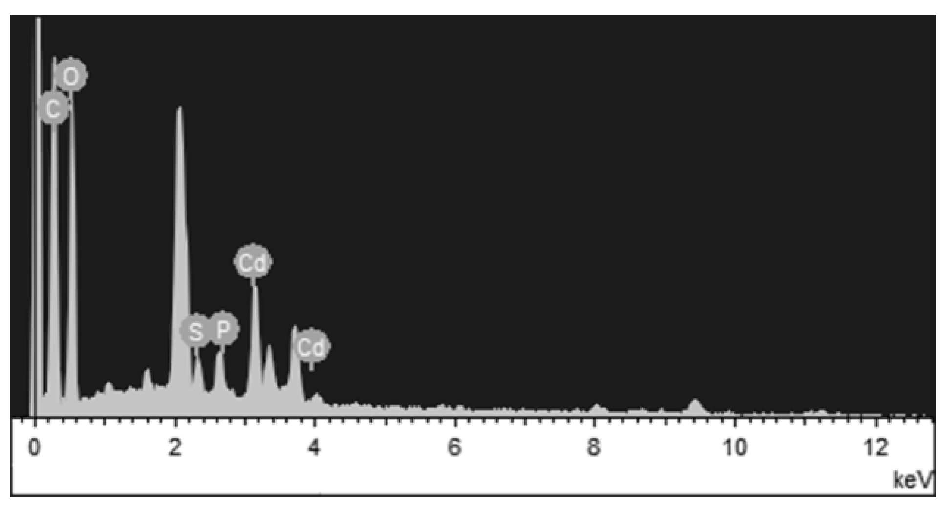
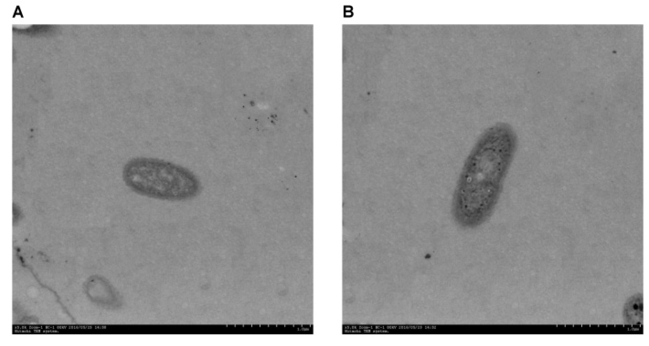
2.5. FTIR, SEM, EDX, and TEM Analysis

The isolated strain was cultured in medium containing 20 mg/L cadmium at 35 °C for 24 h. Ten mL supernatant fluid was collected by centrifugation at 5000 rpm for 15 min and washed four times with phosphate buffer saline solution (pH 7.3). Then, bacterial pellets were freeze-dried for 24 h and analyzed by FTIR spectrometer (Bruker, Karlsruhe, Germany). FTIR spectra were collected in the 400 to 4000 cm−1 region. Bacterial pellets were fixed by 2.5% (v/v) glutaraldehyde for 2 h. After that, bacterial pellets were dehydrated in 25%, 50%, 75%, 90%, and 100% (v/v) ethanol for 20 min each. Then, samples were freeze-dried for 24 h to observe surface morphologies under emission scanning electron microscope (FE-SEM) (JEOL, Tokyo, Japan) at 5.0 kV, field equipped with an energy-dispersive X-ray spectrometer (Oxford, UK). In TEM analysis, bacterial pellets were washed three times with deionized water and fixed by 1% (w/v) osmium tetroxide for 3 h. After fixing, the sample was dehydrated in 50%, 75%, 90%, and 100% (v/v) ethanol for 15 min each. Then, the sample was incubated in the mixture of acetone and epoxy resin (v/v = 2/1 for 3 h, v/v = 1/2 for 12 h) in epoxy resin for 3 h. The sample was centrifugated, embedded in solid resin blocks for 24 h at 60 °C, and then sectioned on an ultramicrotome. Sections were analyzed by transmission electron microscopy (Hitachi, Tokyo, Japan).

2.6. Cadmium Sorption Experiments

Dried biomass was prepared from washed cells, which were autoclaved at 121 °C for 20 min and dried in an oven at 60 °C for 24 h, and then grounded into powder. For the sorption experiments, 2 mL of 5 × 10⁹ cells/mL living biomass (equal to 10 mg) and 2 mL of dried biomass (10 mg) were added into Erlenmeyer flasks containing 50 mL of 50 mg/L cadmium solutions, respectively. Living Escherichia coli DH5α was used as a negative control. The effects of different pH-values (2 to 7) and temperatures (20 to 40 °C) on the adsorption were also studied. The pH was adjusted to 2, 3, 4, 5, 6, and 7 with 1M nitric acid and 1M sodium hydroxide before adding the adsorbent. Erlenmeyer flasks were placed in a thermostat incubator for 48 h. A portion of 20 mL supernatant fluid was collected and centrifuged at 5000 rpm for 15 min for cadmium analysis by ICP Optima 8000 Inductively coupled plasma (PerkinElmer, Waltham, MA, USA) at 228.8 nm. The Cd2+ sorption rate was calculated using Equation (1). Three biological replicates were used in the experimental design.
Sorption rate = C i C f C i × 100 %
where Ci and Cf are the initial and final Cd2+ concentrations (mg/L), respectively.

2.7. Adsorption Kinetic Models

The adsorption kinetic models experiments were conducted by adding the dried biomass (10 mg) into 50 mL of 50 mg/L cadmium solutions and then adsorbing for 48 h at 30 °C. Dried biomass adsorption kinetic models were analyzed by using Lagergren pseudo-first-order (Equation (2)) and pseudo-second-order models (Equation (3)):
1 / Q t = K 1 / ( Q m t ) + 1 / Q m , and
t / Q t = 1 / K 2 Q m 2 + t / Q m
where Q m t is the metal concentrations at each time point, and Q m (mg/g) is the maximum mass. K 1 is kinetic rate constant (/min), and K 2 is pseudo-second kinetic rate constant (g∙mg/min).

2.8. Statistical Analysis

Our data are expressed as mean ± S.D. and use IBM SPSS Statistics 22.0 (IBM Software Inc., New York, NY, USA) to analyze significant differences. The value of p < 0.05 were considered statistically significant.

3. Results

3.1. Metal Contents of Sediment and Screening of Cd2+-Resistant Bacteria

Heavy metal contents in the sediment sample are presented in Table 1. The pH of the river in Hejia station was 8.62 ± 0.16, which is alkaline. Cadmium (2.51 ± 0.21 mg/kg) in sediment samples was 11.61 times higher than the local background value (0.199 mg/kg). Other metal ions were lower than the local background value. The total heterotrophic bacterial (THB) level was 5.6–6.4 × 10⁵ CFU/g. A strain of bacteria showed the highest resistance against Cd2+ (150 mg/L) after increasing Cd2+ concentration from 20 to 200 mg/L, which was dubbed as H5 and selected for further studies.

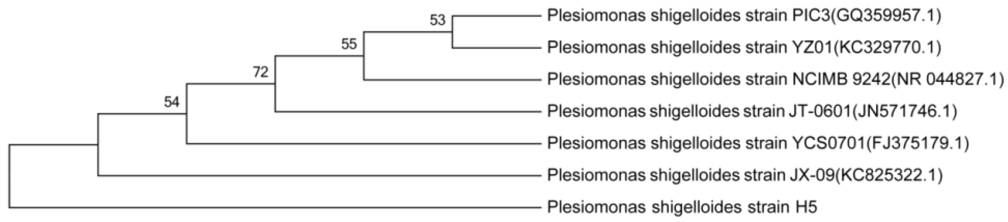
3.2. Characterization and Identification of Cd2+-Resistant Bacteria

Isolated strain H5 was identified by morphology, biochemical characteristics, and the 16SrDNA ribotyping test. Biochemical and morphological characteristics of the strain H5 are summarized in Table 2. The strain H5 exhibited activities of lysine decarboxylase, galactosidase, cytochrome oxidase, arginine dihydrolase, and indol production. It could produce acid from sugars such as glucose and inositol, other than sucrose, mannitol, sorbitol, rhamnose, melibiose, amy-gdalin, and arabinose. It did not show activities of gelatin hydrolase, urease, or tryptophan decarboxylase, nor the production of H2S and Voges–Proskauer. The fractional sequence of the 16SrDNA gene of strain H5 was a 99% identical match with P. shigelloides JT-0601 according to the NCBI Reference Sequence Database. The resultant tree topologies were evaluated by bootstrap analysis of neighbor-joining data sets based on 1000 resamplings, and the phylogenetic tree is shown in Figure 1.

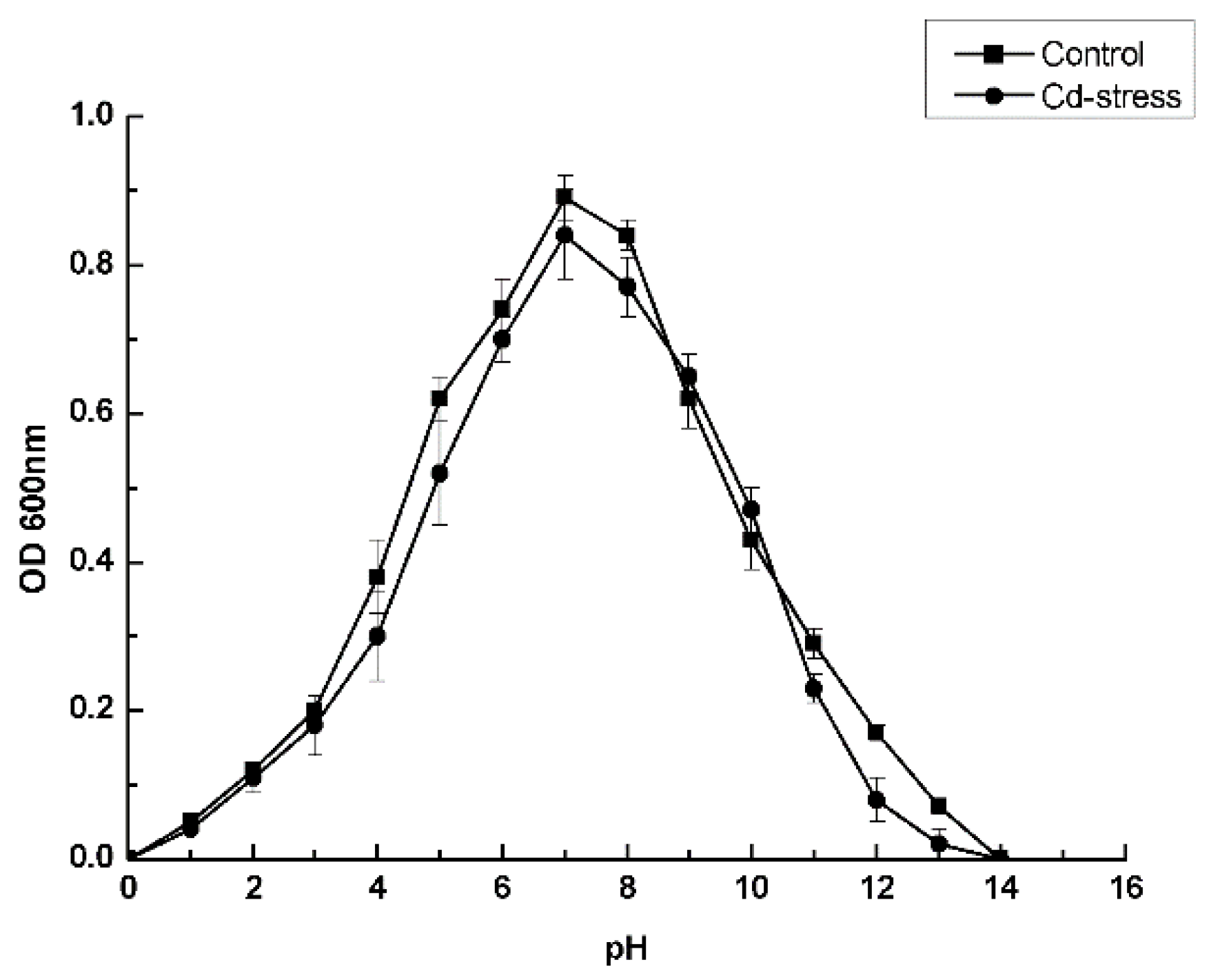
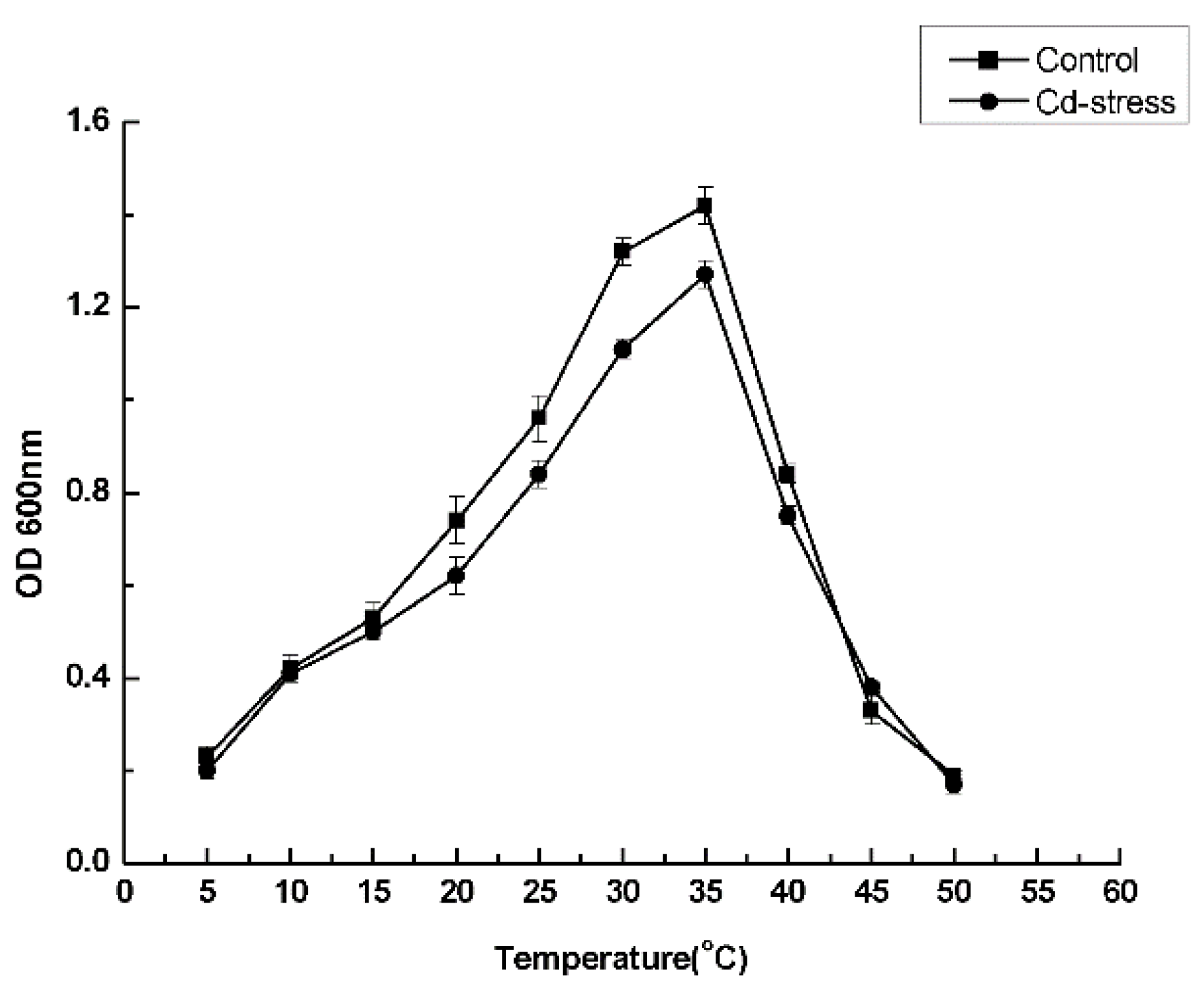
3.3. Optimal Culture Conditions

Beef extract and peptone, as the nitrogen source and carbon sources, were used to determine the optimum growth conditions of strain H5. Optimal culture conditions of strain H5 were pH 7.0 and 35 °C in BEP medium and are presented in Figure 2 and Figure 3.

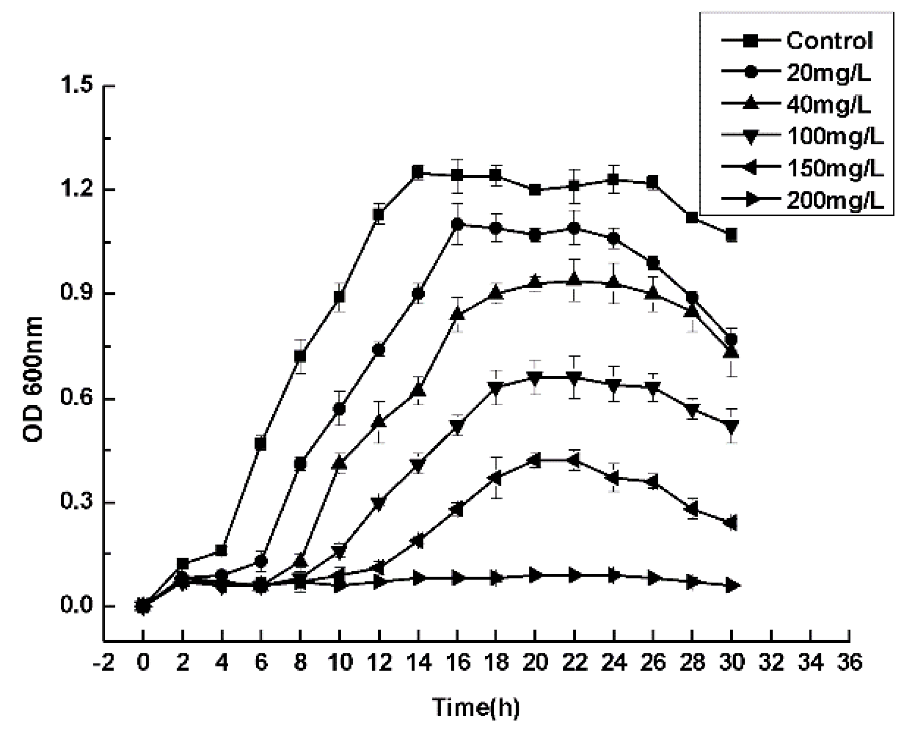
3.4. Maximum Tolerance Concentrations

Compared with the control group, the growth rate of strain H5 decreased gradually with the increase of Cd2+ concentration in the experimental group. Figure 4 shows that higher Cd2+ concentration had an important influence on the growth rate of bacteria. Maximum tolerance concentration of strain H5 is 150 mg/L. In the control group, the bacteria reached the stationary phase at 14 h. However, in the experimental group with Cd2+ stress, the strain H5 reached the stationary phase at 16 to 20 h.

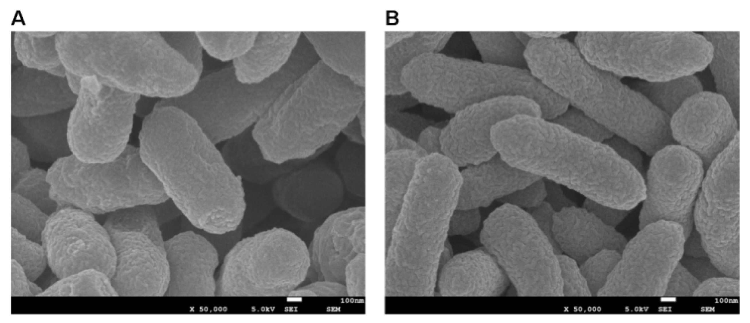
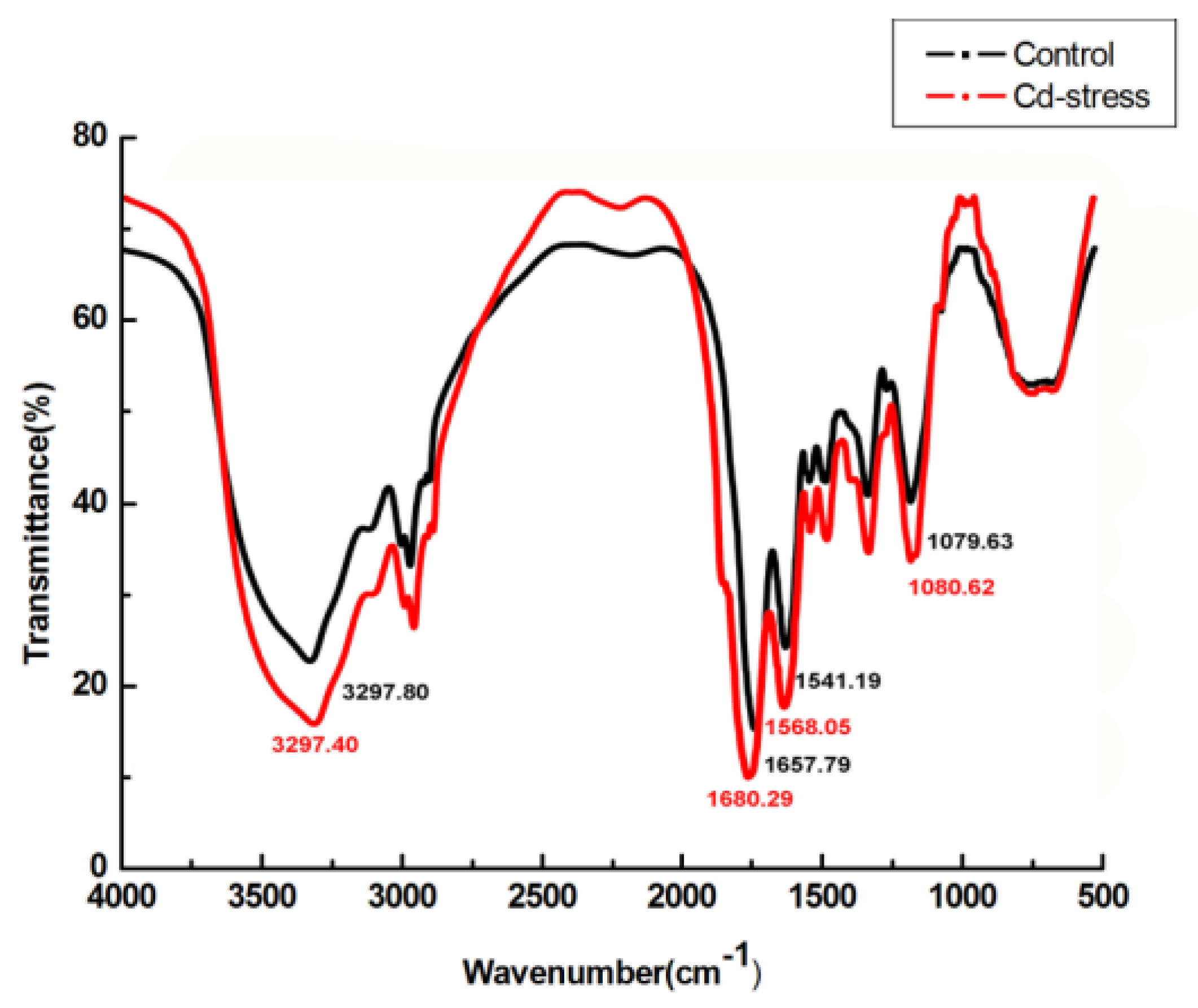
3.5. FTIR, SEM, EDX, and TEM Analysis

In FTIR analysis, characteristic peaks of carboxyl, amino, and phosphate groups were observed, which confirmed the presence of these moieties in strain H5. Figure 5 shows that, in the presence of Cd2+, the amide linkage peaks appearing at 1657.79 and 1541.19 cm−1 were shifted to 1680.29 and 1568.05 cm−1, respectively. In SEM analysis, the surface morphologies of the adsorbed Cd2+ strain H5 was distinct from the wild-type strain H5. Figure 6B shows that the outer membrane of strain H5 after Cd2+ biosorption became rougher, and the volume became bigger than the wild-type strain H5 as shown in Figure 6A. EDX result shows that Cd2+ was along with the surface of bacteria (Figure 7). TEM results show that some precipitates formed in cells after they were cultured in Cd2+ solution (Figure 8B) and is compared with the control group (Figure 8A).

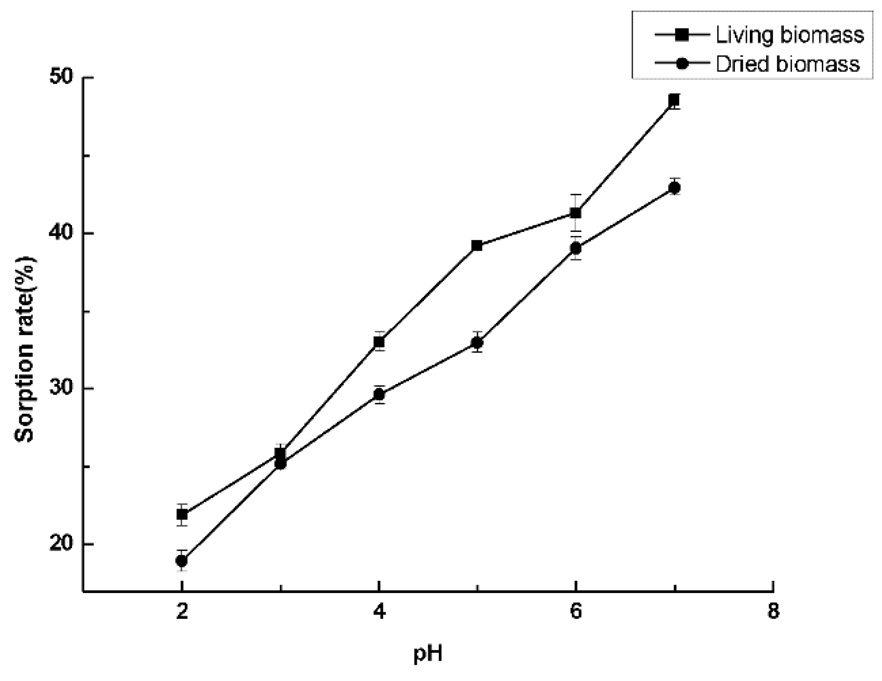
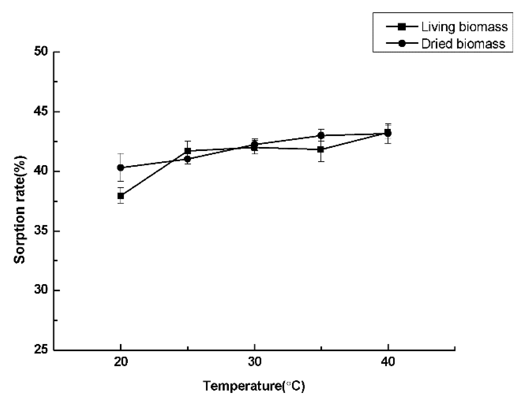
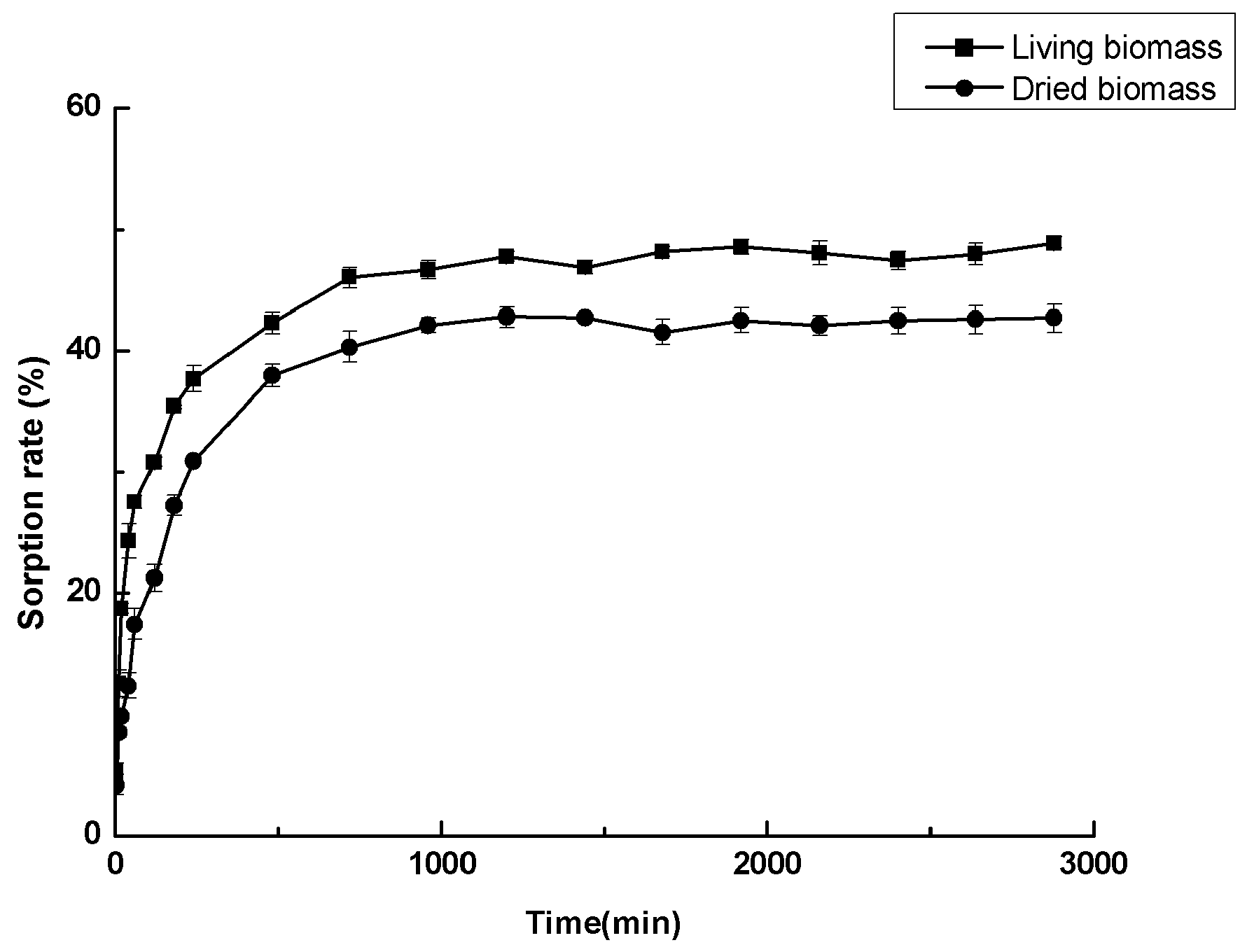
3.6. Cadmium Sorption Experiments

As shown in Figure 9 and Figure 10, pH has a great impact on the sorption rate. With the increase of pH, the sorption rate was obviously improved. Maximum adsorption rate occurred at pH 7. However, temperature has no significant impact on the sorption efficiency. The sorption rate of strain H5 is illustrated in Figure 11. The equilibrium sorption rate of living biomass and dried biomass were 48.91% ± 0.46% and 42.71% ± 0.88%, respectively. Cd2+ was rapidly combined with P. shigelloides H5 during the initial 60 min, and both biomass sorption patterns then reached a stable condition at 480 min. The equilibrium sorption amounts of P. shigelloides H5 were 122.275 ± 1.15 mg/g and 106.775 ± 2.325 mg/g. The maximum Cd2+ sorption rate of E. coli DH5α was 11.42% ± 0.16%. This result indicated that the sorption rate of P. shigelloides H5 was four times that of the control E. coli DH5α (Table 3).

3.7. Adsorption Kinetic Models

The biosorption of Cd2+ by dried biomass was a rapid process and occurred in three periods. During the initial period, the adsorption rate increased rapidly; after 60 min, Cd2+ adsorption began to slow down and then stabilized as it reached equilibrium after 480 min. The fitting results show that the adsorption process of dried biomass was in accordance with Lagergren pseudo-second-order models (Table 4).

4. Discussion

Many indigenous species of microorganisms have the capacity to tolerate heavy metals, for example, Enterococcus faecalis, Escherichia coli, Bosea sp., and Bacillus catenulatus [12,20,21,22,23]. However, mutations can occur in some microorganisms due to the contamination of heavy metals in the environment. These evolved microorganisms have different mechanisms to tolerate or adsorb heavy metal ions, such as active efflux of toxic ions and enzymatic detoxification [24,25,26,27]. In previous studies, scientists discovered that P. shigelloides caused fish and human diseases. Affected fish showed a blackening of body color, a hemorrhage on the surface, and fin rotting, and P. shigelloides can also cause gastroenteritis in human [28,29]. However, no researchers have found that P. shigelloides can adsorb cadmium ions in a polluted environment. In this research, we isolated P. shigelloides from cadmium-polluted river sediment and discovered that it has the ability to adsorb cadmium ions from aqueous solutions. The isolated strain was very similar to P. shigelloides JT-0601, based on the fractional 16SrDNA gene sequences alignment, and the morphological and biochemical characteristics tests. Therefore, isolated strain was designated as Plesiomonas shigelloides H5.
pH and temperature are two important factors in affecting the growth of P. shigelloides H5. Although P. shigelloides H5 was obtained from river sediment having an alkaline pH at 8.62, results showed that its optimum pH is 7.0. Just like other bacteria, the P. shigelloides H5 cannot grow in extreme acidic and alkaline pH in BEP medium. Studying the effect of different temperatures revealed that 35 °C was the optimum temperature for P. shigelloides H5 because, as the temperature rises, the enzymes of bacterium speed up metabolism, but, when the temperature is too low or too high, enzymes are inactivated in the bacterial organism, which finally leads to the stoppage of growth. Furthermore, with the increase in cadmium ion concentration, the bacteria growth rate slows significantly, and OD600 values clearly decrease, which might be due to cadmium ions’ considerably toxic effect on Gram-negative bacteria [30]. The maximum tolerance concentration of P. shigelloides H5 is 150 mg/L. Statistical analysis results are p < 0.05, which indicates that the Cd-stress group is significantly different from the control group. This implies that Cd2+ can affect the growth of bacteria. FTIR results show N–H bending and C–O stretching, which means that the protein amide band II was active in the adsorption of Cd2+. The intensity of the bands varied in different regions after the interaction with Cd2+. These results are similar to the findings by Khan [23]. SEM results revealed that P. shigelloides H5 adapts to high concentrations of cadmium ions in the environment by increasing the volume, which is similar to findings by Wu and Sun [22]. EDX results indicate that the outer membrane of P. shigelloides H5 was involved in the adsorption of heavy metals, as Kim reported [31]. One previous study found that bacteria can transport metal ions into the cells by passive transport or by an outmembrane’s Mn2+/Zn2+/Ca2+ transporter [22]. The intracellular Cd2+ can combine with phosphate to perform precipitate, or Cd2+ toxicity can cause a cadmium efflux pump CzcCBA to efflux Cd2+ in order to reduce damage [23]. In this study, it was found that living biomass can adsorb more cadmium ions than dried biomass and that some precipitates formed in P. shigelloides H5 after being cultured in Cd2+ solution, which indicated that living biomass can absorb Cd2+ in cells, but adsorption amounts were much lower than adsorption amounts (Figure 8 and Figure 11). Therefore, we will focus on the absorption mechanism and precipitate constituents in future research. In the experiment of the pH effect, the maximum sorption rate occurred at pH 7, which indicates that, with the decrease in hydrion, living biomass and dried biomass can effectively remove cadmium ions in solutions. The statistical analysis result was p = 0.009 (p < 0.01), which indicated that living biomass is extremely different from dried biomass under different pH-values. As for the TEM results, it is hypothesized that living biomass can absorb partial Cd2+ into the cells and cause a difference. This experiment did not study biological sorption in alkaline solutions because cadmium ions and hydroxyl ions can form a cadmium hydroxide precipitant in solutions. Therefore, an aqueous solution should be adjusted in the neutral pH to improve sorption efficiency in future practical applications. However, the temperature has a slight effect on the sorption efficiency experiment, which can be applied to actual pollution control in the future. There are no significant differences between both sorption patterns (p = 0.328). We consider sometimes that the adsorption capacity of living biomass may be weak in affecting the whole entire process. On the basis of the plot in Figure 11 and of Table 4, both biomass sorption patterns can reach a stable condition at 480 min. There are extremely significant differences between both biomass patterns (p = 0.000). Dried biomass was in accordance with Lagergren pseudo-second-order models. The sorption process was composed of three periods. The first period was attributed to the large number of vacant active sites on the dried biomass surface, to electrostatic adherence cadmium ions. The second period was attributed to the fact that the adsorption rate gradually decreases as vacant active sites decrease. The third period is the final equilibrium in which the vacant active sites were all combined with cadmium ions. Similar results are also reported in a previous study [21]. Furthermore, the sorption rate of P. shigelloides H5 was four times that of the control E. coli DH5α, which can verify its effectiveness.

5. Conclusions

To sum up, we have successfully isolated the cadmium-resistant strain Plesiomonas shigelloides H5, which can adsorb cadmium ions in solution. The surface functional groups of P. shigelloides H5 can participate in the adsorption of cadmium ions from aqueous solutions. Adsorption kinetic models of dried biomass were in accordance with Lagergren pseudo-second-order models, and equilibrium adsorption amounts was 106.775 ± 2.325 mg/g. Dried biomass can also achieve adsorption saturation in a short time. A preliminary study found that P. shigelloides H5 can absorb a small amount of Cd2+ into cells to form precipitates. The present study indicated that P. shigelloides H5 can be applied in wastewater treatment in the future.

Acknowledgments

This study was financially supported by the Major Science and Technology Program for Water Pollution Control and Treatment (No. 2013ZX07201007).

Author Contributions

Chao Xue conceived and designed the experiments, performed the analysis, and analyzed the data. Mengsha Li and Yunzhi Liu provided experimental guidance. All authors (Chao Xue, Peishi Qi, Mengsha Li and Yunzhi Liu) contributed to the writing of the paper.

Conflicts of Interest

The authors declared no conflict of interest.

References

  1. Sanità di Toppi, L.; Gabbrielli, R. Response to cadmium in higher plants. Environ. Exp. Bot. 1999, 41, 105–130. [Google Scholar] [CrossRef]
  2. Radotić, K.; Dučić, T.; Mutavdžić, D. Changes in peroxidase activity and isoenzymes in spruce needles after exposure to different concentrations of cadmium. Environ. Exp. Bot. 2000, 44, 105–113. [Google Scholar] [CrossRef]
  3. Zhao, L.; You, W.; Hu, H.; Hong, W.; Liao, X.; Xiao, S.; Wang, R.; Cai, J.; Fan, X.; Tan, Y. Spatial distribution of heavy metals (Cu, Pb, Zn, and Cd) in sediments of a coastal wetlands in eastern Fujian, China. J. For. Res. 2015, 26, 703–710. [Google Scholar] [CrossRef]
  4. AMAP Assessment 2011: Mercury in the Arctic; Arctic Monitoring and Assessment Programme: Oslo, Norway, 2011.
  5. Filipič, M.; Fatur, T.; Vudrag, M. Molecular mechanisms of cadmium induced mutagenicity. Hum. Exp. Toxicol. 2006, 25, 67–77. [Google Scholar] [CrossRef] [PubMed]
  6. Benavides, M.P.; Gallego, S.M.; Tomaro, M.L. Cadmium toxicity in plants. Braz. J. Plant Physiol. 2005, 17, 21–34. [Google Scholar] [CrossRef]
  7. Flora, S.J.S.; Megha, M.; Ashish, M. Heavy metal induced oxidative stress & its possible reversal by chelation therapy. Indian J. Med. Res. 2008, 128, 501–523. [Google Scholar] [PubMed]
  8. Bai, H.; Zhang, Z.; Yang, G.; Li, B. Bioremediation of cadmium by growing rhodobacter sphaeroides: Kinetic characteristic and mechanism studies. Bioresour. Technol. 2008, 99, 7716–7722. [Google Scholar] [CrossRef] [PubMed]
  9. Halttunen, T.; Salminen, S.; Tahvonen, R. Rapid removal of lead and cadmium from water by specific lactic acid bacteria. Int. J. Food Microb. 2007, 114, 30–35. [Google Scholar] [CrossRef] [PubMed]
  10. Roane, T.M.; Pepper, I.L. Microbial responses to environmentally toxic cadmium. Microb. Ecol. 1999, 38, 358–364. [Google Scholar] [CrossRef] [PubMed]
  11. Abbas, S.Z.; Rafatullah, M.; Ismail, N.; Lalung, J. Isolation, identification, characterization, and evaluation of cadmium removal capacity of enterobacter species. J. Basic Microb. 2014, 54, 1279–1287. [Google Scholar] [CrossRef] [PubMed]
  12. Neethu, C.S.; Mujeeb Rahiman, K.M.; Saramma, A.V.; Mohamed Hatha, A.A. Heavy-metal resistance in Gram-negative bacteria isolated from Kongsfjord, Arctic. Can. J. Microb. 2015, 61, 429–435. [Google Scholar] [CrossRef] [PubMed]
  13. Pérez-Rama, M.; Torres, E.; Suárez, C.; Herrero, C.; Abalde, J. Sorption isotherm studies of Cd(II) ions using living cells of the marine microalga Tetraselmis suecica (Kylin) Butch. J. Environ. Manag. 2010, 91, 2045–2050. [Google Scholar] [CrossRef] [PubMed]
  14. Yang, Y.; Xie, Y.; Li, X. Characterization of biological iron sulfide composites and its application in the treatment of cadmium-contaminated wastewater. J. Environ. Biol. 2015, 36, 393–398. [Google Scholar] [PubMed]
  15. Vullo, D.L.; Ceretti, H.M.; Daniel, M.A.; Ramírez, S.A.M.; Zalts, A. Cadmium, zinc and copper biosorption mediated by Pseudomonas veronii 2E. Bioresour. Technol. 2008, 99, 5574–5581. [Google Scholar] [CrossRef] [PubMed]
  16. Kılıç, N.K.; Dönmez, G. Environmental conditions affecting exopolysaccharide production by Pseudomonas aeruginosa, Micrococcus sp., and Ochrobactrum sp. J. Hazard. Mater. 2008, 154, 1019–1024. [Google Scholar] [CrossRef] [PubMed]
  17. Harrison, J.J.; Ceri, H.; Stremick, C.; Turner, R.J. Differences in biofilm and planktonic cell mediated reduction of metalloid oxyanions. FEMS Microbiol. Lett. 2004, 235, 357–362. [Google Scholar] [CrossRef] [PubMed]
  18. Coral, G.; Arιkan, B.; Coral, M.N.Ü. A preliminary study on tellurite resistance in Pseudomonas spp. isolated from hospital sewage. Pol. J. Environ. Stud. 2006, 15, 517–520. [Google Scholar]
  19. Dewhirst, F.E.; Chien, C.C.; Paster, B.J.; Ericson, R.L.; Orcutt, R.P.; Schauer, D.B.; Fox, J.G. Phylogeny of the defined murine microbiota: Altered schaedler flora. Appl. Environ. Microbiol. 1999, 65, 3287–3292. [Google Scholar] [PubMed]
  20. Xie, X.H.; Jin, F.; Wang, H.P.; Liu, J.S. Heavy metal resistance by two bacteria strains isolated from a copper mine tailing in China. Afr. J. Biotechnol. 2010, 9, 4056–4066. [Google Scholar]
  21. Zhang, H.; Liu, L.; Chang, Q.; Wang, H.; Yang, K. Biosorption of Cr(VI) ions from aqueous solutions by a newly isolated Bosea sp. strain Zer-1 from soil samples of a refuse processing plant. Can. J. Microbiol. 2015, 61, 399–408. [Google Scholar] [CrossRef] [PubMed]
  22. Wu, G.; Sun, M.; Liu, P.; Zhang, X.; Yu, Z.; Zheng, Z.; Chen, Y.; Li, X. Enterococcus faecalis strain LZ-11 isolated from Lanzhou reach of the Yellow River is able to resist and absorb Cadmium. J. Appl. Microbiol. 2014, 116, 1172–1180. [Google Scholar] [CrossRef] [PubMed]
  23. Khan, Z.; Nisar, M.A.; Hussain, S.Z.; Arshad, M.N.; Rehman, A. Cadmium resistance mechanism in Escherichia coli P4 and its potential use to bioremediate environmental cadmium. Appl. Microbiol. Biotechnol. 2015, 99, 10745–10757. [Google Scholar] [CrossRef] [PubMed]
  24. Silver, S.; Le, T.P. A bacterial view of the periodic table: Genes and proteins for toxic inorganic ions. J. Ind. Microbiol. Biotechnol. 2005, 32, 587–605. [Google Scholar] [CrossRef] [PubMed]
  25. Nies, D.H. Efflux-mediated heavy metal resistance in prokaryotes. FEMS Microbiol. Rev. 2003, 27, 313–339. [Google Scholar] [CrossRef]
  26. Malin Mejare, L.B. Metal-binding proteins and peptides in bioremediation and phytoremediation of heavy metals. TRENDS Biotechnol. 2001, 19, 67–73. [Google Scholar] [CrossRef]
  27. Piddock, L.J.V. Multidrug-resistance efflux pumps? Not just for resistance. Nat. Rev. Microbiol. 2006, 4, 629–636. [Google Scholar] [CrossRef] [PubMed]
  28. Matsuyama, R.; Kuninaga, N.; Morimoto, T.; Shibano, T.; Sudo, A.; Sudo, K.; Asano, M.; Suzuki, M.; Asai, T. Isolation and antimicrobial susceptibility of Plesiomonas shigelloides from great cormorants (Phalacrocorax carbo hanedae) in Gifu and Shiga Prefectures, Japan. J. Vet. Med. Sci. 2015, 77, 1179–1181. [Google Scholar] [CrossRef] [PubMed]
  29. Novoa-Farias, O.; Frati-Munari, A.C.; Peredo, M.A.; Flores-Juarez, S.; Novoa-Garcia, O.; Galicia-Tapia, J.; Romero-Carpio, C.E. Susceptibility of bacteria isolated from acute gastrointestinal infections to rifaximin and other antimicrobial agents in Mexico. Rev. Gastroenterol. Mex. 2015, 91, 3–10. [Google Scholar] [CrossRef]
  30. Tsuno, T.; Mejido, J.; Schmeisser, H.; Fey, S.; Morrow, A.; Nie, H.; Zhao, T.; Bekisz, J.; Goldman, N.; Zoon, K. Isolation and characterization of heavy-metal resistant microbes from roadside soil and phylloplane. Cotton Sci. 2012, 52, 53–65. [Google Scholar]
  31. Kim, S.Y.; Jin, M.R.; Chung, C.H.; Yun, Y.S.; Jahng, K.Y.; Yu, K.Y. Biosorption of cationic basic dye and cadmium by the novel biosorbent Bacillus catenulatus JB-022 strain(environmental biotechnology). J. Biosci. Bioeng. 2015, 119, 433–439. [Google Scholar] [CrossRef] [PubMed]
Figure 1. Phylogenetic tree for 16SrDNA gene sequences of P. shigelloides H5.
Water 08 00241 g001
Figure 2. The optimum pH of P. shigelloides H5 with and without Cd2+ stress. (p = 0.007).
Water 08 00241 g002
Figure 3. The optimum temperature of P. shigelloides H5 with and without Cd2+ stress. (p = 0.016).
Water 08 00241 g003
Figure 4. Maximum tolerance concentrations test of P. shigelloides H5. (p = 0.000).
Water 08 00241 g004
Figure 5. FTIR image of P. shigelloides H5 with and without Cd2+ stress.
Water 08 00241 g005
Figure 6. SEM images of P. shigelloides H5 without Cd2+ stress (A) and with Cd2+ stress (B).
Water 08 00241 g006
Figure 7. EDX image of P. shigelloides H5 with Cd2+ stress.
Water 08 00241 g007
Figure 8. TEM images of P. shigelloides H5 without absorbing Cd2+ (A) and after absorbing Cd2+ (B).
Water 08 00241 g008
Figure 9. pH effect of both biomass sorption patterns. (p = 0.009).
Water 08 00241 g009
Figure 10. Temperature effect of both biomass sorption patterns. (p = 0.048).
Water 08 00241 g010
Figure 11. The equilibrium sorption curve of both biomass sorption patterns. (p = 0.000).
Water 08 00241 g011
Table 1. Metal concentration (mg/kg) in sampling spots of the study area.
ParametersValue
Latitude45°75’47” N
Longitude126°57’57” E
sampling spots number6
Water temperature (°C)4
Water pH8.62 ± 0.16
Cd2.51 ± 0.21
Ni7.64 ± 0.14
Cu6.14 ± 0.54
Cr14.46 ± 0.32
Pb18.32 ± 1.21
Zn26.86 ± 1.84
THB level (CFU/g)5.6–6.4 × 105
Note: THB: total heterotrophic bacteria.
Table 2. Morphological and biochemical characteristics of P. shigelloides H5.
StrainH5
Morphology
Colony colorYellowish
Gram nature
Cell morphologyStraight rod
Motility+
Colony shapeRound
ElevationRaised
SurfaceSmooth
OpticalOpaque
Biochemical tests
Lysine decarboxylase+
Urease
Arginine dihydrolase+
Indol production+
O-nitrophenyl d-galactoside+
H2S production
Gelatin hydrolysis
Citrate
Inositol+
Glucose+
Sucrose
Mannitol
Sorbitol
Rhamnose
Melibiose
Amy-gdalin
Arabinose
Tryptophan
Voges–Proskauer
Cytochrome oxidase+
Notes: −: negative; +: positive.
Table 3. The sorption ability of P. shigelloides H5 compared to E. coli DH5α.
Living BiomassSorption Rate (%)Sorption Amounts (mg/g)
P. shigelloides H548.91 ± 0.46122.275 ± 1.15
E. coli DH5α11.42 ± 0.1628.55 ± 0.41
Table 4. Simulation of sorption kinetic equations and corresponding parameters.
ModelsK1 or K2QmR2
Lagergren pseudo-first-order0.00170.13920.7577
Lagergren pseudo-second-order0.0003109.8910.9992

